
Study the Effect of the Magnetic Field on the Healing of Bone Fracture after Implant Avian Bone in Femoral Bone in Rabbits


Research Article
Study the Effect of the Magnetic Field on the Healing of Bone Fracture after Implant Avian Bone in Femoral Bone in Rabbits
Mohammed M. Jassim, Mohammed R. Abduljaleel*, Zainab B. Abdulkareem, Noor H. Sanad, Ibrahim M.H. Alrashid
Department of Surgery and Obstetrics, College of Veterinary Medicine, University of Basrah, Basrah, Iraq.
Abstract | Although promotional impacts upon the healing of the bone of the static electro-magnetic fields (PEMF) were well demonstrated, static magnetic fields (SMF) effects had stayed unclear. The effects of a specially constructed magnetic wrap on the histological and radiographic features of bone healing using a femoral rabbit model with an unstable osteotomy space and an implant have been investigated in the current work. After avian bone implantation and mid shaft femorus osteotomy, bone healing has been evaluated within a 22-days-period in control rabbits (n = 5) and the rabbits that have been exposed to the SMF (300 gauss) (n = 5). The healing of the bones has been assessed through the quantitative and qualitative evaluation of the serial radiographs each couple of weeks. The histopathological study has been carried out as well on the osteotomized femor upon the completion of experimental time. The radiographic recovery of the osteotomy sites was significantly improved in the rabbits that had been subjected to the SMF. When comparing to the controls, the SMF group’s cellular morphology ratings were significantly higher (P 0.050). These findings showed that using the osteotomy gap model with SMF enhanced the histological and radiographic features of rabbit bone healing. Rabbits that were under the risk of the delayed fracture healing could take advantage of treatments with the SMF.
Keywords | Rabbits, Bone fracture, Avian implantation
Received | August 30, 2023; Accepted | October 08, 2023; Published | November 06, 23
*Correspondence | Mohammed R. Abduljaleel, Department of Surgery and Obstetrics, College of Veterinary Medicine, University of Basrah, Basrah, Iraq; Email: [email protected]
Citation | Jassim MM, Abduljaleel MR, Abdulkareem ZB, Sanad NH, Alrashid IMH (2023). Study the effect of the magnetic field on the healing of bone fracture after implant avian bone in femoral bone in rabbits. Adv. Anim. Vet. Sci., 11(11):1779-1784.
DOI | https://dx.doi.org/10.17582/journal.aavs/2023/11.11.1779.1784
ISSN (Online) | 2307-8316
Copyright: 2023 by the authors. Licensee ResearchersLinks Ltd, England, UK.
This article is an open access article distributed under the terms and conditions of the Creative Commons Attribution (CC BY) license (https://creativecommons.org/licenses/by/4.0/).
INTRODUCTION
The longest bone in the body and the only one within the thigh is the femur. It may be separated into three segments, proximal, shaft, and distal, and is thought to be the location of origin and attachment for various ligaments and muscles (Boese et al., 2016). The femur shaft descends in slight medial direction. Which results in bringing knees closer to the center of gravity of the body’s, which increases the stability. A shaft cross section in the middle is circular, however, it is posteriorly flattened at distal and proximal aspects. On femoral shaft’s posterior surface, there are roughened bone ridges, which have been referred to as linea aspera (which is Latin for “rough line”). This distally splits in order to form lateral and medial supracondylar lines. Flat popliteal surface is between them (Lazaro et al., 2015).
Bio-magnetics can be defined as inter-disciplinary area, where the biology, magnetism, and medicine are overlapping. Although shepherd magnes first documented the magnetic properties of lodestones around 1000 BCE in what is now Turkey, the use of electro-magnetic fields in therapeutic arts dates back to the 15th century (Ueno et al., 2002; Steyn et al., 2000). The weak magnetic fields are utilized for treating parkinsonism and motor complications of the chronic levodopa therapy (Sandyk et al., 1992). Hazardous effects that are related to the SMFs on the human health became noticeable. Newer researches are necessary for filling in gaps in our knowledge and give an assurance of the fact that new medical technologies won’t lead to causing unwanted health risks (Lezczynski, 2005). The action mechanism whereby those effects could be attained is still elusive, however, one of the hypotheses is that there’s a blood flow increase in the areas that are affected by magnetic field (Steyn et al., 2000). For instance, the use of moderately intense SMF (1mT-1T) in magnetic field therapy may be advantageous for the circulatory diseases such as inflammations, ischemia pains, and hypertension, mostly due to the modification of blood pressure and/or blood flow. In prior studies, it was demonstrated that sub-chronic exposure to the SMF (180 mT) over a period of 3 to 12 weeks sped up the bones’ recovery from the ischemia and surgical invasion that the artery ligation had caused (Xu et al., 2001, 2007). The field of implants and devices encompasses a number of classes, including biomaterials (synthetic or normal non-drug substance(s) used as a whole or as a component of system for the purpose of therapy, augmentation, or substitution of any tissue, function, or organ), combination products (such as anti-microbial coatings for devices), and tissue-engineered constructs (Boretos and Eden,1984).
The implants have a general positive influence on the human health, the patients could have implant-associated complications (which are modeled typically in rabbits and rodents). Pains, immunologic reactions/ hypersensitivity, anatomical malalignments, osteolytic loss of bone, instability/breakage from the implanted, wear debris reactions, and heterotopic ossification might all be adverse implant reactions. More than 100000 implant-related infection and osteomyelitis cases have been reported (Vigorita et al., 2008).
Thyroid stimulating hormone (TSH), which is released by the pituitary, controls the thyroid’s function. Triiodothyronine (T3) and thyroxin (T4), a hormone active in at least 20 enzyme systems and one of whose major effects is increasing protein synthesis, are produced by the thyroid when TSH levels are elevated (Bortkiewicz, 2001).
Study on biochemical changes in liver and kidney exposed to electrical magnetic field was also done. Most of the studies applied magnetic field stimulation or exposure for a shorter period of time (Zare et al., 2007; Sallam and Awad, 2008).
MATERIALS AND METHODS
In the current investigation, fifteen adult rabbits were used. They were randomly separated into three groups: the control group, the group that received a magnetic field treatment, and the group that received no magnetic field treatment. Gaps were formed in the second and third groups, and an avian bone implant was used.
The femoral muscles were separated from the femoral bone by blind dissection, and (0.5X0.25) cm (Figures 1–8) were formed. All of the rabbit was kept in the same circumstances. They were all sedated with ketamine (13 mg/kg bw) and xylazine (5 mg/kg bw) (Aydin and Bezer, 2011), and then implant the piece of avian bone in same size.
Cut the avian femoral bone, preparing a portion of avian bone, placing it in a conical solution of bicarbonate of sodium and sodium chloride, and using a magnetic starrier for an hour are shown in Figure 9 (Jensen, 2007).
After the first day after surgery, 300 gauss of magnetic treatment was started every day for 15 days.
After surgery, blood from a cardiac puncture was collected and examined, and radiographic images were obtained after 21 days. Histopathological pictures were archives after 22 days of surgery (Luna,1968).
Magnetic field therapy pmf has been estimated based on this equation:
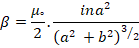
ß=represents magnetic dosage (flux), μ=12.57x10⁻⁷Weber/amp.m, a represents turn radius, i represents current through turn, b represents axial distance from the perpendicular to turn plan Figure 10 (Sears,1977).
RESULTS and Discussion
Clinical finding
Clinically there are many signs was seen after surgical operation such as pain, swelling in the site of operation, and lameness.
All groups showed pain and swelling at the site of operation and began disappear gradually after 10 days in control group and after 7 days in (SMF) groups. Lameness continuous for many days after operation and started disappears after two weeks in the treated groups and after three weeks in control group.
Radiographic results
To evaluate bone fracture healing must be using X-ray (Radiographic examination).
All groups were examined at the third week post operation; the control group demonstrates mild periosteal reaction near the fracture gaps and mild bone formation (Figure 13).
T2 group also showed mild to moderate periosteal reaction with callus bone formation in the site of fracture gap (Figure 12).
T3 group showed best result radiologically the callus formation across the fracture gaps and try to make bridge between the two fragments with shown in an invisible fracture line (Figure 11).
Blood parameters
Blood parameters have been recorded WBCs, RBCs, and HB of animal groups, which had been listed in Table 1.
There aren’t any reports discussing roles of the magnetic fields comprehensively, suggesting this mechanism that simulated magnetic field mechanisms in the fractured bone, in experiment the magnetic field either pulsed or static has been directly exposed to dorsal view of the bones and achilles tendon (fibula and tibia), (Alrashid et al., 2009). Five factors play main role in magneto therapy, animal species, body organs, magnetic field tension, frequency, and exposure period. The present study includes all the above parameters except frequency. Animals species differ from type to other. In the present study used rabbits with three levels of magnetic field tensions 300 gauss. The best results showed with 300 gauss, and, all applications are with period at 15 minutes per day/daily for 15 days, the healing showed at 21 days post operation, other study used 40 gauss per 1hr per day/twice daily, the healing showed after 23 days post operation in mid tibial osteotomy in rabbits (Fredricks et al., 2003; Inoue et al., 1999). Therefore, some animals like dog responses to magneto therapy faster than other animals like rabbits. The causes that effect to faster healing related to bone itself directly and other organs that effect on bone fracture healing indirectly. The health or recovery of an organ with a deficiency depends on its function or vitality. The bone non vitality rather organ compared with other organ like glands, heart, and skin. In present study the bone healing in minimum period 21-23 days post operation and maximum period 33-35 days post operation. This agrees with (Harlandand and Liburday, 1997; Alfano et al., 2001). The idea that bone has an organ life similar to other organs The treated subgroups recover more quickly than the control group, which could be the result of a number of factors, including the magnetic field’s direct impact on microorganisms and its indirect effects on immune function and wound healing through increased vascularization and promotion of fibrillation to strengthen the connections between wound edges (Salzberg, 1995; Patino, 1996). Nevertheless, the differences among groups are related to magnetic field tension, this suggestion agreement with Some cases erupt yellow spots at application of magnetic field, these yellow spots are related to thermal effect of magnetic field (Philips, 2007).
Acknowledgement
We want to thank head of department of surgery and obstetric, Dean of Veterinary Medicine and Basrah university’s president for their support in college laboratory.
Novelty Statement
The novelty of our work entitled( study the effect of the magnetic field on the healing of bone fracture after implant avian bone in femoral bone in rabbits) despite of many cases of fractures but still treatment challenging in veterinary medicine and few researcher highlight on the using of magnetic field to acceleration of bone fracture healing.
Author’s Contribution
Mohammed M. Jassim: Contributed to the bone preparation, surgery operation and monitor post operation period.
Mohammed R. Abduljaleel: Contributed to the surgery operation. Analysis and interpretation of data, as well as writing and revision of the manuscript.
Zainab B. Abdulkareem: Contributed to x-ray image take
Noor H. Sanad: Prepare the surgical site.
Ibrahim MH. Alrashid: Contributed to the bone preparation, surgery operation and give magnetic therapy to the animals.
Conflict of interest
The authors have declared no conflict of interest.
References
Alfano AP, Taylar AG, Foresman PA (2001). Static magnetic field for treatment of fibromyalgia: A randomized controlled trial. J. Altern. Complement. Med., 7(1): 53-55. https://doi.org/10.1089/107555301300004538
Alrashid IMH, Alfaris AA, Emshary CA (2009). The effect of static magnetic field of mid-shaft femoral fractures healing in Rabbits. J. B Sci., 35(1): 21.
Aydin N, Bezer M (2011). The effect of an intramedullary implant with a static magnetic field on the healing of the osteotomised rabbit femur. Int. Orthop., 35(1): 135-141. https://doi.org/10.1007/s00264-009-0932-9
Boese CK, Dargel J, Oppermann J, Eysel P, Scheyerer MJ, Bredow J, Lechler P (2016). The femoral neck-shaft angle on plain radiographs: A systematic review. Skeletal Radiol., 45(1): 19-28. https://doi.org/10.1007/s00256-015-2236-z
Boretos JW, Eden M (1984). Contemporary biomaterials. Material and Host Response, Clinical Applications, New Technology and Legal Aspects. Park Ridge, NJ: Noyes Publications. https://doi.org/10.1115/1.3138526
Bortkiewicz A (2001). A study on the biological effects of exposure mobile-phone frequency EMF. Med. Pr., 52: 101–106.
Salzberg CA (1995). The effect on non-thermal pulsed electromagnetic energy on wound of pressure ulcers in spinal cord –injured patients. A randomized double-blind study. Osteotomy Wound Mange., 41(3): 42-51.
Fredricks DC, Diehle DJ, Abbott J, Nepola JV (2003). Effect of pulsed electromagnetic field stimulation on distraction osteogensis in the rabbits tibial leg lengthening model. Bone healing research laboratory, University of Iowa College of Medicine. J. Pediatr. Orthol., 23(4): 230-233. https://doi.org/10.1097/01241398-200307000-00012
Harland JD, Liburday RD (1997). Environmental magnetic field inhibit the antiproliferaton action of Tamoxifen and melatonin in human breast cell line. Life science division, Lawrence BerkelyNat-Lab. University of California. Bioelectromagnetics, 18: 555-556. https://doi.org/10.1002/(SICI)1521-186X(1997)18:8<555::AID-BEM4>3.0.CO;2-1
Philips JL (2007). Topical review of magnetic field hyperthermia. International, pp. 587-605.
Lazaro LE, Klinger CE, Sculco PK, Helfet DL, Lorich DG (2015). The terminal branches of the medial femoral circumflex artery: the arterial supply of the femoral head. Bone Joint J. 97-B(9): 1204-1213. https://doi.org/10.1302/0301-620X.97B9.34704
Leszczynski D (2005). Rapporteur report: Cellular, animal and epidemiological studies of the effects of static magnetic fields relevant to human health. Prog. Biophys. Mol. Biol., 87: 247-253. https://doi.org/10.1016/j.pbiomolbio.2004.08.014
Luna,L.G.(1968). Manual of histologic staining methods of Armed forces institute of pathology. (pp. xii-258).
Inoue N, Ohnishi I, Chen D, Deitz L, Schwardt J, Chao EY (1999). The effect of pulsed electromagnetic field on long bone belayed fracture union in canine model. Department of Orthopedic Surgery, John’ Hopkins University. 45th Annual Meeting, Orthopedic Research Society. Anaheim, California. pp. 44-50.
Patino O (1996). Pulsed electromagnetic field in experimental cutaneous wound healing in rats. J. Born Care Rehabilit., pp. 528-531. https://doi.org/10.1097/00004630-199611000-00009
Jensen SS, Yeo A, Dard M, Hunziker E, Schenk R, Buser D (2007). Evaluation of a novel biphasic calcium phosphate in standardized bone defects. A histologic and histomorphometric study in the mandibles of minipigs. Clin. Oral Implants Res., 18(6): 752–760. https://doi.org/10.1111/j.1600-0501.2007.01417.x
Xu S, Tomita N, Ikeuchi K, Ikada Y (2007). Recovery of small-sized blood vessels in ischemic bone under static magnetic field. Evid. Based Complement. Altern. Med., 4(1): 59–63. https://doi.org/10.1093/ecam/nel055
Xu S, Tomita N, Ohata R, Yan Q, Ikada Y (2001). Static magnetic field effects on bone formation of rats with an ischemic bone model. Bio-Med. Mater. Eng., 11(3): 257–263.
Sallam SM, Awad AM (2008). Effect of static magnetic field on the electrical properties and enzymes function of rat liver. Roman. J. Biophy Bucharest, 18(4): 337–347.
Sandyk R, Anninos PA, Tsagas N, Derpapas K (1992). Magnetic fields in the treatment of Parkinson’s disease. Int. J. Neurosci., 63: 141-150. https://doi.org/10.3109/00207459208986664
Sears FW (1977). Electricity and magnetism. Dep. of physics. Dartmouth college, Addison-Wesley publishing co. Amsterdam.
Steyn PE, Ramey DW, Kirschvink J, Uhrig J (2000). Effect of a static magnetic field on blood flow to the metacarpus in horses. J. Am.Vet. Med. Assoc., 217: 874-877. https://doi.org/10.2460/javma.2000.217.874
Ueno S, Funamizu H, Ogiue-Ikeda M (2002). Magnetic stimulation and control of neuronal regeneration and cell growth. Proceedings of the 2nd international workshop on biological effects of electromagnetic fields. 7-11 October, Aldemar Paradise Royal Mare Hotel, Rhodes, Greece. pp. 987-989.
Vigorita VJ, Ghelman B, Mintz D (2008). Orthopaedic pathology. Philadelphia, PA: Lippincott Williams & Wilkins.
Zare S, Alivandi S, Ebadi AG (2007). Histological studies of the low frequency electromagnetic fields effect on liver, testes and kidney in Guinea pig. World Appl. Sci., 2(5): 509–511.
To share on other social networks, click on any share button. What are these?

