Study the Effect of the Magnetic Field on the Healing of Bone Fracture after Implant Avian Bone in Femoral Bone in Rabbits
Study the Effect of the Magnetic Field on the Healing of Bone Fracture after Implant Avian Bone in Femoral Bone in Rabbits
Mohammed M. Jassim, Mohammed R. Abduljaleel*, Zainab B. Abdulkareem, Noor H. Sanad, Ibrahim M.H. Alrashid
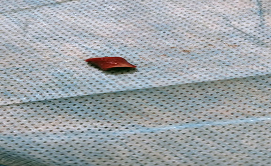
Avian bone.
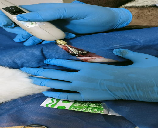
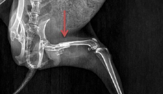
Site of gap fracture.
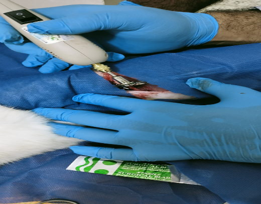
Use drill to made gap in femoral bone.
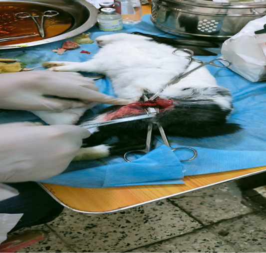
Site of femoral bone.
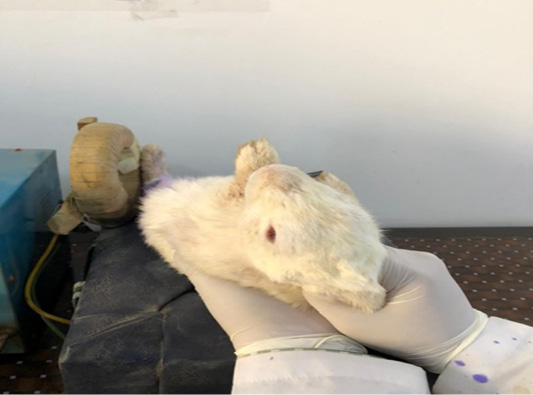
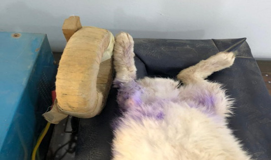
Correct position of magnetic therapy.
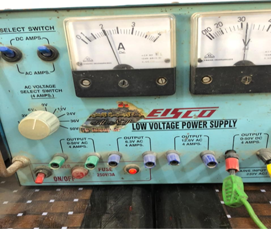
Power supply of magnetic therapy system.
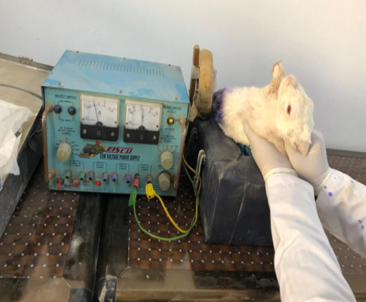
Power supply, magnetic coil, rabbit patient bed.
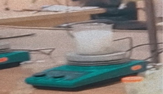
Magnetic starrier.
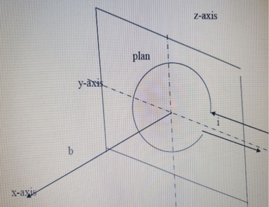
x=axis, i= current, plan (x, y, z,) magnetic coil plan, b represents axial distance perpendicular to the turn plan.
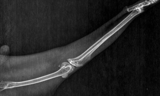
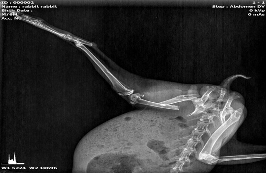
T1 group after 21 day there are complete fracture healing.
T2 group after 21 day there are semi complete fracture healing.
C group after 21 day there are nonunion fracture.
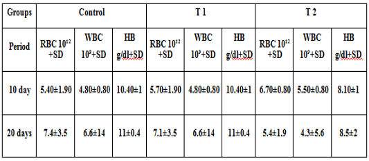
Blood picture after 21 days.


