
Taxonomic Data of New Recorded Plant Parasitic and Soil Nematodes from Different Vegetation of Thar Desert, Sindh Pakistan

Taxonomic Data of New Recorded Plant Parasitic and Soil Nematodes from Different Vegetation of Thar Desert, Sindh Pakistan
Ramzan Ali1, Erum Iqbal1*, Muhammad Ismail Bhatti2 and Saboohi Raza1
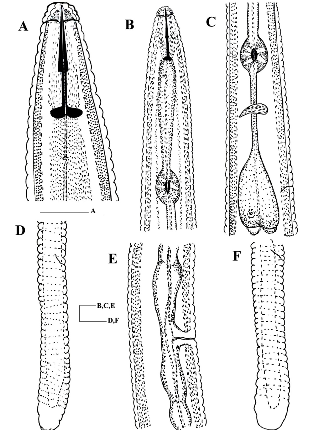
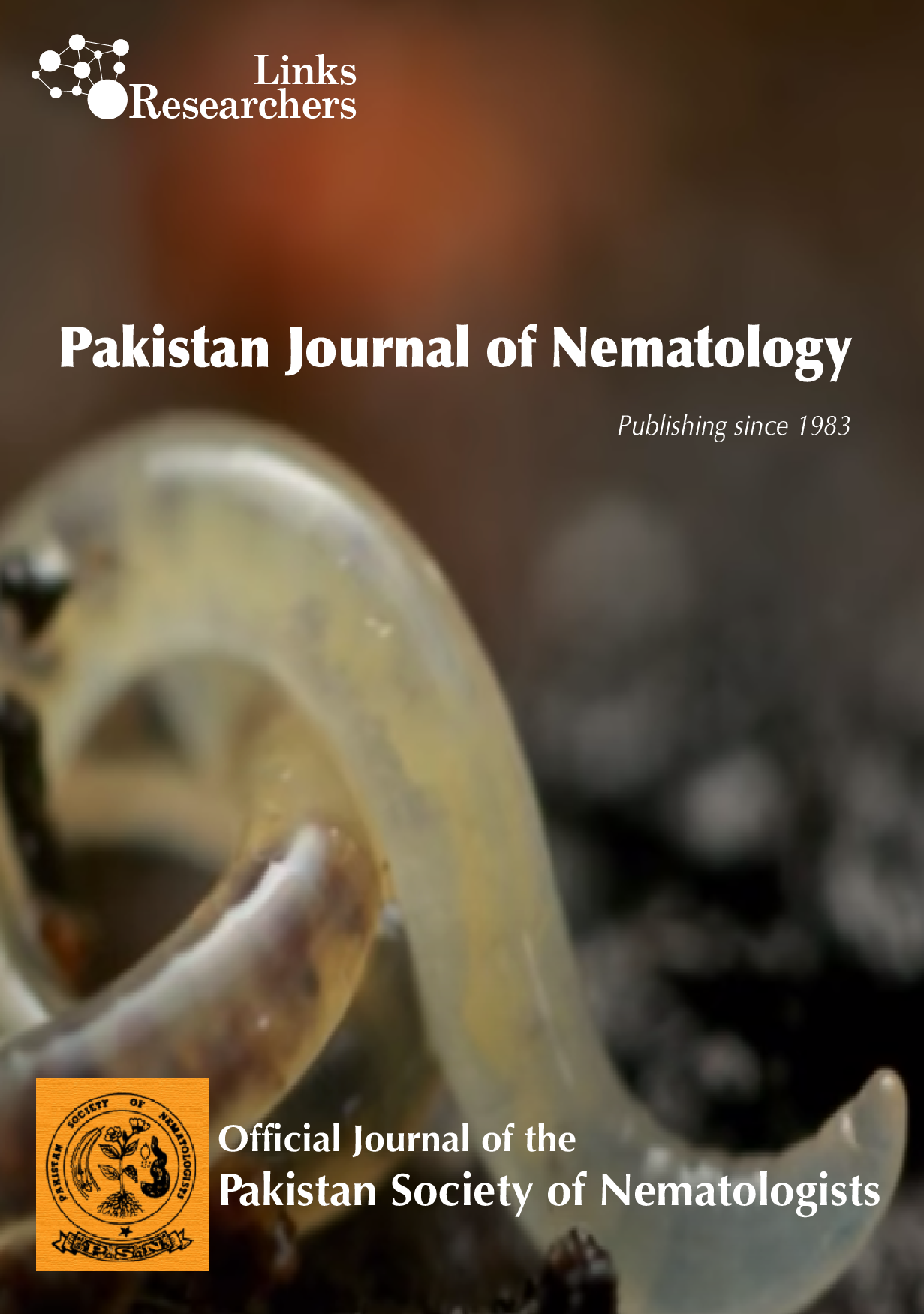
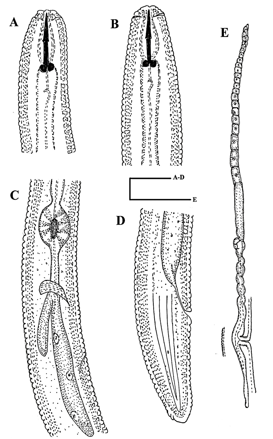
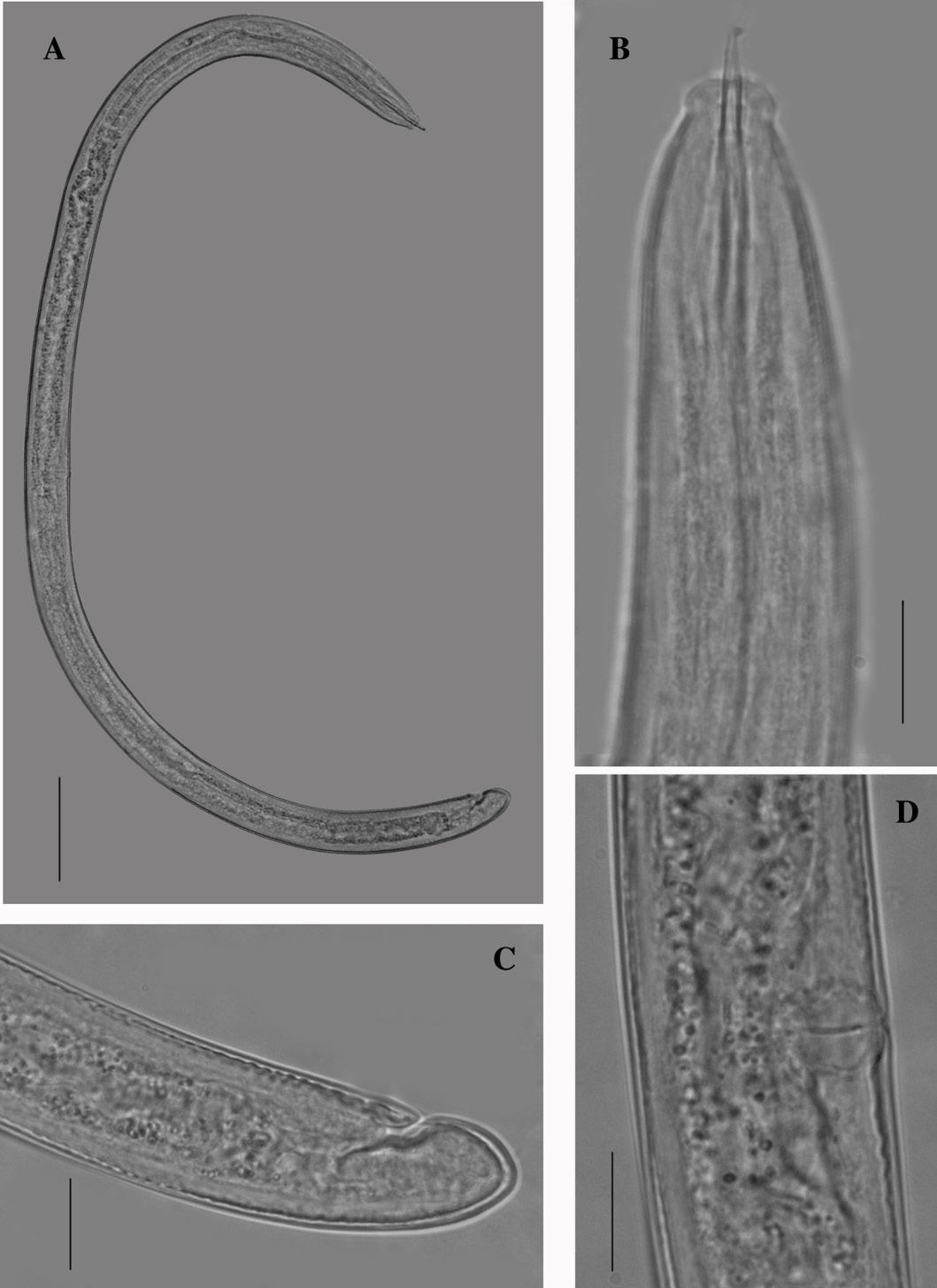
Tylenchorhynchus neoclavicaudatus (Mathur et al., 1978). A: Head (50µm); B: Anterior end (20µm); C: Oesophageal part (20µm); D, F: Female tail (20µm), E: Vulva (20µm).

Tylenchorhynchus neoclavicaudatus (Mathur et al., 1978). A and B: Anterior end (50µm); C: Median bulb and Oesophageal bulb (20µm); D and F: Female tail (20µm); E: Vulva (20µm).

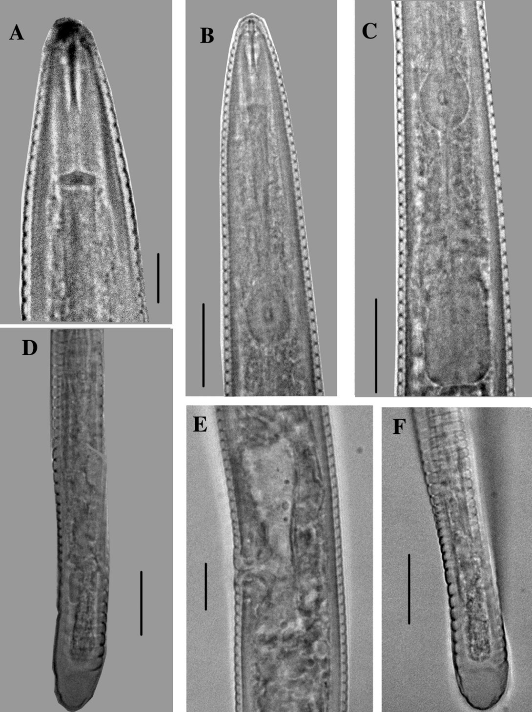
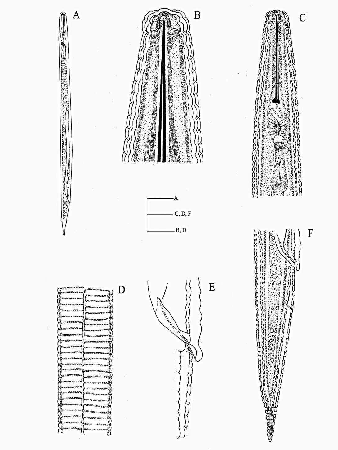
Tylenchorhynchus mashhoodi (Siddiqi and Basir, 1958). A: Whole body (100µm); B: Anterior end (20µm); C: Vulva (20µm); D: Male tail (20µm); E: Female tail (20µm).
Tylenchorhynchus mashhoodi (Siddiqi and Basir, 1958). A: Whole body (100µm); B: Anterior end (20µm); C: Vagina (20µm); D: Male tail (20µm); E: Female tail (20µm).

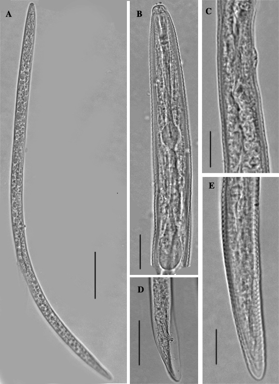
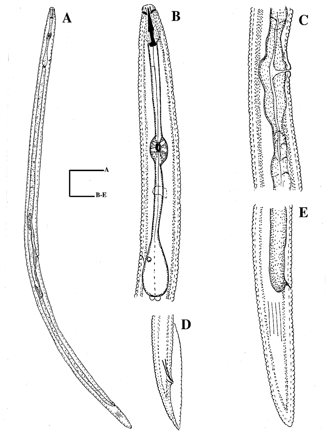
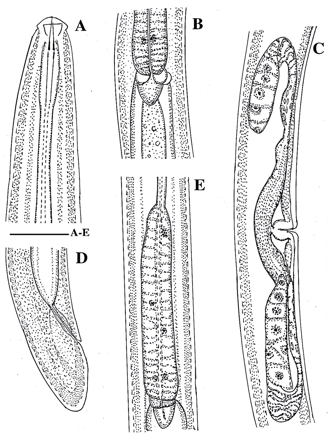
Pratylenchus curvicauda Siddiqi et al. (1991). A: Whole body (100µm); B: Anterior end (20µm); C: Head and median bulb (20µm); D: Vulva and post uterine sac (20µm); E: Female tail (20µm).
Pratylenchus curvicauda Siddiqi et al. (1991). A and B: Anterior end (20µm); C: Median bulb and oeophageal glands (20µm); D: Whole body(100µm); E: Post uterine sac(20µm); F: Female tail (20µm).

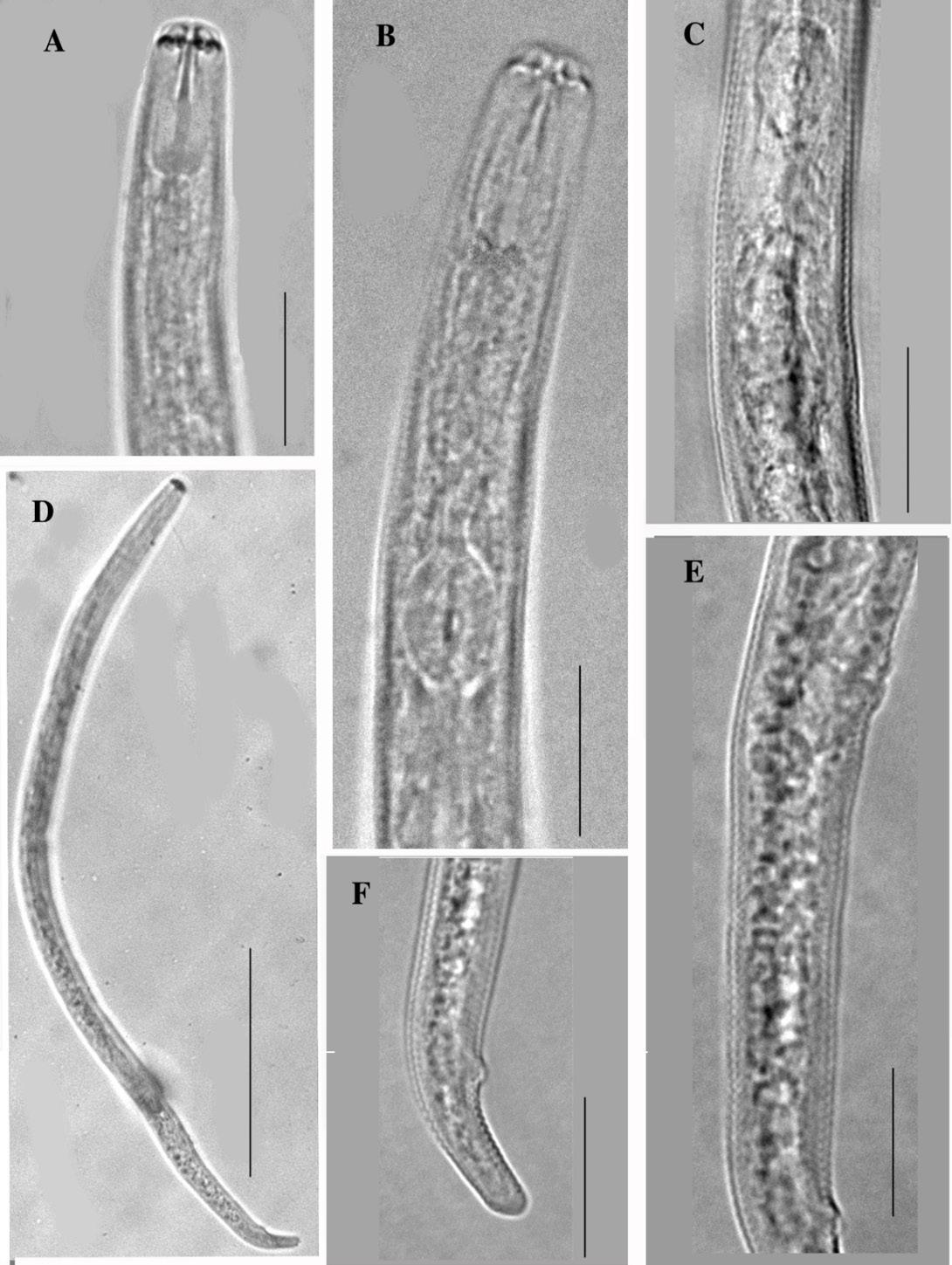
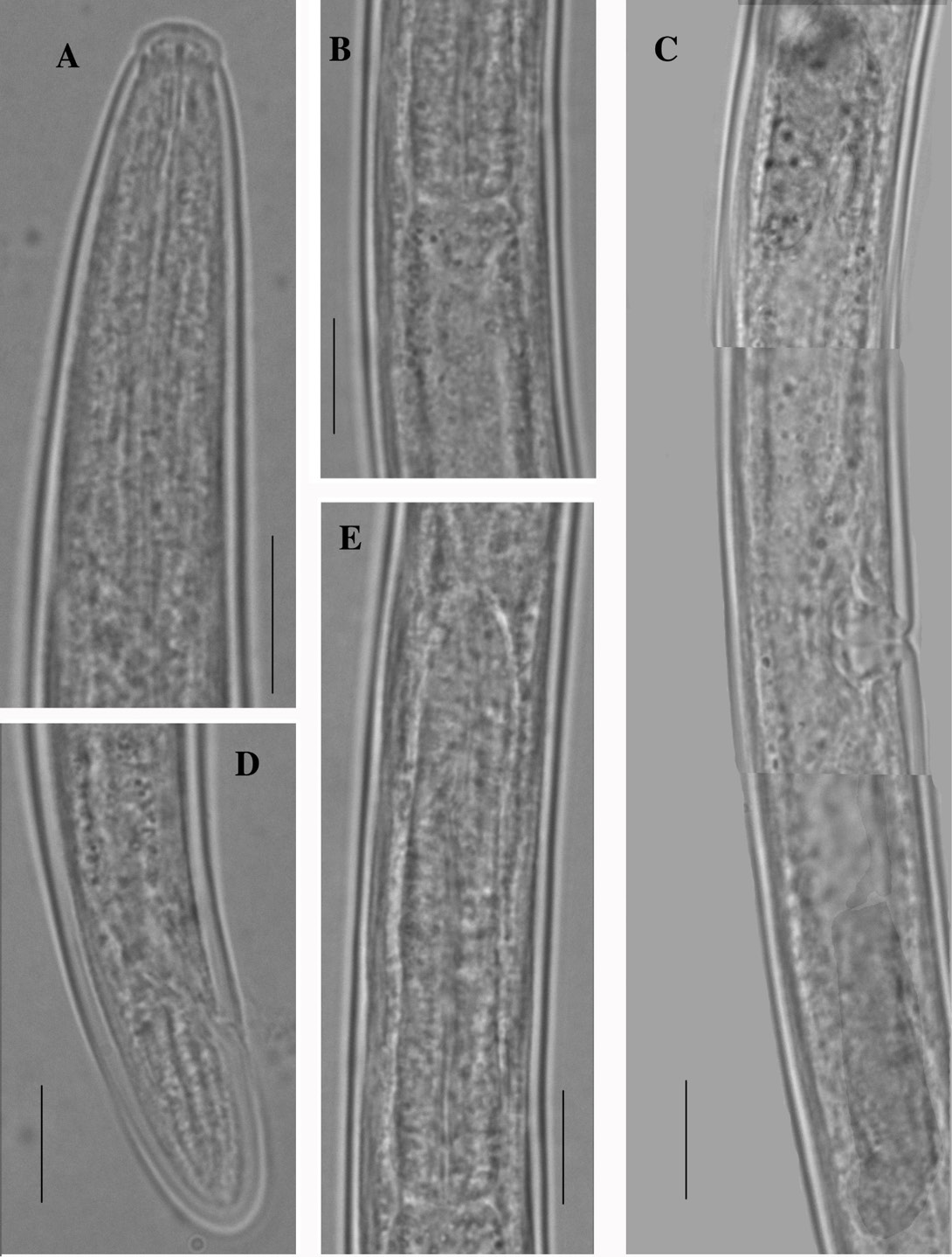
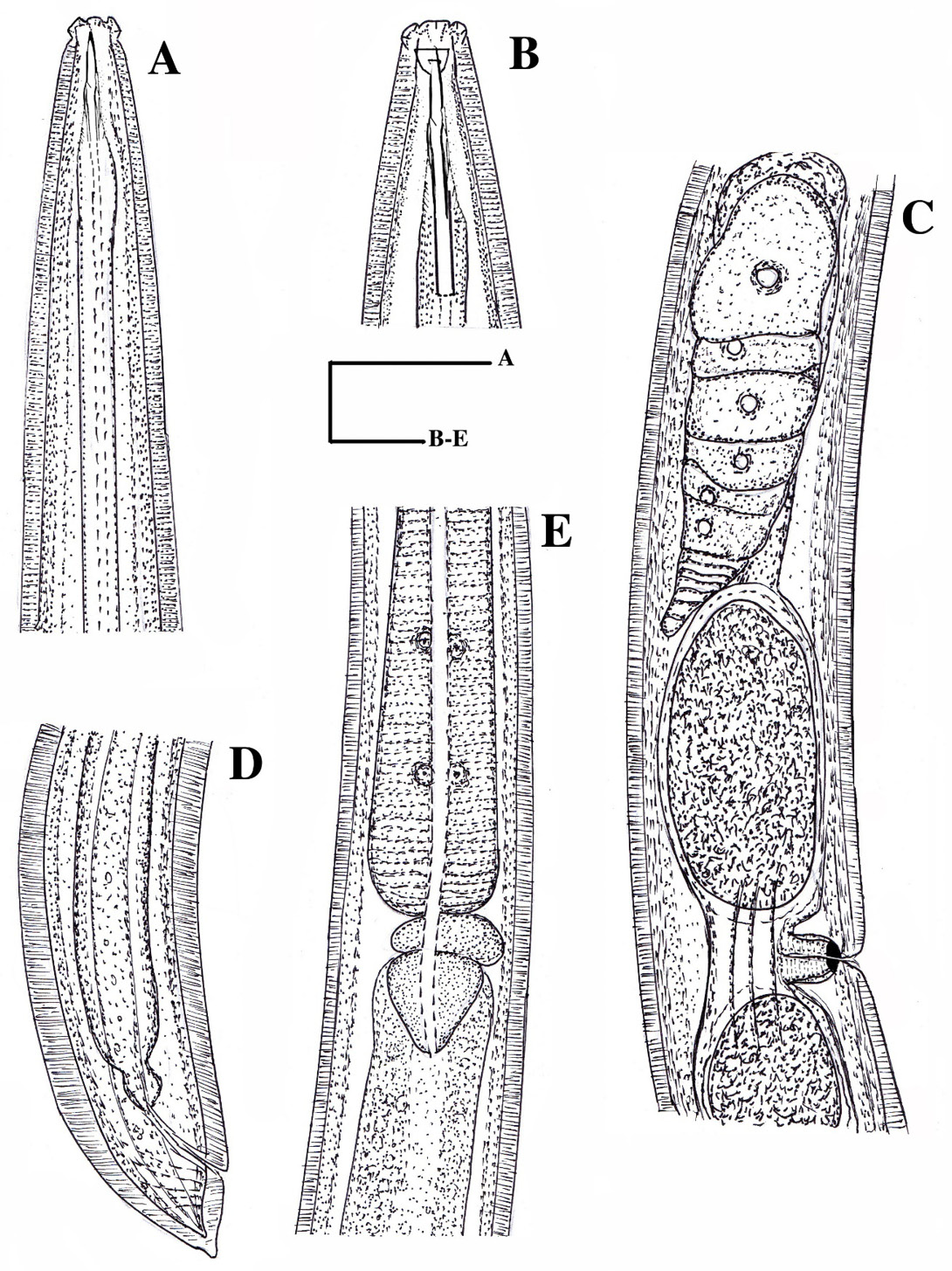
Pratylenchus elamini Zeidan and Geraert (1991). A and B: Anterior end (20µm); C: Oesophageal part (20µm); D: Female tail (20µm); E: Reproductive system(10µm).
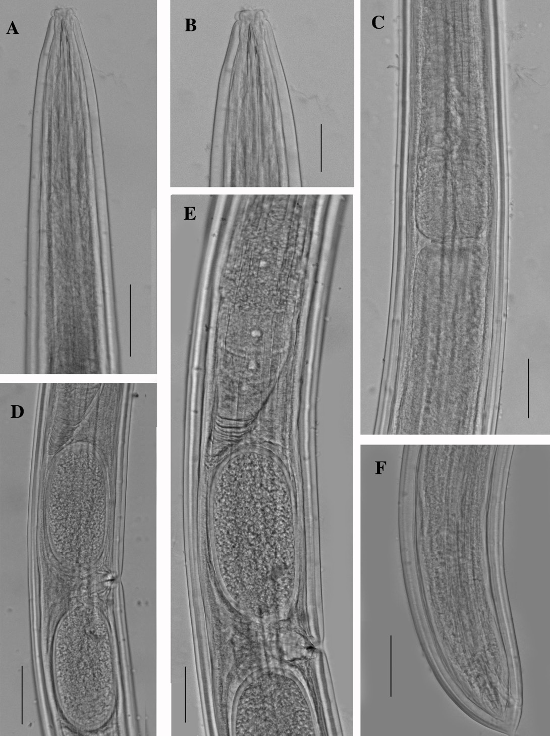
Pratylenchus elamini Zeidan and Geraert (1991). A and B: Anterior end (20µm); C: Vulva and post uterine sac (20µm); D: Oesophageal gland(20µm); E: Female tail (20µm). F and G: Hemicyclophora punensis Darekar and Khan (1981). F: Anterior end (50µm); G: Female tail (50µm).

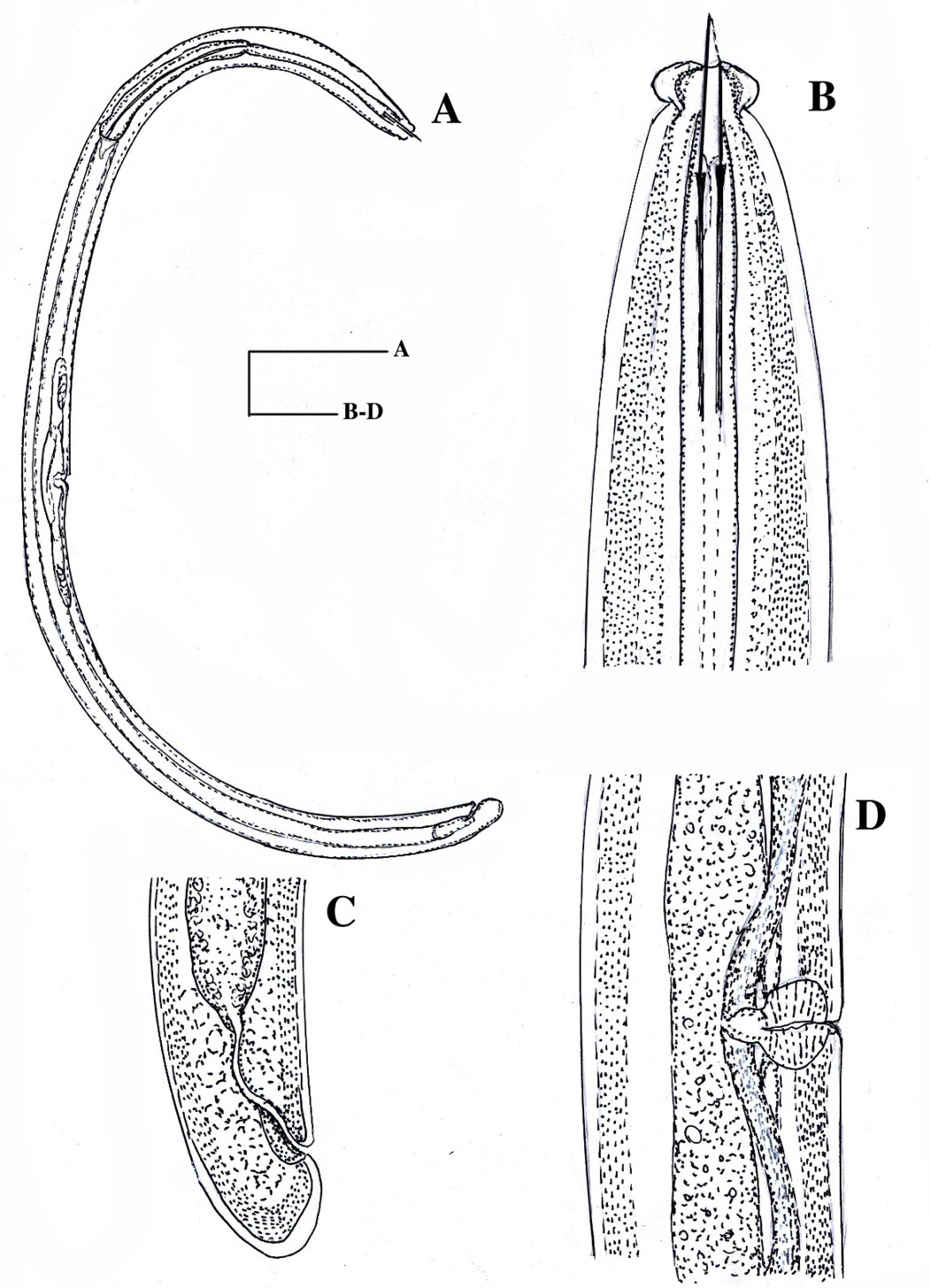
Hemicyclophora punensis Darekar and Khan (1981). A: Whole body (100µm); B (40µm) and C: Anterior end (20µm); D: Lateral field (20µm); E: Anus (40µm); F: Female tail (20µm).

Tylencholaimus shamimi Islam and Ahmed (2021). A: Anterior end (20µm); B: Cardia (20µm); C: Reproductive system (20µm); D: Female tail (20µm); E: Oesophagus posterior expension (20µm).
Tylencholaimus shamimi (Islam and Ahmed, 2021). A: Anterior end (20µm); B: Cardia(20µm); C: Reproductive system(20µm); D: Female tail(20µm); E: Oesophagus posterior expension (20µm).
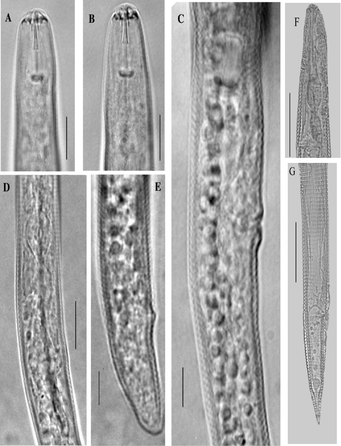
Makatinus punctatus Heyns (1965). A (30µm) and B: Anterior end (20µm); C: Vulva and anterior ovary (20µm); D: Female tail (20µm); E: Cardia (20µm).
Makatinus punctatus Heyns (1965). A (30µm); and B: Anterior end (20µm); C: Oespphageal part (20µm); D: Vulva (20µm); E: Anterior ovary (20µm); F: Female tail (20µm).
Labronema mangalorense Ahmad and Ahmad (2002). A: Whole body (50µm); B: Anterior end (20µm); C: Female tail (20µm); D: Vulva (20µm).

Labronema mangalorense Ahmad and Ahmad (2002). A: Whole body (100µm); B: Anterior end (20µm); C: Female tail (20µm); D: Vulva (20µm).

