

Skeletal Ontogeny and Anomalies in Larval and Juvenile Crimson Snapper, Lutjanus erythropterus Bloch, 1790


Skeletal Ontogeny and Anomalies in Larval and Juvenile Crimson Snapper, Lutjanus erythropterus Bloch, 1790
Dachuan Cheng1,2, Md Mahbubul Hassan3, Zhenhua Ma1, 2, 3,*, Qibin Yang1 and Jian G. Qin3
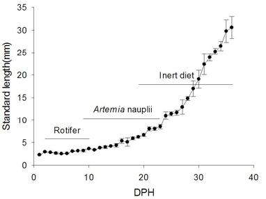
Standard length of crimson snapper reared under different feeding protocol from 1 to 36 day post hatch (DPH). Feeding protocol was presented above the growth curve.

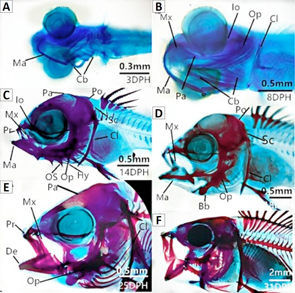
Formation and ossification of the skull in larval crimson snapper. A, Larvae on 3 DPH show developing mandible (Ma) and ceratobranchial (Cb); B, Larvae on 8 DPH show developing maxillary (Mx), cleithrum (Cl), operculum (Op), parietal (Pa), infraorbital (Io) and cartilaginous mandible and ceratobranchial; C, Larvae on 14 DPH show developing premaxillary (Pr), posttemporal (Po), supracleithrum (Sc), hyoid (Hy), opercular spine (OS) and cartilaginous maxillary, parietal, infraobital, cleithrum; D, Larvae on 18 DPH show cartilaginous basibranchial and ossification of mandible, operculum, cleithrum; E, Larvae on 25 DPH show ossification of dentary, maxillary, parietal; F, Larvae on 31 DPH show ossification of neurocranium.
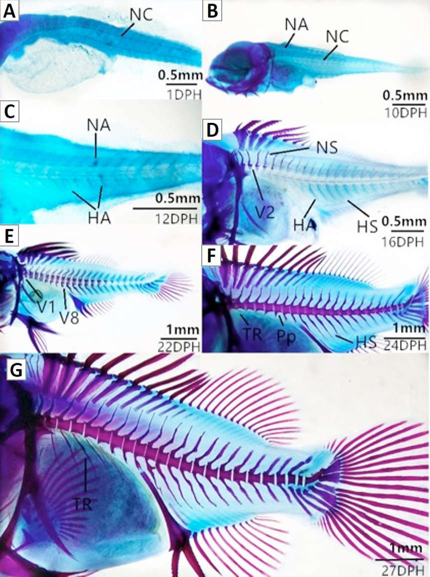
Formation and ossification of vertebral column in larval crimson snapper. A, Larvae on 1 DPH show developing notochord (NC); B, Larvae on 10 DPH show developing neural arch (NA); C, Larvae on 12 DPH show developing haemal arch (HA); D, Larvae on 16 DPH show developing neural spine, vertebral centra, 1st vertebra to 5th vertebra (V1 to V5) and haemal spine (HS); E, Larvae on 22 DPH show cartilaginous of vertebra centra, (V1 to V16). F) Larvae on 24 DPH show developing parapophysis (Pp), thoracic rib (TR) and ossification of vertebral centra, haemal spine; F, Larvae on 27 DPH show ossification of vertebral column.

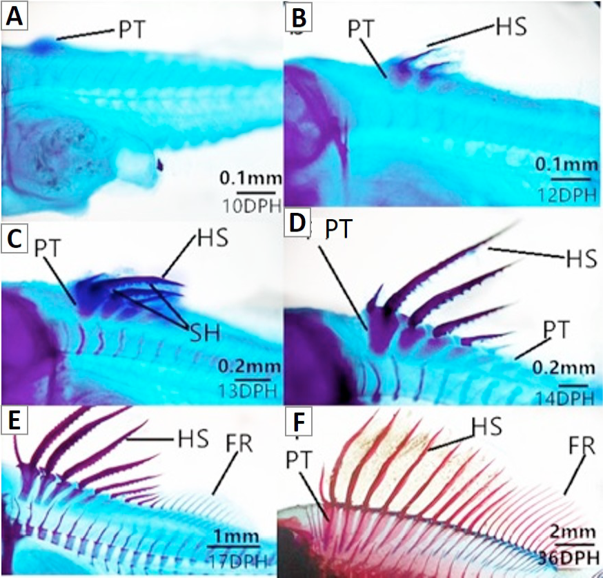
Formation and ossification of dorsal fins in larval crimson snapper. A and B, Larvae on 10 DPH show developing dorsal pterygiophores (PT); C, Larvae on 12 DPH show developing hard spine (HS) and cartilaginous of pterygiophores; D, Larvae on 13 DPH show developing sharp hooks (SH); E, Larvae on 14 DPH show first five hard spine and cartilaginous 6th spine; F, Larvae on 17 DPH show ossification of fin rays and pterygiphores.

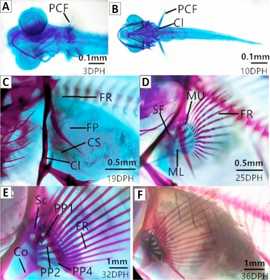
Formation and ossification of pectoral fins in larval crimson snapper. A, Larvae on 3 DPH show developing pectoral fins (PCF); B, Larvae on 10 DPH show pectoral fins and cleithrum; C, Larvae on 19 DPH show developing fin rays (FR), fin plate (FP), coracoid-scapula (CS) and ossification of cleithrum; D, Larvae on 25 DPH show developing scapular foramen (SF), metacleithrum lower (ML), metacleithrum upper (MU) and ossification of fin rays; E, Larvae on 32 DPH show cartilaginous of coracoid (Co), scapula (Sc), proximal pterygiophore (PP), 1st proximal pterygiophore to 4th proximal pterygiophore (PP1-PP4); F, Larvae on 36 DPH show ossification of pectoral fins.
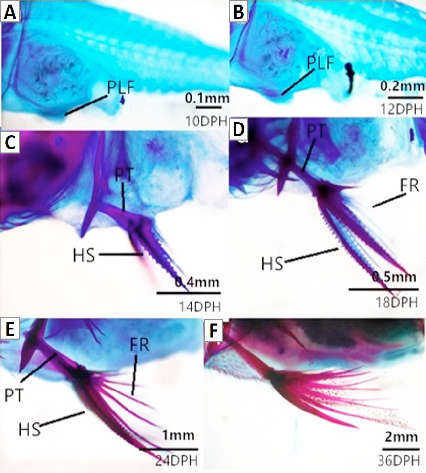
Formation and ossification of pelvic fins in larval crimson snapper. A and B, Larvae on 10-12 DPH show developing pelvic fins (PLF); C, Larvae on 14 DPH show cartilaginous of pterygiophores (PT), hard spine (HS) and sharp hooks; D, Larvae on 18 DPH show developing fin rays (FR); E, Larvae on 24 DPH show ossification of pterygiophores and hard fins; F, Larvae on 36 DPH show ossification of pelvic fins.
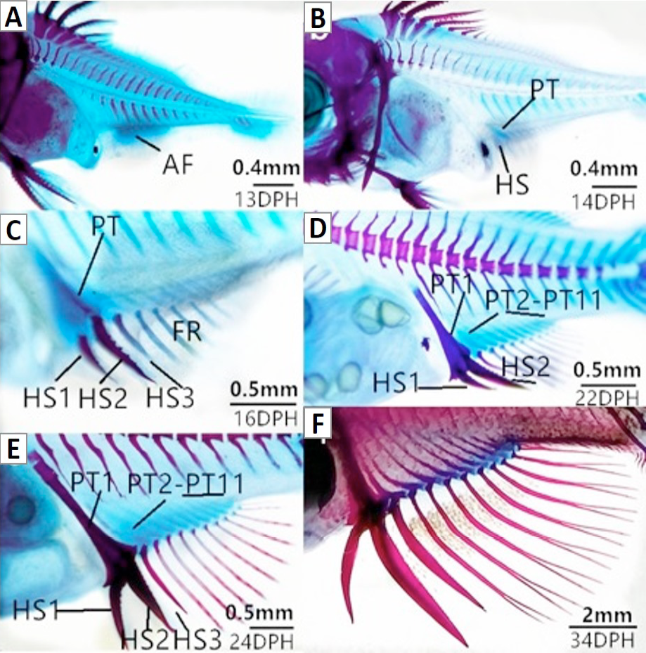
Formation and ossification of anal fins in larval crimson snapper. A, Larvae on 13 DPH show developing anal fins (AF); B, Larvae on 14 DPH show developing pterygiophores (PT) and hard spine (HS); C, Larvae on 16 DPH show cartilaginous of pterygiophores, three hard spine and fin rays; D, Larvae at 22 DPH show 2nd to 11th pterygiophores; E, Larvae on 24 DPH show ossification of pterygiophores, hard spine and fin rays; F, Larvae on 34 DPH show ossification of anal fins.
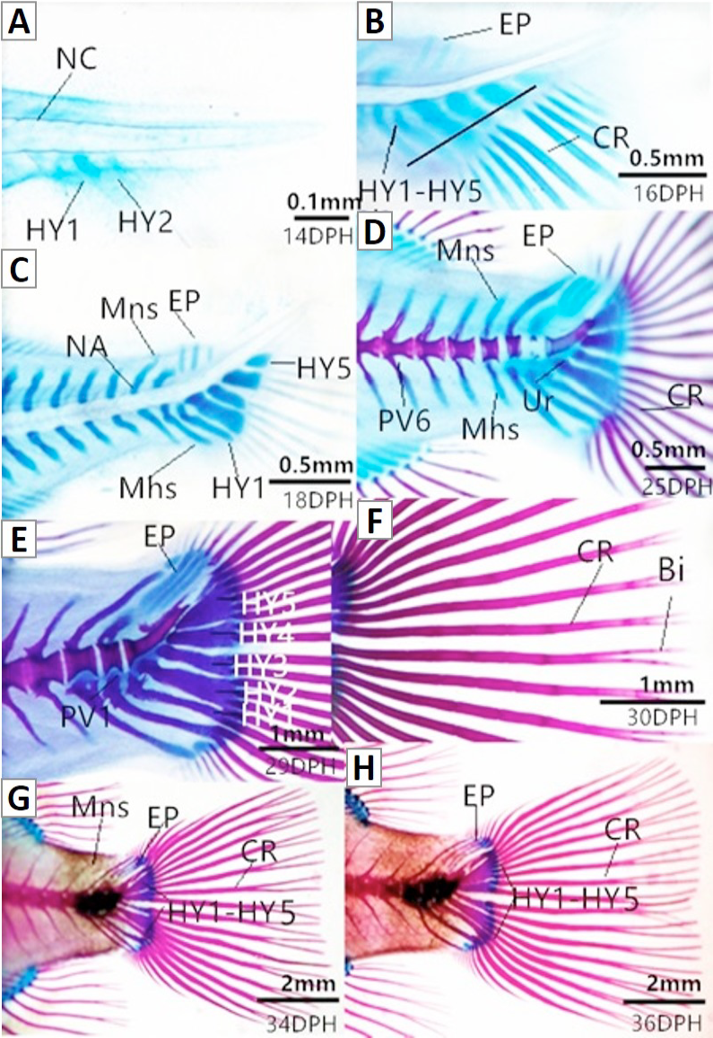
Formation and ossification of caudal complex in larval crimson snapper. A, Larvae on 13 DPH show two cartilaginous hypurals (HY) and Notochord (NC); B, Larvae on 16 DPH show developing caudal fin rays (CR), three epurals (EP), five hypurals, 1st hypural to 5th hypural (HY1 to HY5); C, Larvae on 18 DPH show five cartilaginous hypurals, three cartilaginous epurals, neural arch (NA), modified neural spines (Mns) and modified haemal spines; D, Larvae on 25 DPH show cartilaginous urostyle (Ur) and preural vertebra (PV); E, Larvae on 29 DPH show ossification of urostyle; F, Larvae on 30 DPH show ossification of fin rays and fin rays end bifucated (Bi); G, Larvae on 34 DPH show ossification of hypurals, modified neural spines and modified neural spines; H, Larvae on 36 DPH show ossification of caudal complex.
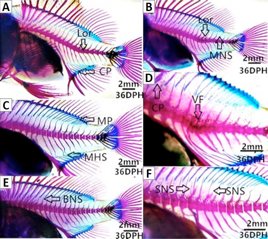
Skeletal anomalies in crimson snapper at 36 DPH. A, Lor, lordosis. CP, connection of adjacent pterygiophores; B, MNS, malformed neural spines; C, MHS, malformed haemal spines. MP, malformed pterygiophores; D, VF, vertebral fusion; E, BNS, bifurcated neural spines; F, SNS, supernumerary of neural spines.


