
Selenium Synergize Levothyroxine in Restoring Leukocytes Cluster Differentiation Expression in Methimazole Induced Hypothyroidism


Selenium Synergize Levothyroxine in Restoring Leukocytes Cluster Differentiation Expression in Methimazole Induced Hypothyroidism
Aryaf Mahmood Sabea1*, Majida A. Al-Qaiym2

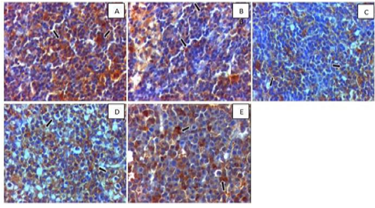
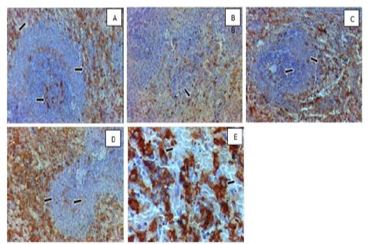
Photomicrograph of mesenteric (paracortex area) lymph node. Positive expression of T-lymphocyte helper receptor (CD4) (arrow) is shown in (A) control group, (B) hypothyroidism group, (C) SC-SeNPs, (D) levothyroxine group, and (E) SC-SeNPs+levothyroxine group. Hematoxylin & DAB. 100x. Note that the lymphoid follicle (arrowhead) in (D and E) did not show CD4 expression.
Photomicrograph of mesenteric (paracortex area) lymph node. Positive expression of T-lymphocyte helper receptor (CD4) (arrow) is shown in (A) control group, (B) hypothyroidism group, (C) SC-SeNPs, (D) levothyroxine group, and (E) SC-SeNPs+levothyroxine group.
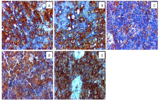
Photomicrograph of mesenteric (paracortex area) lymph node. Positive expression of T-lymphocyte helper co-receptor (CD8) (arrow) is shown in (A) control group, (B) hypothyroidism group, (C) SC-SeNPs, (D) levothyroxine group, and (E) SC-SeNPs+levothyroxine group. Hematoxylin & DAB. 100x.
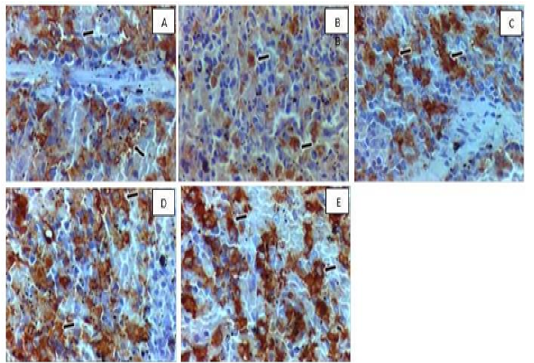
Photomicrograph of mesenteric (paracortex area) lymph node. Positive expression of T-lymphocyte helper co-receptor (CD8) (arrow) is shown in (A) control grou , (B) hypothyroidism group, (C) SC-SeNPs, (D) levothyroxine group, and (E) SC-SeNPs+levothyroxine group. Hematoxylin & DAB. 400x.
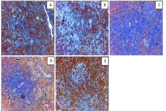
Photomicrograph of mesenteric (cortex area) lymph node. The expression of B-lymphocyte antigen (CD19) (black arrow) has shown (A) positive expression in control group, (B) weak expression in follicle area of hypothyrodism group, (C) moderate expression in the SC-SeNPs group, (D) positive expression in the levothyroxine group, and (E) positive expression in the SC-SeNPs+levothyroxine group.
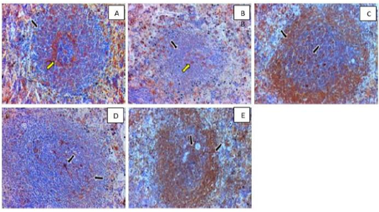
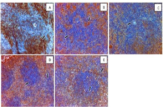
Photomicrograph of lymphatic follicle of white pulp in spleen. The expression of B-lymphocyte antigen (CD19) (black arrow) has shown (A) positive expression with intensive quantity (yellow arrow) at the center in control group, (B) weak expression in follicle area with intensive quantity (yellow arrow) at the center of hypothyrodism group, (C) positive expression in the SC-SeNPs group, (D) low-intensity expression in the levothyroxine group, and (E) positive expression in the SC-SeNPs+levothyroxine group. Hematoxylin & DAB. 100x.

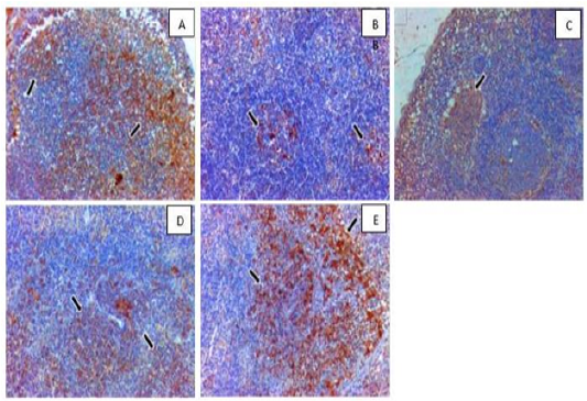
Photomicrograph of lymphatic follicle of red pulp in spleen. The expression of anti-CD68 primary antibody (arrow) has shown (A) positive expression in control group, (B) weak expression in hypothyrodism group, (C) moderate expression in the SC-SeNPs group, (D) strong expression in the levothyroxine group, and (E) strong expression in the SC-SeNPs+levothyroxine group. Hematoxylin & DAB. 100x.
Photomicrograph of lymphatic follicle of red pulp in spleen. The expression of anti-CD68 primary antibody (arrow) has shown (A) positive expression in control group, (B) weak expression in hypothyrodism group, (C) moderate expression in the SC-SeNPs group, (D) strong expression in the levothyroxine group, and (E) strong expression in the SC-SeNPs+levothyroxine group. Hematoxylin & DAB. 400x

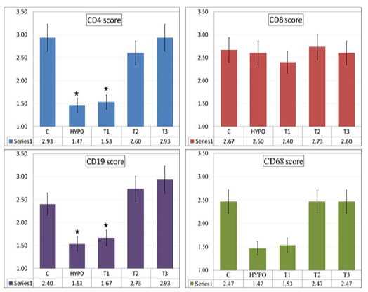
Scoring for CD4, CD8, CD19 and CD68

