
Selection of A New Site for Epidural Analgesia in Egyptian Goat (Capra Hircus) Based on Anatomy, Ultrasonography and Computed Tomography


Selection of A New Site for Epidural Analgesia in Egyptian Goat (Capra Hircus) Based on Anatomy, Ultrasonography and Computed Tomography
Ashraf Sayed Awaad1 and Mohamed Zaki Fathy2*

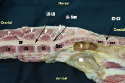
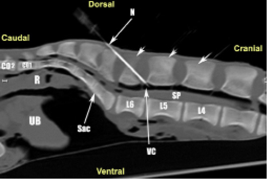
Photograph of sagittal section of the lumbosacropelvic part of goat cadaver showing, L4,4th lumbar vertebra; L5, 5th lumbar vertebra; L6, 6th lumbar vertebra; L5-L6, last interlumbar space; Sac, Sacrum; L6-S, lumbosacral space P, spinous processes; Co1, 1st coccygeal vertebra; 2nd cooygeal vertebral; Co1-Co2, 1st intercoccygeal space; SP, spinal cord; R, Rectum, UB, urinary bladder.
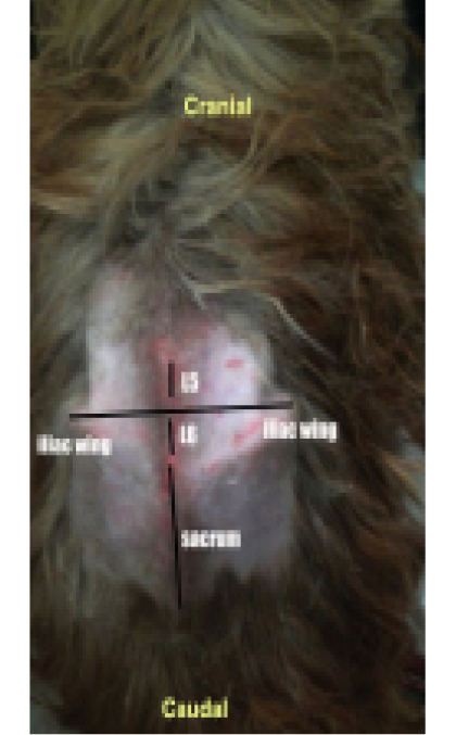
Photograph of the lumbosacropelvic part of goat showing the last inter lumbar space; L5, 5th lumbar vertebra; L6, 6th lumbar vertebra; L5-L6, last interlumbar space; Sac, Sacrum.

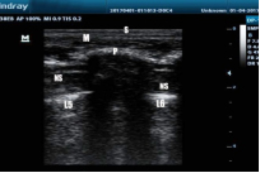
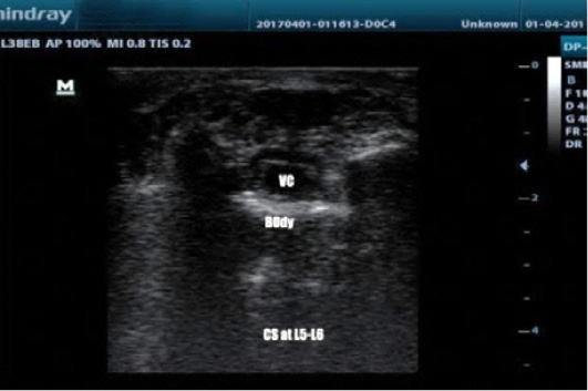
Parasagittal ultrasonogram of epidural space of goat at L5- L6 showing hypoechoic muscles and the echoic lumbar fascia (M), vertebral canal as a hypoechoic round area (VC) and hyper-echoic vertebral body of L5 and L6. Hyper-echoic line of ligamentum flavum. (P). Nervous structures appeared as multiple, small hyperechoic rods. Skin (S).
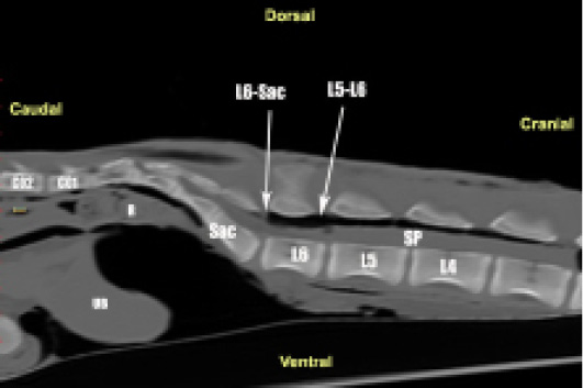
Sagittal CT scan section of the lumbosacropelvic part of goat cadaver showing; L4, 4th lumbar vertebra, L5, 5th lumbar vertebra, L6, 6th lumbar vertebra, L5-L6, last interlumbar space; Sac, Sacrum; L6-S, lumbosacral space P, spinous processes; Co1, 1st coccygeal vertebra; SP, spinal cord; R, Rectum, UB, urinary bladder.
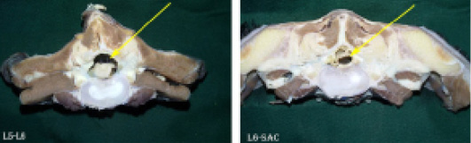
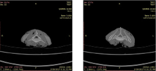
Photograph of cross sectional anatomy at the levels of; A) last inter lumbar space; B) lumbo-sacral space of goat cadavers showing, (arrow) the lumen of the spinal canal.

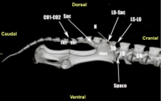
Volume 3D dimension CT scan of a lateral view of the lumbosacropelvic part of goat cadaver showing; L4,4th lumbar vertebra; L5, 5th lumbar vertebra; L6, 6th lumbar vertebra; L5-L6, last interlumbar space; Sac, Sacrum; L6-S, lumbosacral space P, spinous processes; N, needle; Co1, 1st coccygeal vertebra; 2nd cocygeal vertebral; Co1-Co2, 1st intercoccygeal space.

Sagittal CT scan section of the lumbosacropelvic part of goat cadaver with the needle inserted in the L5-L6 showing; L4,4th lumbar vertebra; L5, 5th lumbar vertebra; L6, 6th lumbar vertebra; L5-L6, last interlumbar space; Sac, Sacrum; L6-S, lumbosacral space P, spinous processes; Co1, 1st coccygeal vertebra; N, needle; VC, vertebral canal; SP, spinal cord; R, Rectum, UB, urinary bladder.

Parasagittal ultrasonogram of the epidural space in the goat at L5- L6 showing hypoechoic muscles and the echoic lumbar fascia (M), vertebral canal as a hypoechoic round area (VC) and hyper-echoic vertebral body of L5 and L6. Hyper-echoic line of ligamentum flavum. (P). nervous structures appeared as multiple, small hyperechoic rods. Skin (S).
Parasagittal ultrasonogram in epidural space in goat at L5- L6 showing hypoechoic muscles and the echoic lumbar fascia (M), hyper-echoic vertebral body of L5 and L6 posteriorly by an-echoic body of sacrum (Sac). Hyper-echoic line ligamentum flavum. (P). nervous structures appeared as multiple, small hyperechoic rods (NS). Skin (S)
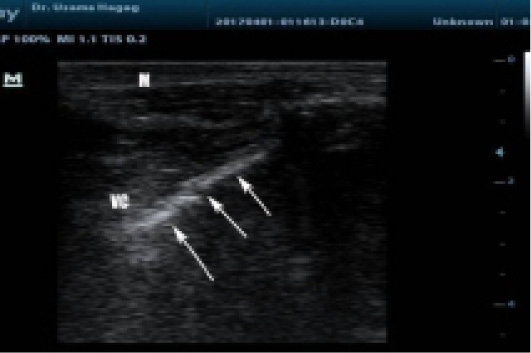
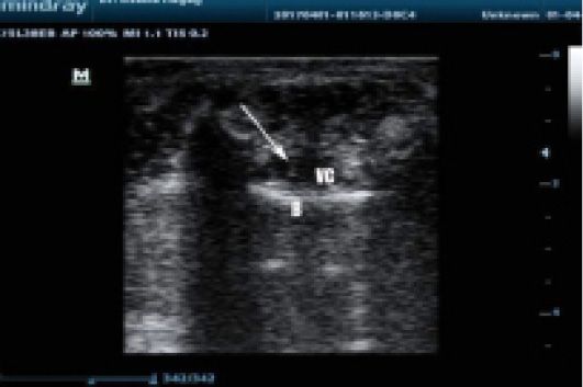
Parasagittal ultrasonogram of the epidural space in the goat at L5- L6 showing hypoechoic muscles and the echoic lumbar fascia (M), vertebral canal as a hypoechoic area (VC) and hyper-echoic needle (white arrow).

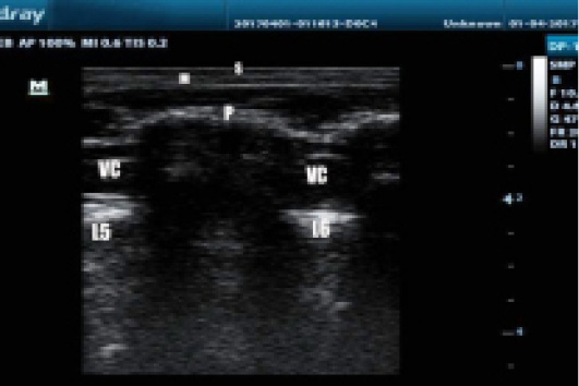
Cross ultrasonogram of the epidural space in the goat at L5- L6 showing hyper-echoic vertebral body and vertebral canal appear as a hypoechoic round area (VC).

Cross ultrasonogram of epidural space in goat at L5- L6 showing vertebral canal as round a hypoechoic area (VC) and hyper-echoic needle (white arrow) and hyper-echoic vertebral body (B).

Coronal CT scan sections at the levels of; A) last inter lumbar space; B) lumbosacral of goat cadaver showing, the lumen of the spinal canal.


