Quantitative Evaluation of Methylmercury Bioaccumulation in Rotifer Brachionus plicatilis by AIEgen
Quantitative Evaluation of Methylmercury Bioaccumulation in Rotifer Brachionus plicatilis by AIEgen
Hangyu Lin1, Minhui Chen1, Hao Yang1, Xinying Xia1, Huaying Zhou1, Xi Song1 and Tao He1,2*
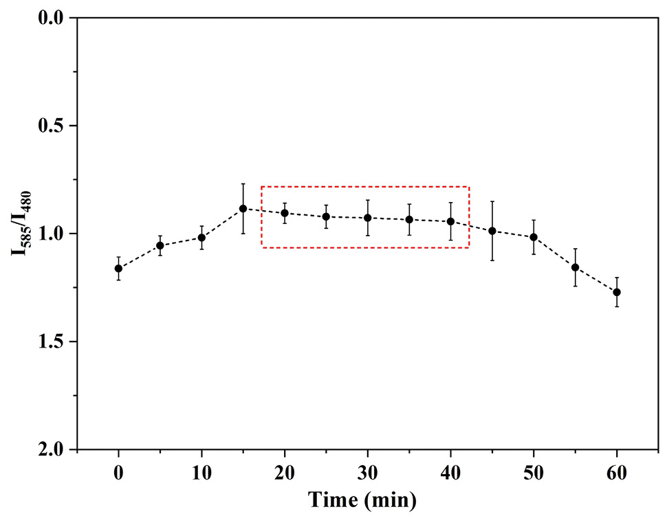
The ratio of photoluminescence (PL) intensity of AIE concentration of 1 µmol L-1 and MeHg concentration of 1 µmol L-1 at different time elapse of 0, 5, 10, 15, 20, 25, 30, 35, 40, 45, 50, 55.
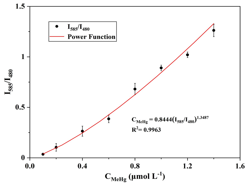
The ratio of photoluminescence (PL) intensity of MeHg (0.1, 0.2, 0.3, 0.4, 0.5, 0.6, 0.7, 0.8, 0.9, 1.0, 1.1, 1.2, 1.3, 1.4 and 1.5 μmol L-1) at the AIE concertation of 1 μmol L-1 at time.
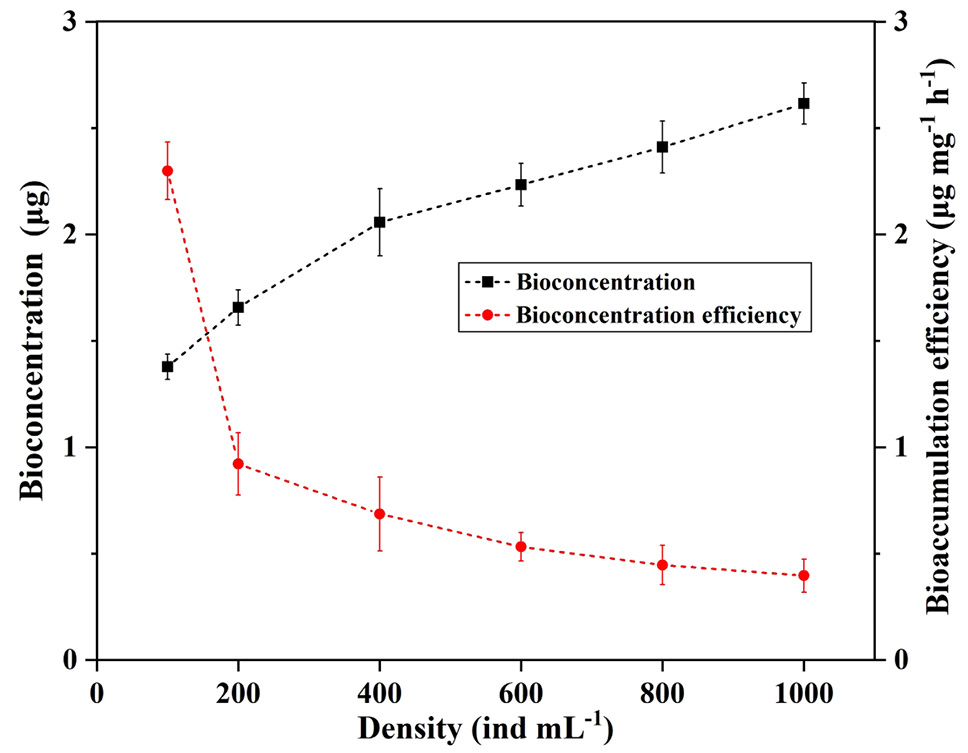
Bioaccumulation and bioaccumulation efficiency of MeHg (1 µmol L-1) at different rotifer densities (100, 200, 400, 600, 800 and 1000 ind mL-1) within 30 min.
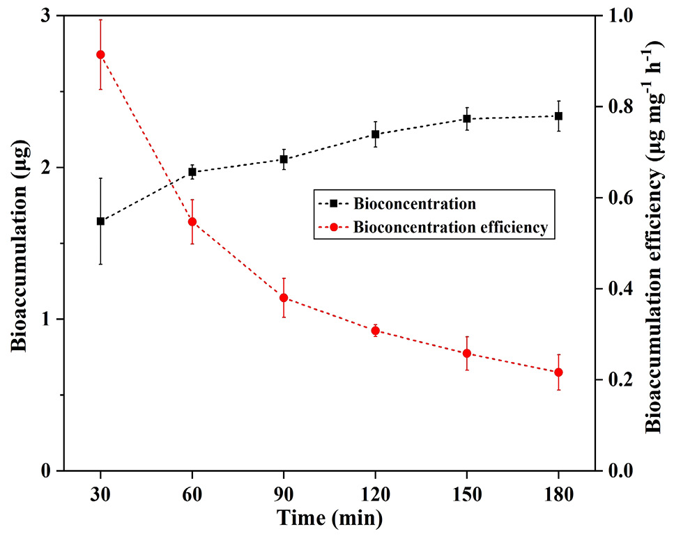
Bioaccumulation and bioaccumulation efficiency of MeHg (1 µmol L-1) in rotifers (400 ind ml-1) at different times (30, 60, 90, 120, 150 and 180min).
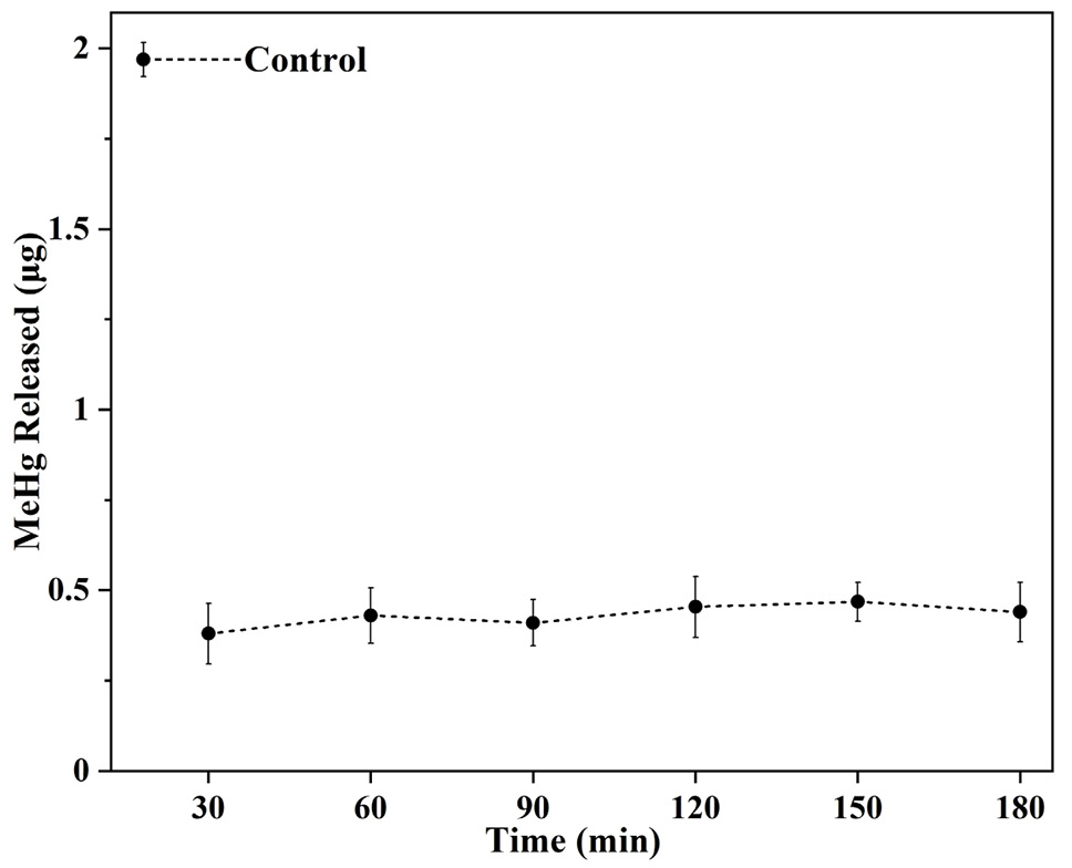
Release of MeHg (1 µmol L-1) in rotifers (400 ind ml-1) at different times (30, 60, 90, 120, 150 and 180 min).


