
Pyometra Treatment in Bitches with Different Protocols


Pyometra Treatment in Bitches with Different Protocols
Sulake Fadhil Al-Zubaidi1*, Ghusoon A.A. Alneamah2, Ali Saleh Mahdi1, Abdulraheem Abduljalil Wali3
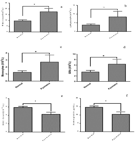
Figure 1:
Hematological parameters of normal bitches and bitches with pyometra.

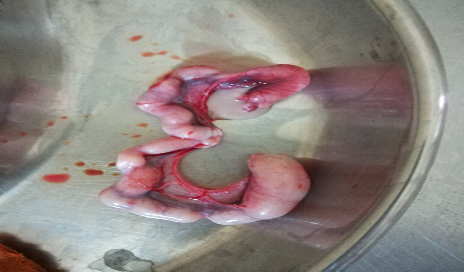
Figure 2:
Uterus of bitch with pyometra.

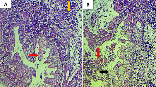
Figure 3:
Histopathological section of bitch uterus (A) showing cystic endometrial hyperplasia (CEH) appeared as papillary growth complex with pyometra (red arrow), also extensive inflammatory reaction seen in the endometrial stroma (yellow arrow) (B) showing sloughing of epithelial cells (red arrow), the epithelial cells appear large foamy cytoplasm (black arrow) (H & E, 400X).

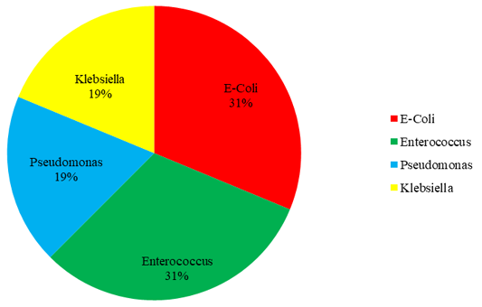
Figure 4:
Distribution of isolated bacteria associated with pyometra in 16 bitches.

January 2024
Vol. 12, Iss. 1, pp. 1-193

