Prevalence of the Peste Des Pettitis Ruminants in Goat and Sheep in District, Sanghar, Sindh, Pakistan


Research Article
Prevalence of the Peste Des Pettitis Ruminants in Goat and Sheep in District, Sanghar, Sindh, Pakistan
Abdul Latif Rind1*, Shahid Hussain Abro1, Rameez Raja Kaleri2,3, Ghulam Mustafa Solangi4, Raza Ali Mangi5, Muhammad Anees Memon6, Depeesh Kumar Bhuptani7, Sana Noor8, Zainab Lanjar9, Zahid Ali Mangrio10 and Abdul Wahid Solangi1
1Department of Veterinary Microbiology, Sindh Agriculture University, Tandojam, Pakistan; 2Department of Animal Breeding and Genetics, Sindh Agriculture University, Tandojam, Pakistan; 3Poultry Production, Tando Allahyar, Livestock and Fisheries Department Government of Sindh, Pakistan; 4Department of Veterinary Pathology, SBBUVAS, Sakrand, Pakistan; 5Department of Veterinary Anatomy and Histology, SBBUVAS, Sakrand, Pakistan; 6Department of Veterinary Physiology and Biochemistry, SBBUVAS, Sakrand, Pakistan; 7Department of Meat Technology, SBBUVAS, Sakrand, Pakistan; 8Department of Veterinary Parasitology, SBBUVAS, Sakrand, Pakistan; 9Department of Veterinary Microbiology, SBBUVAS, Sakrand, Pakistan; 10Pakistan Agriculture Research Council-Arid Zone Research Center (PARC-AZRC), Umarkot, Pakistan.
Abstract | The goal of the research was to assess the occurrence of the Peste des pettitis ruminant PPR among various breeds of goat and sheep in district, Sanghar, Sindh, Pakistan. Two hundred samples of oculo-nasal discharge were gathered and subjected to Ic-ELISA testing to identify PPR antigens. The analysis of PPR prevalence was carried out according to breed, sex, age, and Taluka. The overall PPR prevalence was found to be 20%, with the highest prevalence rate (50%) observed in Jam Nawaz Ali Taluka and the lowest (7.5%) in Shahdadpur Taluka. The prevalence was higher in goats (30%) than in sheep (10%). Female animals had a higher prevalence than males, with a prevalence of 25% in female goats and 7% in female sheep. The prevalence in male goats and sheep was 5% and 3%, respectively. The highest prevalence was observed in animals aged two years, with 56.66% in goats and 50% in sheep. The study found that infected animals exhibited various clinical signs and symptoms such as diarrhea, coughing, fever, pneumonia, stomatitis, and occulo-nasal discharge. Furthermore, no instances of pregnancy were reported during the research. These findings underscore the high prevalence of PPR in sheep and goats within District Sanghar, emphasizing the urgent requirement for effective control measures to curb its propagation.
Received | May 26, 2023; Accepted | August 02, 2023; Published | October 28, 2023
*Correspondence | Abdul Latif Rind, Department of Veterinary Microbiology, Sindh Agriculture University, Tandojam, Pakistan; Email: rindlatif@gmail.com
Citation | Rind, A.L., Abro, S.H., Kaleri, R.R., Solangi, G.M., Mangi, R.A., Memon, M.A., Bhuptani, D.K., Noor, S., Lanjar, Z., Mangrio, Z.H. and Solangi, A.W., 2023. Prevalence of the peste des pettitis ruminants in goat and sheep in District, Sanghar, Sindh, Pakistan. Journal of Innovative Sciences, 9(2): 192-197.
DOI | https://dx.doi.org/10.17582/journal.jis/2023.9.2.192.197
Keywords | Goat, Sheep, PPR, Prevalence, Sanghar
Copyright: 2023 by the authors. Licensee ResearchersLinks Ltd, England, UK.
This article is an open access article distributed under the terms and conditions of the Creative Commons Attribution (CC BY) license (https://creativecommons.org/licenses/by/4.0/).
1. Introduction
The Peste des pettitis ruminant PPR disease is severely contagious viral disease that mainly affects the young small ruminant of goat and sheep with most common symptoms of high fever, pneumonia, oculo-nasal discharges, ulceration on mucous membrane. The disease occurs in an epizootic form and can have devastating consequences, with mortality rates ranging from 50-80% and morbidity rates reaching 80-90% (Diallo et al., 2007). This disease commonly known as goat plague, in Asia and Africa (Shaila et al., 1989; Robert et al., 1994; Awa et al., 2000) as it mostly affect the young kids of sheep and goat with significant animal population. The disease was considered a relatively confined issue in western part of Africa, but after that this disease was spread in various parts of south, east and central Asia (FAO, 1999; Ali, 2004). Given that there have been several cases of PPRV reported in both sheep and goats; this study aims to evaluate the presence of PPRV in flocks of these animals in the district of Sanghar in the Sindh province.
2. Materials and Methods
2.1 Study area
Sanghar District, has a latitude of 26°2’39.91”N and a longitude of 68°57’13.96”E or 26.044419 and 68.953879 respectively. Based on population, the area is ranked 111th in Pakistan (Figure 1).
The history of PPR suspected sheep and goats was taken by using Proforma. A total of 200 hundred oculo-nasal discharge samples were collected by sterile nasal swab from sheep and goat flocks from the District Sanghar. The collected specimens were brought and processed at Central Veterinary Diagnostic Laboratory, Tandojam for detection of PPR antigens using Ic-ELISA Immuno-capture Enzyme Linked Immuno-sorbent Assay. The Ic-ELISA was used as a test of screening for observing the presence antigens against the PPRV.
2.2 (Immuno-capture Enzyme Linked Immuno-sorbent Assay) Ic-ELISA
The detection of nucleoprotein antigens of PPRV was performed using an Immuno-capture ELISA kit. For this purpose polyclonal anti-PPR antibodies were utilized to observe the antibody after diluting in 7.2 to 6 for using to coat of enzyme linked immune sorbet assay plates. After that all these plates were placed under the incubator for 1 hour at 37oC and thoroughly washed. Next, samples were added at a volume of 50 µl per well, followed by the addition of specific PPR Monoclonal Antibodies that were diluted to 1/500 in blocking buffer. An enzyme conjugate, consisting of Streptavidin-peroxidase, was added, and the Plates were placed for incubated again being washed for 1 hour at 37oC. In this way to develop the color chromo genes like hydrogen peroxide and orthophenyl diamine substrate were mixed during incubation time for 10 minutes. After that the reading of OD value sample were considered as positive and negative control by utilizing the ELISA meter reading. In this reading any sample having reading more than twice mean OD value for black control were recorded as positive reference.
2.3 Sampling and preparation
The procedures used to analyze the oculo-nasal discharge samples were as follows:
First, a 200 µL dilution of buffer-13 was mixed with all swab and placed for incubation at the room temperature for overnight.
In this study the test was conducted with method such as (25µL) buffer 13 dilutions was mixed in both (A1 and B1) negative control well as well as in (C1 and D1) positive control well. After that another amount of (25µL) buffer 13 dilution was also mixed in negative control well as well as in (C1 and D1) positive control well. Finally (50µL) buffer 13 dilutions were also included left over wells.
Plates were kept for 2 minutes at 21oC than placed under the incubator at 37oC till 45 minutes. After it each plate was cleaned using (300 µL) XI washing solution for 6 times.
After washing a (100 µL) amount of conjugate was mixed than each plate was placed under the incubator at 21oC till 30 minutes. After completion of incubation process again each plate was cleaned using (300 µL) XI washing solution for 6 times.
Then, a 100 µL substrate was added to After all that again plates were placed under the incubator at room temperature under the dark area. After overall process a (100 µL) was mixed of stop solution in all and then mixed thoroughly, and the results were recorded by measuring the O.D. value at 450nm wavelength.
2.4 Calculation was done as under
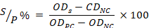
2.5 Data analysis
The data regarding the prevalence of PPR in various animal species, sex, age and area–wise, the mean percentage was adopted throughout study for analysis of the data.
3. Results and Discussion
A study was conducted to determine the prevalence of PPR peste Des Petites Ruminants in small ruminant sheep goat at the district Sanghar using the Ic-ELISA kit method. A total 200 samples, 100 each from sheep and goats, were collected to detect the occurrence Peste Des Petitis small ruminants sheep and goat population in mentioned districts. In this study, it was also observed the prevalence of the disease in male and female animals and different age groups (<1 year, <2 years, and >2 years) as well as the clinical manifestation of the disease.
3.1 Prevalence of peste des petitis ruminants in small ruminants sheep and goat
Results showed that 20% (40 out of 200) of the serum samples tested positive for peste peste des petitis ruminant virus antibodies among the population of sheep and goat district Sanghar (Table 1). Among the 40 antibody-positive samples, 30 were from goats and 10 were from sheep. It was observed that ratio of PPR was observe maximum in goat as compared with sheep (30%) and (10%) in district Sanghar.
To study the prevalence of PPR in population of sheep and goat district Sanghar in Sindh province, 45, 30, 30, 40, and 55 discharge samples were collected from small animals in Sanjhoro, Jhol, Jam Nawaz Ali, Shahdadpur, and Khipro Talukas of Sanghar. The samples were then tested using Ic-ELISA.
Out of the collected samples, 5, 10, 15, 3, and 7 were detected positive for Peste Des Petitis Ruminants in Sanjhoro, Jhol, Jam Nawaz Ali, Shahdadpur, and Khipro Talukas, respectively. The percentage prevalence in these areas was found to be 11.11%, 33.33%, 50.00%, 7.5%, and 12.72%, respectively (Table 2).
Table 1: Prevalence of peste des petitis ruminants in small ruminant sheep and goat.
|
Animal species |
Number of samples examined |
Number of positive samples |
% of positive samples |
|
Goat |
100 |
30 |
30 |
|
Sheep |
100 |
10 |
10 |
|
Overall |
200 |
40 |
20 |
*Percentages obtained from total no. of samples examined
Table 2: Area-wise prevalence of PPR in population of sheep and goat in district Sanghar of Sindh.
|
Locations |
Animal species |
Samples tested by Ic-ELISA |
No. of positive samples determined by Ic-ELISA |
% of positive samples |
|
|
Goat |
Sheep |
||||
|
Sanjhoro |
10 |
35 |
45 |
5 |
11.11 |
|
Jhol |
15 |
15 |
30 |
10 |
33.33 |
|
Jam Nawaz Ali |
20 |
10 |
30 |
15 |
50.00 |
|
Shahdadpur |
20 |
20 |
40 |
3 |
7.5 |
|
Khipro |
35 |
20 |
55 |
7 |
12.72 |
|
Total |
100 |
100 |
200 |
40 |
114.66 |
*Percentages obtained from samples tested by Ic-ELISA.
The study found that the highest prevalence of the PPR was recorded in sheep and goat from Taluka Jhol, while the second-highest occurrence was observed in sheep and goats from Khipro.
The lower percentage prevalence of the disease was noted in other Talukas as compared to Jhol and Khipro. Furthermore, on the other hand, the goats were found more susceptible to sheep. This might be resistant from sheep due to natural immunity or the samples collected from the goats are higher in numbers that made this difference. Otherwise, there are no any clear reasons that might be discussed here. Generally, a mixed trend of infection was noted irrespective of role of animal species and area. However, a variation in the occurrence of disease is evidence there. But from this, one cannot make a clear conclusion for the prevalence of disease among animal species and areas where from the samples collected, tested and results obtained during present survey.
A study was conducted to investigate the occurrence of PPR among sheep goat and male female population. The total 200 number of samples were examined, and 5 samples from male goats and 25 samples from female goats tested positive for PPRV antibodies. The prevalence of PPR in male goats was recorded as 5.00%, while it was noted as 25.00% in female goats. In addition, 10 positive samples from male and female sheep were tested for PPRV antibodies. Of these, 7 female sheep samples were found positive, and the percentage prevalence was detected as 7.00%. Meanwhile, 3 samples of male sheep were positive for PPRV antibodies, and the percentage prevalence was observed as 3.0% (Table 3).
Table 3: The presence of PPR recorded across various age groups of animals.
|
Breed |
Sex |
Total samples examined |
Positive samples (%) |
Negative samples (%) |
|
Goat |
Male |
100 |
5 (5.00%) |
45 (45.00%) |
|
Female |
25 (25.00%) |
25 (25.00%) |
||
|
Sheep |
Male |
100 |
3 (3.00%) |
47 (47.00%) |
|
Female |
7 (7.00%) |
43 (43.00%) |
*Percentages obtained from total no. of samples examined.
3.2 The presence of PPR recorded among different age stages of sheep and goat
In this study (Table 4 and 5), the incidence of PPR was recorded across various age groups. The survey revealed a fluctuation in PPRV infection among various age groups of sheep and goat. Goats with the age of <1, <2 and > 2 years showed 20.00%, 23.33% and 56.66% infection of peste des petitis ruminants PPRV respectively. Similar pattern of PPRV infection was noted in sheep population. The sheep with the age of <1, <2 and >2 years, the prevalence of PPRV was detected in 20.00%, 30.00% and 50.00%, respectively. The higher prevalence of peste des petitis ruminants was recorded in age of >2 years followed by <2 years for both, goats and sheep population. However, the lower prevalence of PPRV infection was recorded in age of <1 year in both, goats and sheep population.
3.3 Clinical signs of PPR were recorded among sheep and goat in this survey
After conducting a study on animals, it was observed that the signs and symptoms of the disease including pneumonia, diarrhea, coughing, anorexia, occulo nasal discharge, fever and stomatitis among sheep and goat.
Table 4: The presence of peste des petitis ruminants in different age groups of animal species.
|
Breed |
Age (years) |
Positive samples (%) |
Chi-square |
|
Goat |
<1 |
6 (20.00%) |
0.9076 |
|
<2 |
7 (23.33%) |
||
|
>2 |
17 (56.66%) |
||
|
Sheep |
<1 |
2 (20.00%) |
|
|
<2 |
3 (30.00%) |
||
|
>2 |
5 (50.00%) |
*Percentages obtained from total no. of samples examined.
Table 5: Clinical signs/ symptoms observed in peste des petitis ruminants in animals during survey.
|
Breeds of animals |
Signs/ symptoms |
|
Goats |
Anorexia, high temperature, stomatitis, ulceration, diarrhoea, oculo-nasal discharges, coughing and pneumonia |
|
Sheep |
High temperature, stomatitis, anorexia, ulceration of mouth part, oculo-nasal discharges, coughing and pneumonia, etc. |
The disease now known as PPR was initially recognized in the Ivory coast in western part of Africa at the time of II world war (Bundza et al., 1988). During that period this disease was known as the pneumoentrirtis, pseudo rinder pest and pneumoentrirtis syndrome (Gargadennec et al., 1942). This disease was 1st recognized in Pakistan during the year of 1991 in Punjab province with some clinical signs and post mortem findings by laboratory confirmation was not obtained in the report by (Amjad et al., 1996). However, subsequent documentations reported (Hussain et al., 2003) after the did confirm the presence of the disease, while others were based on clinico-epidemiological observations. Abubakar et al. (2008a), Abubakar and Munir (2014), and Khan et al. (2008) have since reported significant economic losses due to the continuing presence of the virus.
To investigate the occurrence of PPRV among population of sheep and goat at district Sanghar, 200 oculo-nasal discharge samples (100 from each species) were collected. The study found that 40 of these samples tested positive for peste des petitis ruminants, with more prevalence among goat as compared with sheep (30%) and (10%). It was also examined prevalence by sex and age, as well as clinical manifestations of the disease in the suspected populations.
The results of this study align with previous research on the prevalence of PPRV in different parts of the world. For instance, (Abubakar et al., 2009), observed 40.98% prevalence of PPR in Pakistan with more severity in goat as compared with sheep animal. Taylor and Barrett (2003) conducted study in Nigeria and observed more number of prevalence 57% in sheep and 44% in goat in a field study. Another study was conducted by Singh et al. (2004) who recorded same result for prevalence 32.4% and 36.3% in goat and sheep, respectively in India. It was observed during the current study that severity of PPR was highly affected by environmental conditions as well as animal species. In Pakistan number of outbreaks of PPRV in goat and sheep were observed having with more number of infected animals in north and south areas of Pakistan. It is real fact that these both species are highly succeptable for PPR and show symptoms. As higher rate of infection was observed goat under different parts of African countries, whereas in south and west parts of Asia higher infection rate was observed in sheep animal as compared with goat.
Conclusions and Recommendations
On the basis of findings, it was concluded that PPR prevalence were higher in sheep population as compared to goat in district Sanghar. The prevalence was found higher in male compared to female in goat and sheep. The high rate of PPR was observed in >2 years age than other age group animals.
Acknowledgement
Authors are thankful to Department of Veterinary Microbiology, Faculty of Animal Husbandry and Veterinary Sciences, Sindh Agriculture University, Tando Jam for providing platform for this research stu.
Novelty Statement
The findings of this study have significant implications for the local economy and food security. Goats and sheep are important sources of livelihood and nutrition in many rural areas, and PPR can have devastating effects on their populations.
Author’s Contribution
Conceived & Designed the Experiment: SH Abro, DK Bhuptani & RA Mangi. Performed Experiment: AL Rind, ZA Mangrio & Z Lanjar. Collected the Data: AW Solangi, S Pahanwar & MA Memon Solangi. Analyzed the data: RR Kaleri & GM Solangi. Wrote the Paper: RR Kaleri & AL Rind.
Conflict of interest
The authors have declared no conflict of interest.
References
Abubakar, M., Jamal, S.M., Khan, M.A. and Ali, Q., 2008. Peste des petitis ruminants outbreak in small ruminants of Northern areas of Pakistan. Res. J. Vet. Sci., 1(1): 56–61. https://doi.org/10.3923/rjvs.2008.56.61
Abubakar, M. and Munir, M., 2014. Peste des petitis ruminants virus: An emerging threat to goat farming in Pakistan. Transbound. Emerg. Dis., 61(1): 1–4. https://doi.org/10.1111/tbed.12192
Abubakar, M., Jamal, S.M., Arshed, M.J., Hussain, M. and Ali, Q., 2009. Peste des petitis ruminants virus (PPRV) infection; Its association with species, seasonal variations and geography. Trop. Anim. Health Prod., 41: 1197–202. https://doi.org/10.1007/s11250-008-9300-9
Ali, Q., 2004. National policy for control of peste des petitis ruminants in Pakistan. Islamabad: GCP/ PAK/ 088-EC, FAO; 2004.
Awa, D.N., Njoya, A. and Tama, A.C., 2000. Economics of prophylaxis against peste des petits ruminants and gastrointestinal helminthosis in small ruminants in north Cameroon. Trop. Anim. Health Prod., 32: 391–403.
Athar, M., Muhammad, G., Azim, F., Shakoor, A., Maqbool, A. and Chaudhry, N.I., 1995. An outbreak of peste des petitis ruminants like disease among goats in Punjab (Pakistan). Pak. Vet. J., 15: 140–143.
Amjad, H., Islam, Q.U., Forsyth, M., Barrett, T. and Rossiter, P.B., 1996. Peste des petitis ruminants in goats in Pakistan. Vet. Rec., 139(5): 118–119. https://doi.org/10.1136/vr.139.5.118
Bundza, A., Afshar, A., Dukes, T.W., Myers, D.J., Dulac Susi, G. and Becker, A.W.E., 1988. Experimental PPR (goat plague) in goats and sheep. Can. J. Vet. Res., 52: 46–52.
Diallo AC, Minet CL, Goff G, Berhe E, Albina G, Barrett LT. 2007. The threat of peste des petitis ruminants: progress in vaccine development for disease control. Vaccine. 25:5591–7.
Gargadennec, L. and Lalanne, A., 1942. La peste des petitis ruminants. Bull. Serv. Zoo AOF., 5: 15–21.
Hussain, M., Afzal, M., Muneer, R., Ashfaque, M. and Haq, E.U., 1998. An outbreak of peste des petitis ruminants in goats in Rawalpindi. Pak. Vet. J., 18(4): 224–226.
Hussain, M., Muneer, R., Jahangir, M., Awan, A.H., Khokhar, M.A., Zahur, A.B., Zulfiqar, M. and Hussain, A., 2003. Chromatographic strip technology: A pen-side test for the rapid diagnosis of peste des petitis ruminants in sheep and goats. J. Biol. Sci., 3: 1–7. https://doi.org/10.3923/jbs.2003.1.7
Kaleri, R.R., Kaleri, H.A., Leghari, A., Solangi, G. M., Mangi, R.A., Chandio, M.A., and Baloch, M.I. 2023. Study on correlation estimates between carcass traits of sheep breeds of Baluchistan. Pure Appl. Biol., (PAB), 12(2), 1242-1247.
Kaleri, R.R., Kaleri, H.A., Kaleri, A., Shah, R.A., Kumar, R., Kumar, D., and Marri, G.M. 2018. Short communication correlation and regression coefficient estimates between some growth performance traits of Harnai sheep. Biol. Sci. PJSIR, 61(2): 112-114.
Khan, H.A., Siddique, M., Abubakar, M., Arshad, M.J. and Hussain, M., 2008. Prevalence and distribution of peste des petitis ruminants virus infection in small ruminants. Small Rumin. Res., 79: 152–157. https://doi.org/10.1016/j.smallrumres.2008.07.021
Pervez, K., Ashfaq, M., Khan, M.S., Hussain, M. and Azim, E., 1993. A rinderpest like disease in goats in Punjab, Pakistan. Pak. J. Livest. Res., 1: 1–4.
Roeder, P.L., Abraham, G., Kenfe, G. and Barrett, T., 1994. Peste des Petitis Ruminants in Ethiopian goats. Trop. Anim. Health Prod., 26: 69–73. https://doi.org/10.1007/BF02239901
FAO, 1999. Recognizing Peste des petitis ruminants. A field manual. FAO Animal Health Manual No. 5. Rome: Food and Agriculture Organization of the United Nations. pp. 1–27.
Roeder, P.L. and Ubi, T.U., 1999. Recognizing peste des petites: A field manual. Animal health manual. Rome: Food and Agricultural Organization. pp. 5–28.
Singh, R.P., B.P. Screeniva, P. Dhar and S.K. Bandyopadhya. 2004. A Sandwich-ELISA for the diagnosis of Peste des petitis ruminants infection in small ruminants using anti-nucleocapsid protein monoclonal antibody.Arch.Virol. 149: 2155-2170.
Shaila, M.S., Purushothaman, V., Bhasavar, D., Venugopal, K. and Venkatesan, R., 1989. Peste des petitis ruminants in India. Vet. Rec., 125: 602.
Shaila, M.S., Purushothaman, V., Bhavasar, D., Venugopal, K. and Venkatesan, R.A., 1989. Peste des petits ruminants of sheep in India. Vet. Rec., 125: 602
Soomro, I. H., Mughal, G.A., Rajput, N., Kaleri, R. R., Bhuptani, D.K., Mangi, R.A., and Soomro, Z. P. 2023. Effect of silage feeding on the growth performance and body confirmation of Tapri goats under intensive management system. J. Innovative Sci., 9(1): 51-55.
Taylor, W.P. and Barrett, T., 2007. Diseases of sheep. 4. Oxford, UK: Blackwell publishing. pp. 460–469. https://doi.org/10.1002/9780470753316.ch61
Zahur, A.B., Ullah, A., Hussain, M., Irshad, H., Hameed, A., Jahangir, M. and Farooq, M.S., 2011. Sero-epidemiology of peste des petitis ruminants (PPR) in Pakistan. Prev. Vet. Med., 102: 87–92. https://doi.org/10.1016/j.prevetmed.2011.06.011
To share on other social networks, click on any share button. What are these?