Potential Role of Recombinant Capsid Protein of Dengue Virus Serotype 2 in the Development of an In-House Elisa Based Detection of Infection
Potential Role of Recombinant Capsid Protein of Dengue Virus Serotype 2 in the Development of an In-House Elisa Based Detection of Infection
Rakhtasha Munir1, Shazia Rafique2*, Iram Amin1,2, Sameen Ahmed2, Ayesha Vajeeha2, Amjad Ali3, Muhammad Shahid2, Muhammad Umer Khan4 and Muhammad Idrees2
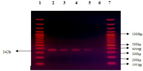
PCR Amplification of dengue C Gene fragment, Lane 2-5: dengue DNA positive of 342bp and Lane 6 showing negative control (containing all PCR reagents but without Template DNA), Lanes 1 and 7: DNA marker of 100bp.
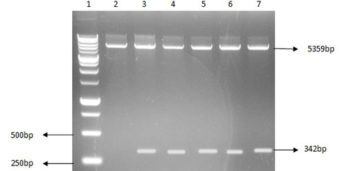
Restriction digestion analysis of clone in expression vector: Lane 1: DNA marker of size 1Kb, Lane 2: negative control (empty vector), Lanes 3 to 7: First band digested of peT28a vector of size 5359bp and second band of dengue C gene fragment size 342bp.
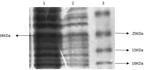
SDS-PAGE of protein showing expression of 24KDa protein fragment. Lane 1: Induced protein sample, Lane 2: uninduced protein sample, Lane 3: Prestained Protein marker.
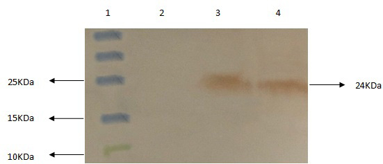
Western Blot representing 24KDa protein fragment of C protein. Lane 1: Prestained Protein marker, Lane 2: uninduced protein sample, Lanes 3-4: Induced protein samples.
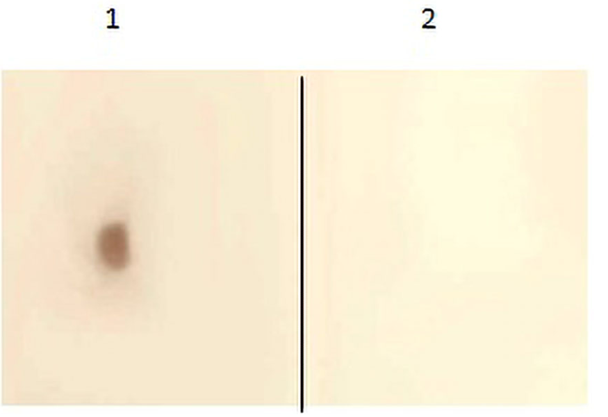
Dot blot representing C protein of dengue. Lane 1: Induced protein sample, Lane 2: uninduced protein sample.


