

Pattern of Ovarian Follicular Development and Steroid Hormone Concentrations during Estrous Cycle of Lohi Sheep


Pattern of Ovarian Follicular Development and Steroid Hormone Concentrations during Estrous Cycle of Lohi Sheep
Muhammad Younis1, Muhammad Irfan-ur-Rehman Khan1*, Mustansar Abbas1, Ali Murtaza1, Imran Mohsin2, Muhammad Shahzad3 and Muhammad Zahid Tahir1

Follicular development of three (n = 16; A) and four-wave (n = 2; B) cycles in Lohi sheep. Diameters (mean ± SEM) of the largest (F1) and subordinate (SF) follicles were monitored daily for two consecutive ovulations via transrectal ultrasonography. The frequency of 3-wave cycles was relatively greater (p<0.05) than the 4-wave cycles i.e., 87% vs. 13%, respectively.

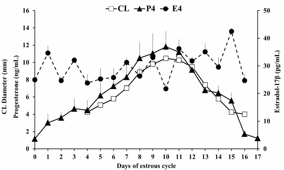
Changes (mean ±SEM) in diameter of corpus luteum, plasma concentrations of progesterone (P4; n= 4) and estradiol-17β (E2; n= 4). The CL became visible by Day 4, reaching a plateau on Day 9.0±0.1 and luteolysis began by Day 12.2 ± 0.2 after the ovulation. The CL diameter was directly associated with the plasma progesterone concentration during the cycle (r = 0.93; p<0.05). Multiple low peaks of plasma E2 during the luteal phase (Days 1-14) and a preovulatory peak was observed during the follicular phase (Days 14-15). Arrow indicates the ovulation.
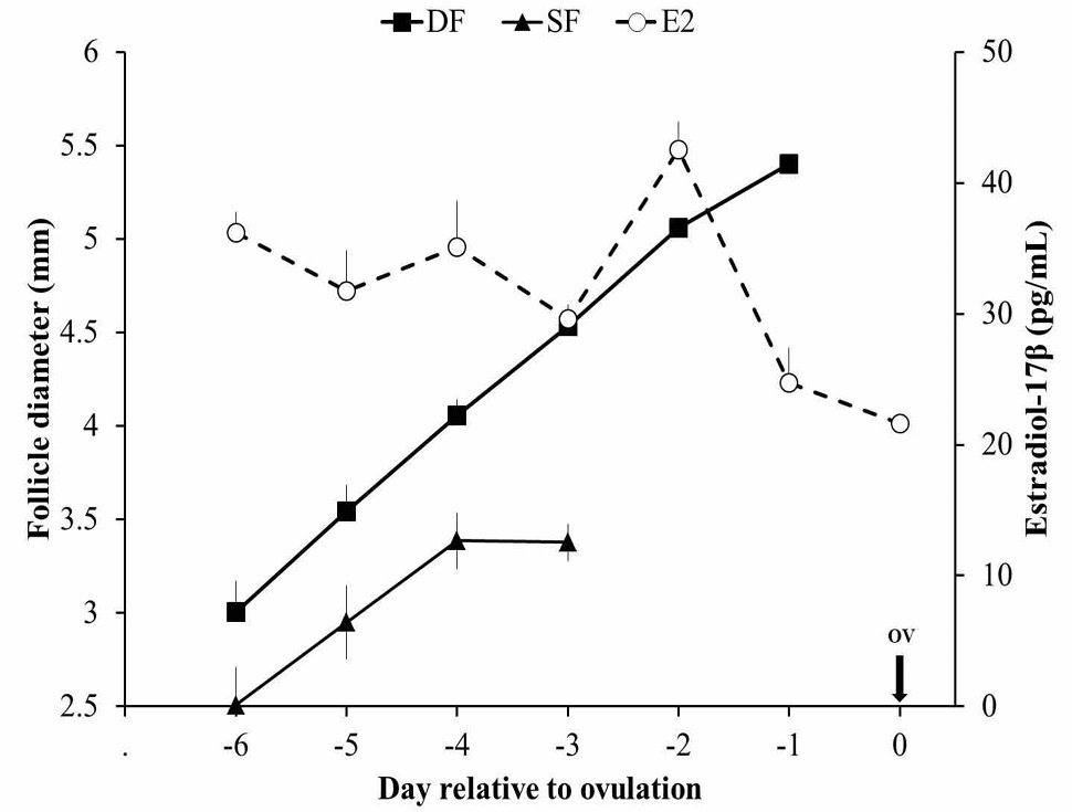
Relationship of the largest follicle (F1) and subordinate (SF) follicle (diameter; mean±SEM) with that of plasma estradiol-17β concentration (E2) during the preovulatory period in Lohi sheep. The plasma E2 increased with the diameter of preovulatory follicles (r = 0.84; p<0.05) and reached at maximum concentration 48h before ovulation.

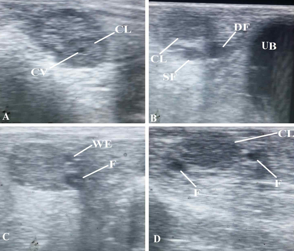
Representative ovarian ultrasound images of Lohi sheep. A) Ovary having a corpus luteum (CL) with cavity (CV). B) Ovary detected cranial to the urinary bladder (UB) showing a CL, dominant follicle (DF), and a subordinate follicle (SF). C) Ovary having multiple small follicles (F) at the time of follicular wave emergence (WE). D) Ovary showing mid-luteal phase CL and follicles (F).


