
Optimizing Bone Healing in Rabbit Models: A Comparative Study of Lidocaine Hydrochloride and Diclofenac: Histological Study


Optimizing Bone Healing in Rabbit Models: A Comparative Study of Lidocaine Hydrochloride and Diclofenac: Histological Study
Qamer J. Jadoaa, Raffal A. Omar*

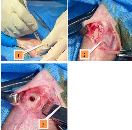
The surgical stages,1: Skin incision, 2: Expose the medial aspect of the proximal end of the Tibia, 3: Induce a 3.5 mm hole.

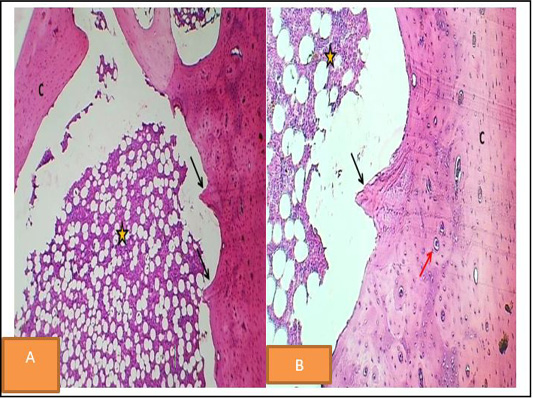
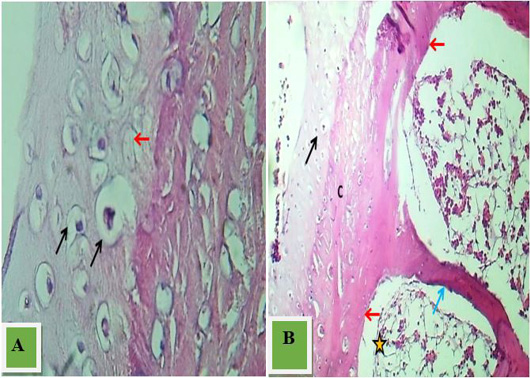
Histopathological of bone Section at the end of 1stwk p. o. control group revealed little bone formation (black arrow) and bone marrow (yellow star). H & E stain A40x. B.100x

Section at the end of 1stwk p. o -2 mg/kg B.W lidocaine hcl shows normal cortical bone (C), active osteogenic tissue (Red arrow), and thick formed bone layer (Black arrows), Red marrow (blue arrows). H & E stain A
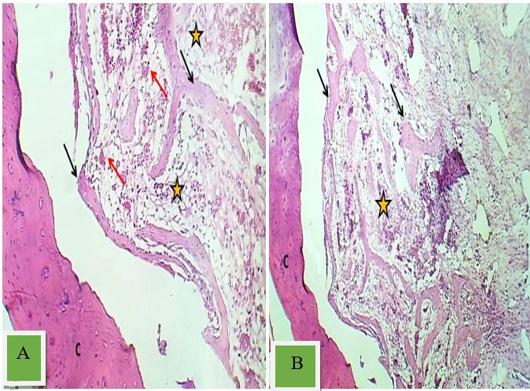
the Section at the end of 1stwk p. o Diclofenac shows thin cortical bone (C), osteogenic tissue (Asterisk), and a network of new trabecular bone formation (Black arrows). H & E stain A. 40x. and B.100x
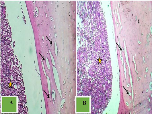
Section at the end of 2nd wk p. o -control) shows normal cortical bone (C), new bone formation (Black arrow), and osteogenic tissue (Asterisk). H & E stain.40x.and B.100x

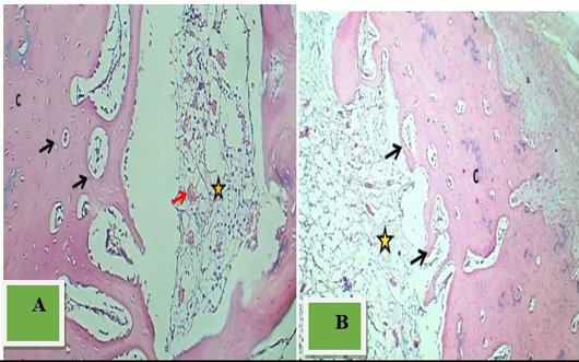
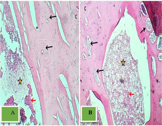
Section at the end of 2ndwk p. o -2 mg/kg B.W lidocaine hcl) shows well- developed mature trabecular bone (Black arrows), active osteogenic tissue (Asterisk), and heavy formation of trabecular bone (Red arrows). H & E.
Section at the end of 2ndt wk p. o -diclofenac shows a thick remodeled bone layer (Arrows), Red marrow (Asterisk), and normal cortical bone (C). H & E stain A. 40x.and B.100x.

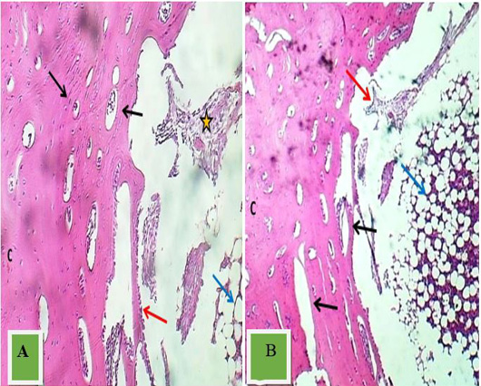
Section at the end of 3rd p. o control shows cortical bone with active endochondral ossification (Black arrow), layer of new bone formation (Red arrows), trabecula (Blue arrows), and osteogenic tissue (Asterisk). H & E stain. A. 40x. and B.100x.
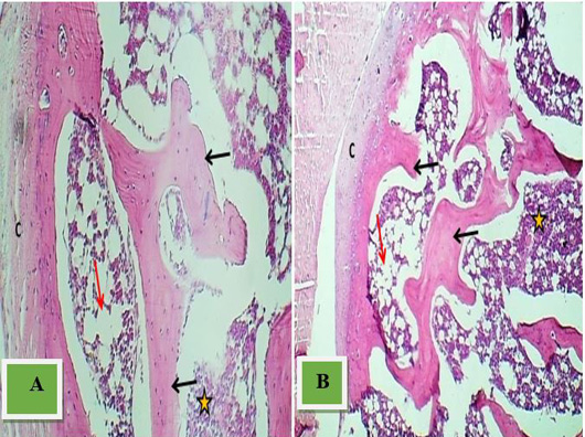
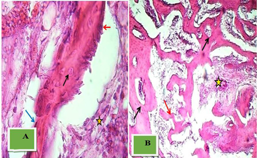
Section at the end of 3rd p.o. 2 mg/kg B.W lidocaine hcl shows red marrow (Asterisk) with little adipocytes (Red arrow) and thick mature trabecular bone (Black arrows). H & E stain. A. 40x. and B.100x

Section at the end of 3rd p. o -Diclofenac shows thick remodeled bone layer (Black arrows), marrow tissue (Red arrow) within osteogenic tissue (Asterisk), and normal cortical bone (C). H & E stain. A. 40x. and B.100x

