Obtaining Osteological Material using Dermestes maculatus De Geer, 1774 (Coleoptera: Dermestidae) in Veterinary Anatomy
Obtaining Osteological Material using Dermestes maculatus De Geer, 1774 (Coleoptera: Dermestidae) in Veterinary Anatomy
Sedef Selviler Sizer1, Semih Kurt1*, Burcu Onuk1, Gokmen Zafer Pekmezci2 and Murat Kabak1
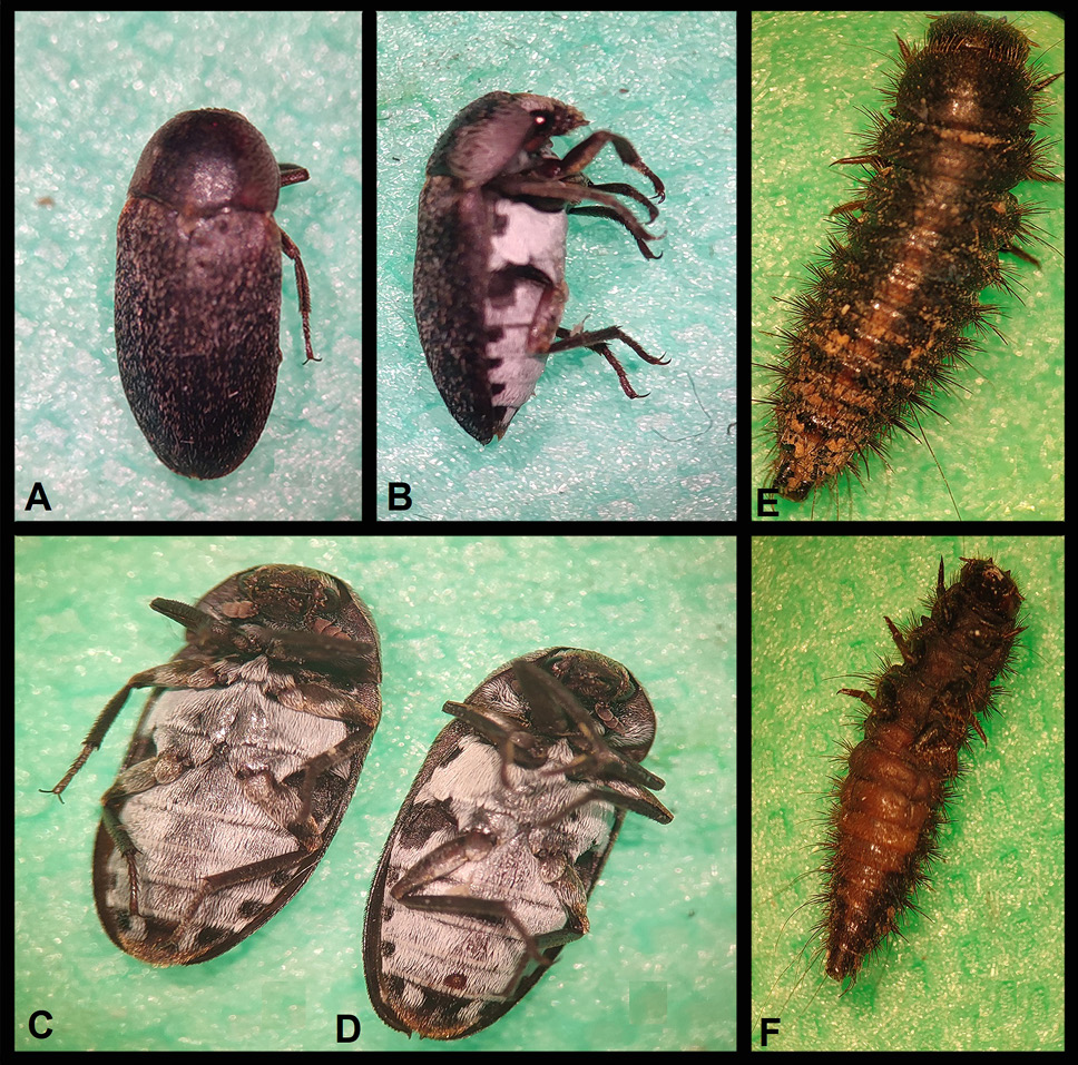
View of adults and larvae of D. maculatus under the stereomicroscope. Adult (A, B), adult female (C), adult male (D) and larvae (E, F).
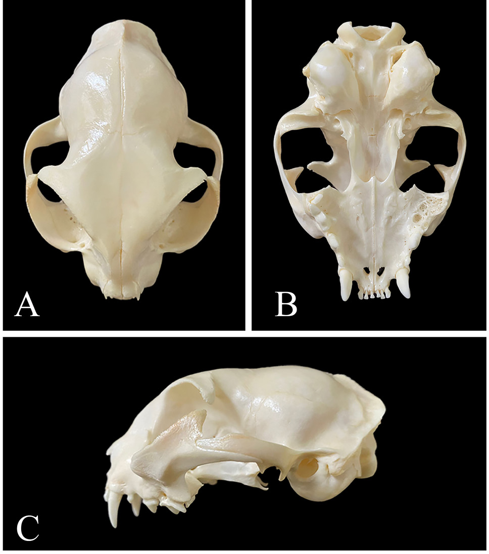
Cat head cleaned by D. maculatus. A, dorsal view of cat head; B, ventral view of cat head; C, lateral view of cat head.
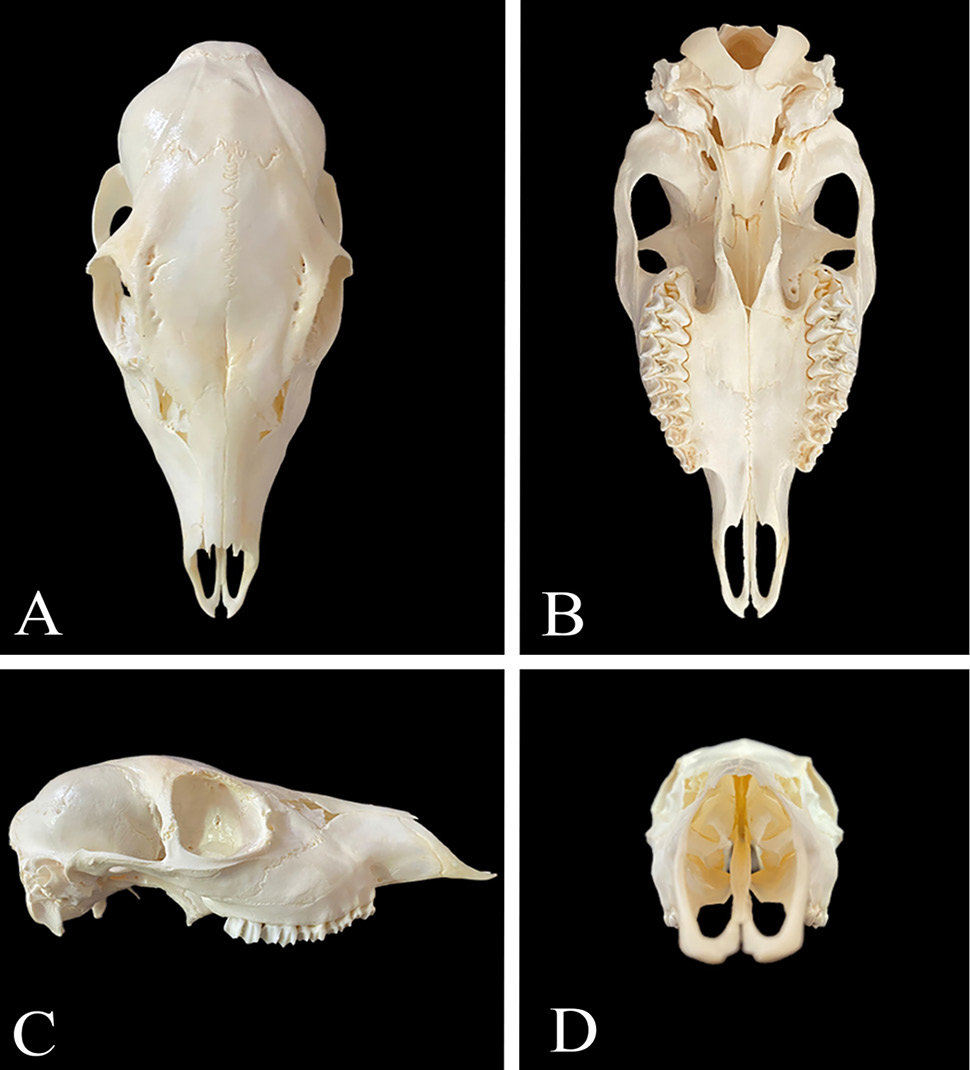
Roe deer head cleaned by D. maculatus. A, dorsal view of a roe deer head; B, ventral view of a roe deer head; C, lateral view of a roe deer head, D: anterior view of a roe deer head.
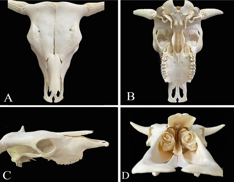
Cow head cleaned by D. maculatus. A, dorsal view of cow head; B, ventral view of cow head; C, lateral view of cow head; D, anterior view of the cow head.
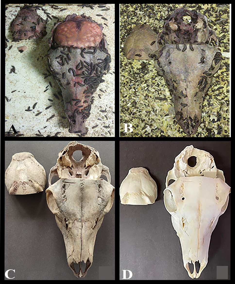
Cleaning stages of the roe deer head (A, B, C), whose brain was found by D. maculatus. Roe deer head whitened in a 5% hydrogen peroxide solution (D).
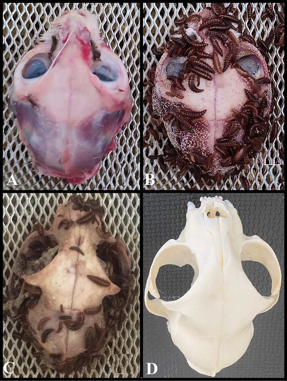
Cleaning stages of the cat head (A, B, C), whose eye was found by D. maculatus. Cat head whitened in a 5% hydrogen peroxide solution (D).


