Morphological and Histological Study of Testis in Hooepoe Bird (Upupa, Epops)
Morphological and Histological Study of Testis in Hooepoe Bird (Upupa, Epops)
Ahlam J.H. Al-Khamas, Sabreen M. Al-Janabi*, Osama Murtadha, Jafar Ghazi Abbas Al-Jebori, Amina Imad Jawad, Dunia M. Al-Rubaie*, Salim Salih Ali Al-Khakani, Baneen, Aqeel Khazal
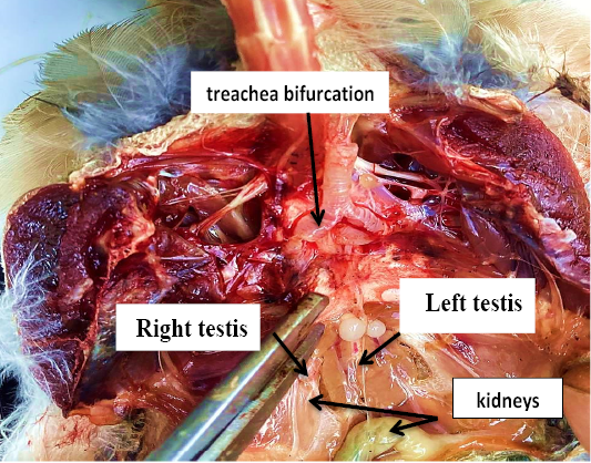
The testis of Iraqi hooepoe bird.
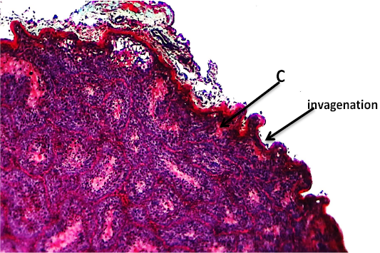
Histological section showing the testis of hoopoe bird (C) cortex (Masson, 4 X).
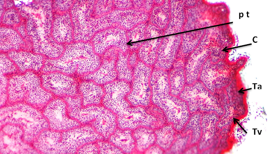
Histological section showing the testis of hoopoe bird (C) cortex. (pt).pretubular tissue (Ta) tunica albugenia (Tv) tunica vasculosa. (Masson, 10X).
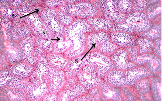
Histological section showing the testis of hoopoe bird; Bv: blood vessel; st: semeniferous tubules; s: spermatozoa. (Masson, 10X).
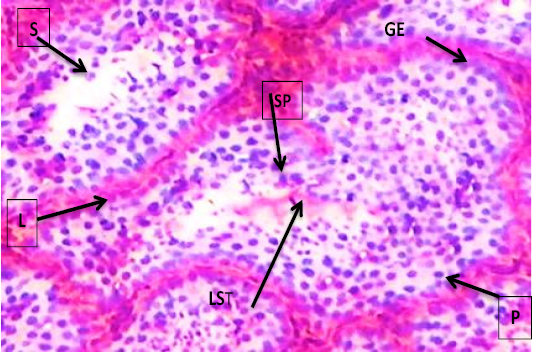
Histological section showing the testis of hoopoe bird; GE: germinal epthelium; LST: lumen semieferos tubules; L: lyding cell; S: sertoli cell; sp: spermatocyte; P: primary spermato cyte. (Masson, 40X).
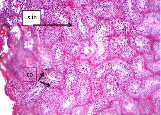
Histological section showing the testis of hoopoe bird; s.in: seminiferous tubules inconvulation; co: convulation; (Masson, 10X).


