Molecular Analysis of Interleukin-10 Gene for -1082 G/A Polymorphism in Breast Cancer Patients
Molecular Analysis of Interleukin-10 Gene for -1082 G/A Polymorphism in Breast Cancer Patients
Muhammad Aijaz1, Nasir Mahmood2, Ghulam Mujtaba3 and Imran Riaz Malik1*
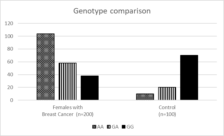
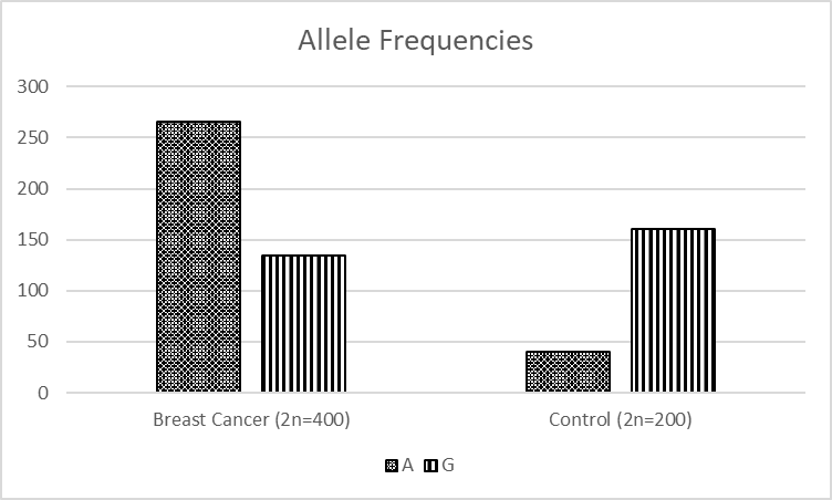
Comparison of genotype AA, GA, and GG in a study group.
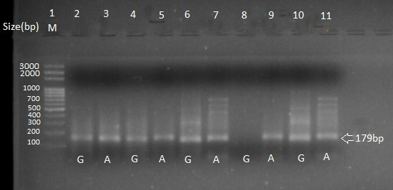
Agarose gel electrophoresis (2%) showing IL10 gene polymorphism. Lane 1 represents 100 bp ladder, Lanes 2 and 3 contain heterozygous samples 1, Lanes 4 and 5 contain heterozygous sample 2, Lanes 6 and 7 contain heterozygous sample 3, Lanes 8 and 9 contain homozygous (AA) sample 4 and, Lanes 10 and 11 contain heterozygous sample 5.
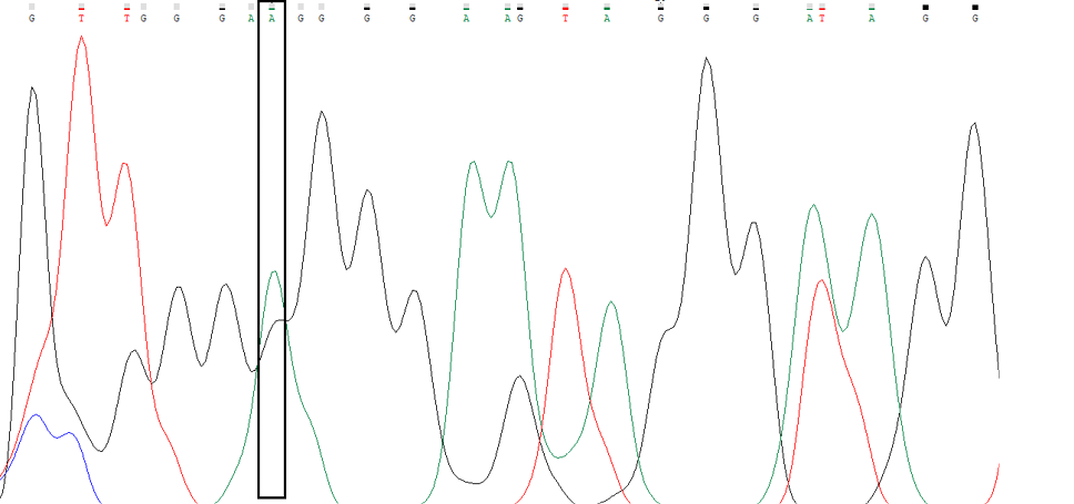
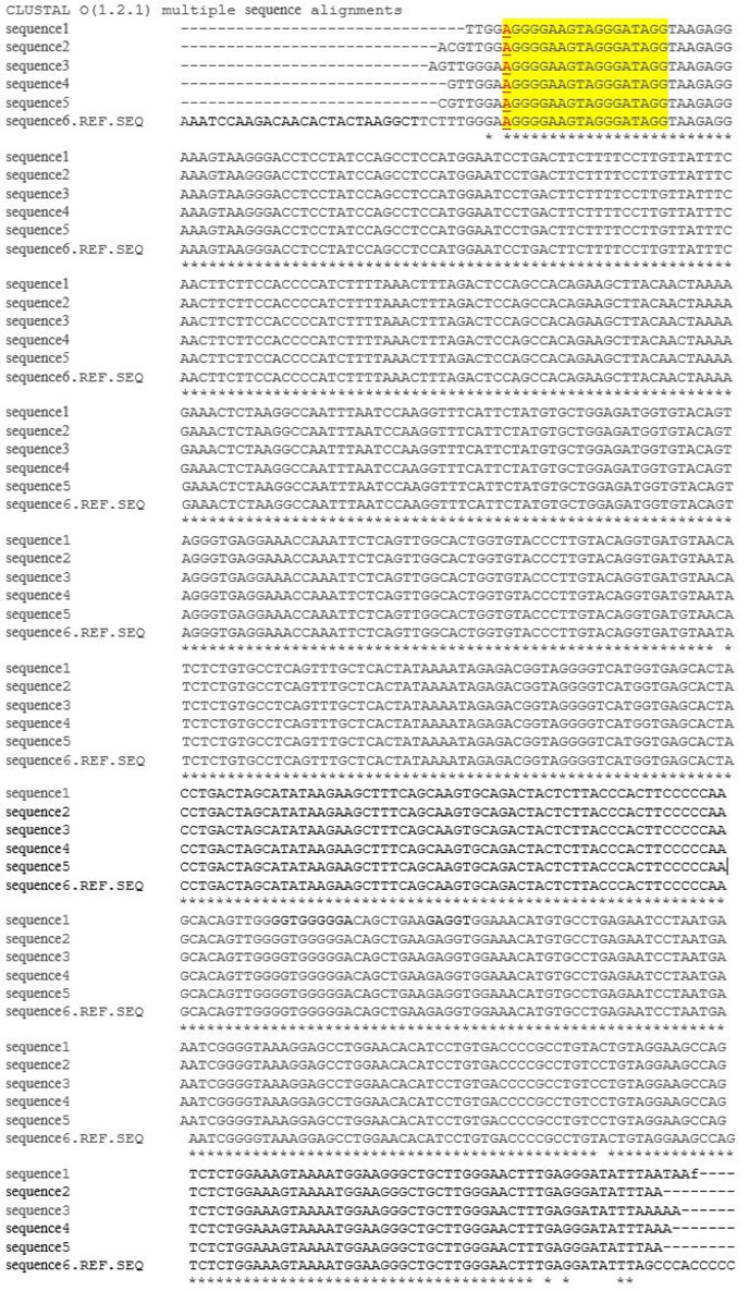
The presence of single nucleotide polymorphism (A nucleotide) in breast cancer patient sample at -1082 position of IL10 gene.
ClustalW analysis results are showing. Molecular analysis of IL10 gene (-1082 G/A) polymorphism is highlighted with red color representing the A allele. Allele-specific primers are yellow highlighted.


