

Larvicidal Potential and Phytochemical Analysis of Garcinia mangostana Extracts on Controlling of Culex pipiens Larvae


Larvicidal Potential and Phytochemical Analysis of Garcinia mangostana Extracts on Controlling of Culex pipiens Larvae
Nael Abutaha, Fahd A. AL-Mekhlafi*, Khalid Elfaki Ibrahim,
Mohammed S. Al-Khalifa and Mohamed A. Wadaan
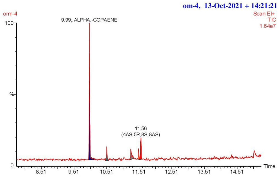
Chromatogram of the hexane extract of Garcinia mangostana pericarp generated using gas chromatography-mass spectrometry.
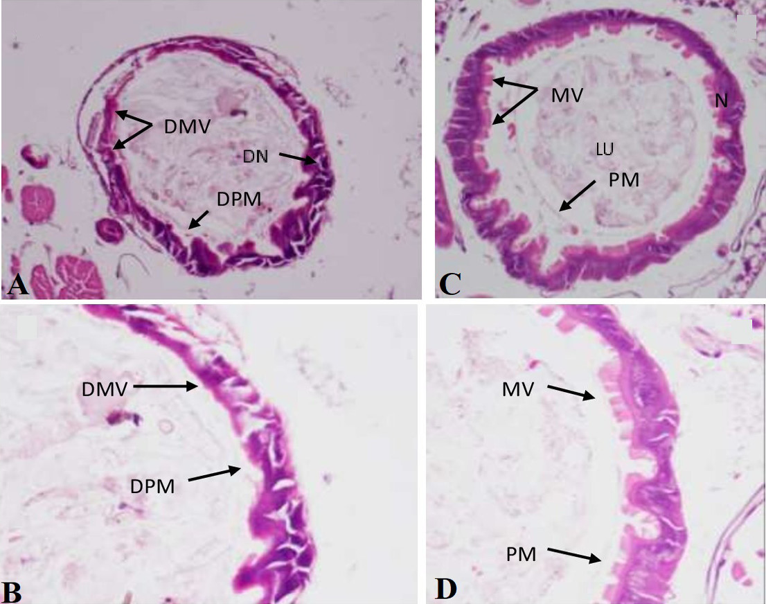
Histology of the 24 h post-treatment midguts of Culex pipiens larvae treated with hexane extract of Garcinia mangostana. (A, B) Cross-sections of the midgut of hexane extract-treated larvae. (C, D) Cross-sections of the midgut of the control (0.01% DMSO) larvae. DMV, Degraded microvilli; MV, Microvilli; DPM, degenerating peritrophic membrane; PM, Peritrophic membrane; DN, degenerating nuclei; N, Nuclei; LU, lumen.
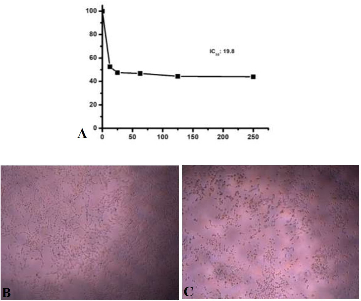
Cytotoxicity effects of the Garcinia mangostana hexane extract against normal human umbilical endothelial cells (HUV-EC) after 24 h of treatment using MTT assay (A). Morphological changes of the HUV-EC cells after treatment with the hexane extract. (B) control cell and C, treated cells.


