

Isolation and Characterization of Virulent Citrobacter freundii Associated with White Muscle Disease in Farmed Red Swamp Crayfish, Procambarus clarkia


Isolation and Characterization of Virulent Citrobacter freundii Associated with White Muscle Disease in Farmed Red Swamp Crayfish, Procambarus clarkia
Hongsen Xu*, Xiaoni Wang, Tie Tian, Changyu Zhao, Denghang Yu and Jun Liu

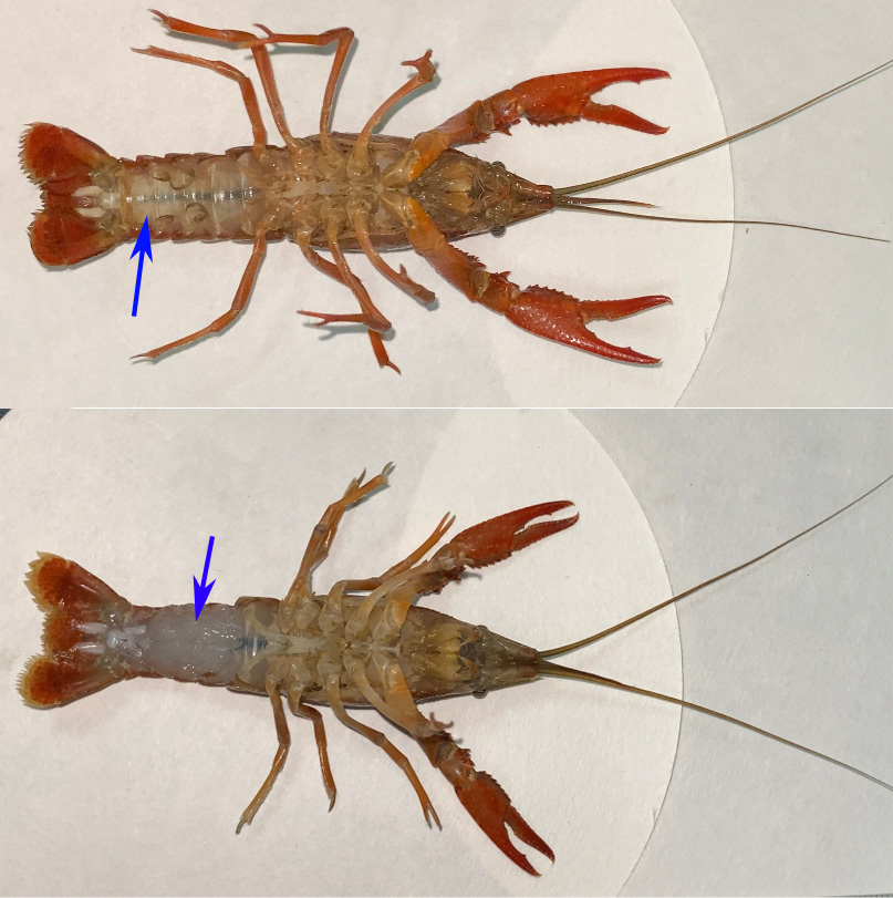
Pathological features of diseased crayfish. A: Diseased crayfish lateral caudal muscle; B: Diseased crayfish inner tail muscle.

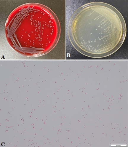
Morphologies of C. freundii FSN1. A: Colony morphologies of FSN1 on LBA plate; B: Colony morphologies of FSN1 on 5% sheep blood agar; C: Gram staining of bacterium FSN1 (100 ×). Scale bars = 20 μm.

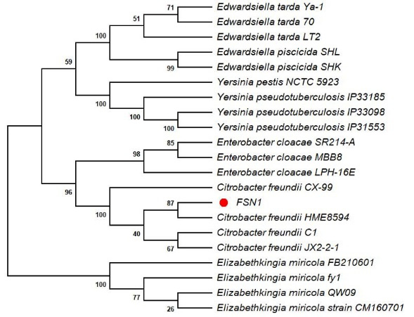
Phylogenetic tree of isolated FSN1 as determined by 16S rRNA gene sequence.

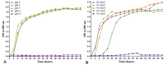
Growth (OD600) of the strain FSN1 at different pH (A) and NaCl concentration (B).
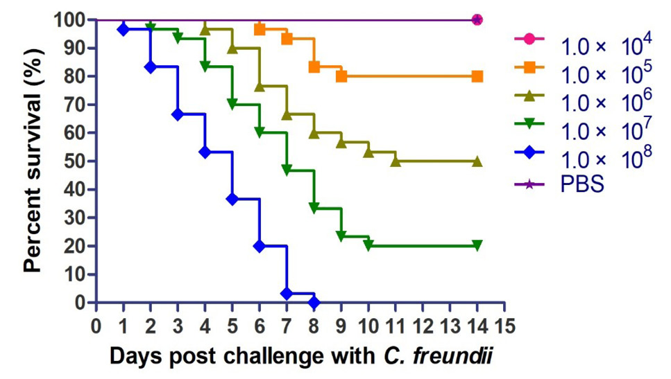
The survival rates of crayfish challenged by various dose of FSN1 during 14 days post infection.

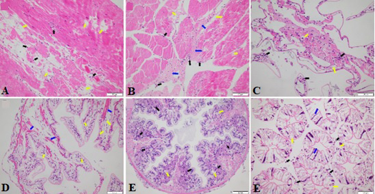
Histopathological observation of various organs in diseased P. clarkia. A-B: The diseased crayfish muscle tissue showed inflammatory cell infiltration (black arrow), cell necrosis (yellow arrow) and fibrinoid degeneration (blue arrow). C: A large number of inflammatory cells infiltrate into the gill fila of the diseased crayfish (black arrow), vacuolar degeneration (yellow arrow). D-E: A large number of inflammatory cells infiltrate the intestines of the diseased crayfish (black arrow), showing vacuolar (yellow arrow) and hyaline degeneration (blue arrow). F: The lumen of the hepatic tubule dilates, the star-shaped structure disappears (the blue arrow), hepatocyte structure collapse, vacuolization (the yellow arrow), interstitial inflammatory cell infiltration (the black arrow).


