Influence of Explant Sources on in vitro Callogenesis and Regeneration in Maize (Zea mays L.)
Zulekha Zameer, Samreen Mohsin, Ammarah Hasnain, Asma Maqbool* and Kauser Abdulla Malik
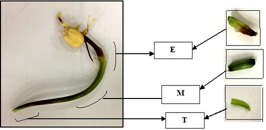
Segments of germinated seedlingas explants for callus induction.
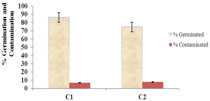
Sterilization efficiency for seeds.
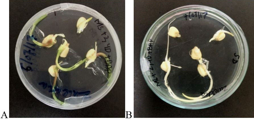
Comparison of maize seed germination. (A) Seeds germinated in light; (B) Seeds germinated in dark.
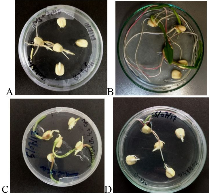
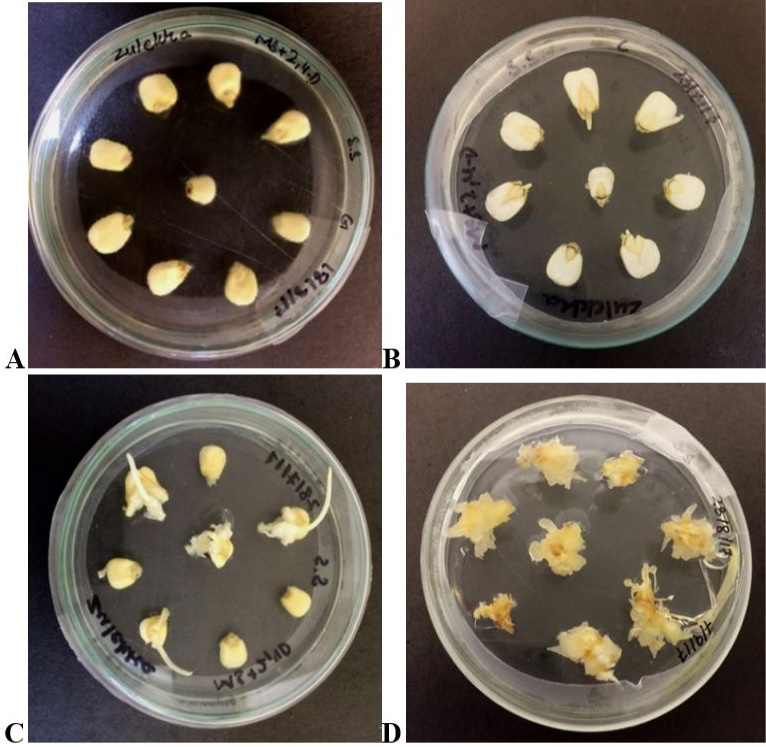
Maize seeds germination. (A) Between two moistened filter papers; (B) On M1 medium; (C) On M2 medium; (D) On M3 medium. The image for each germinating media was captured after 7 days.
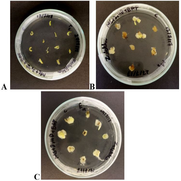
Callogenesis from three parts of 22 days old germinated seedling. (A) Tip (T); (B) Middle part (M); (C) Bulged inter node (E).
Callogenesis by ‘split seed technique’. (A) Sterilized seeds on germination medium; (B) Four days old germinated seeds split longitudinally and placed on callus induction medium; (C) Initial callus formation observed after 7 days in split seeds; (D) Final calli derived from split seed technique after 26 days.
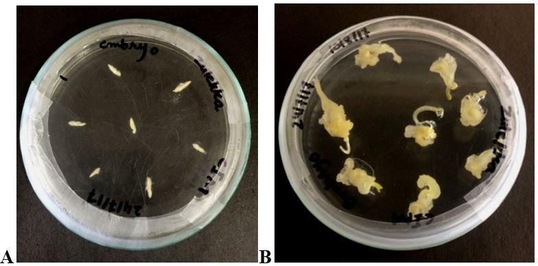
Callogenesis from mature embryos on CIM-3. (A) Day 1; (B) Day 26.
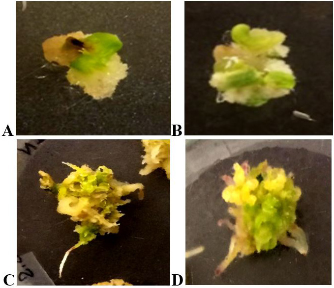
Regeneration response of calli derived from different explants. (A) Middle part of shoot, M; (B) Bulged Internode, E; (C) Split Seed; (D) Mature Embryo.
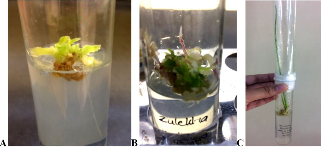
Shoot and root elongation. (A) Buldged internode, E; (B) Split Seed; (C) Mature Embryo.
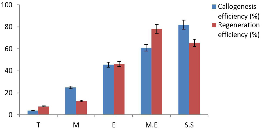
Callogenesis efficiency (%) and regeneration efficiency (%) of different explant sources.


