Infection of Bone Marrow-Derived Mesenchymal Stem Cells with Virulent Newcastle Disease Virus Maximizes Cytokine Production: A Step Toward vNDV Immunotherapy
Infection of Bone Marrow-Derived Mesenchymal Stem Cells with Virulent Newcastle Disease Virus Maximizes Cytokine Production: A Step Toward vNDV Immunotherapy
Tarek A. Wrshana1, Yousry A. Dowidar1, Bahgat A. El-Fiky2, Ali M. El-Rify1, Walaa A. El-Sayed3, Basem M. Ahmed4*
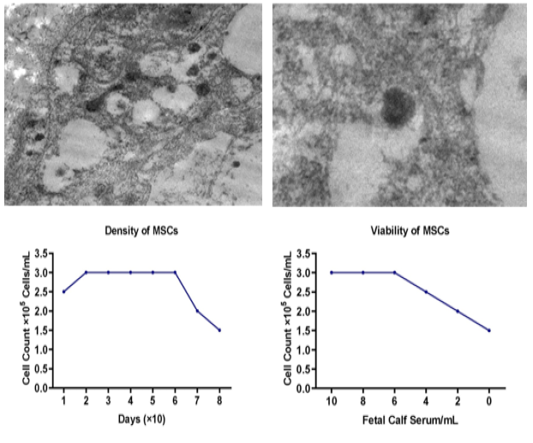
A & B) NDV by TEM examination by thin and ultrastructure examinations in 15000 nm and 3000nm magnification power, C) the density of stem cells with the progression in time, D) viability of stem cells and the effect of FCS on MSCs-CD105.
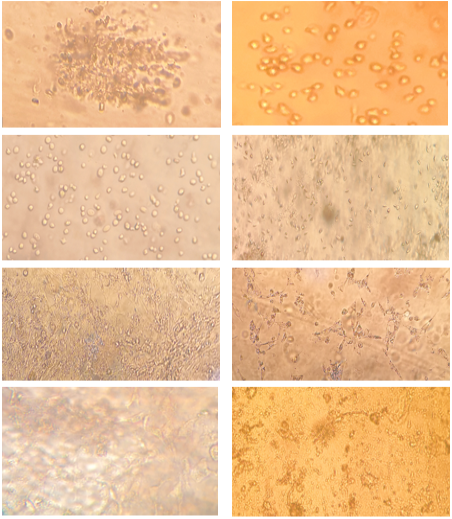
A) First trypsinization, B) second trypsinization, C) third separation shows cell elongation, D) CEFs, E) CEFs maturation, F) CEFs propagation, G) CEFs shows a full cells elongation and forming mono layer, H) CEFs after virus inoculation that from infected CEFs cell, magnification power equal 40x.
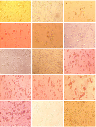
A) Mononuclear cells after separation, B) mononuclear cells after one week shows cell proliferation and cell shape was rounded. C) mononuclear cells after two weeks from separation shows the onset of cell elongation and beginning of the appearance of the bumps entertainment, D) mononuclear cells after three weeks from separation shows a full cells elongation and forming mono layer from MSCs cell which attached on surface and cells was spindle or fibroblastoid, E) mononuclear cells after four weeks from separation shows a full cells elongation and forming monolayer from MSCs cell which attached on surface and cells was spindle or fibroblastoid, F) mononuclear cells after five weeks from separation shows a full cells elongation and forming mono layer from MSCs cell which complete attached and conformation shapes on surface and cells was spindle or fibroblastoid. G) full differentiation to MSCs-CD-105 showed by elongation and connective conjunction between cells like a colonies attached on down surface to adhesive propriety in tissue culture flask, H) Un infected MSCs-CD105 showed by elongation and connective or conjunction between cells like a colonies attached on down surface to adhesive propriety in tissue culture flask with normal growth without any CPEs in MSCs-CD105, I) after inoculation of the virus and attacking cells for beginning of the emergence of the immune and internal response to the virus as first passage infected MSCs-CD105 by NDVs, J) attacking stem cells, and the emergence of giant cells caused by the virus as second passage at infected MSCs-CD105 by NDVs, K) the middle stage of infection with the virus and the spread of infection to most cells as third passages and formation of dendritic shaped cells in MSCs-CD105, L) tourth passage showed complete cythopatic effect (CPE) plaque formation in MSCs-CD105 monolayer, M) the final stage of infection with the virus and the deterioration of the cytological and physiological status of stem cells as fifth passages lysis and degradation, N) restore validity, O) MSCs-CD105 after death by inhibition with NDVs and restore proliferation to generate new resistances cells to NDVs, magnification power equal 40x.
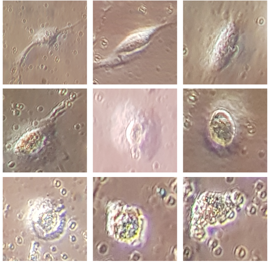
Micro-photos for vNDV effect on MSCs-CD105 during inhibition process, A) single cell shown, a complete external membrane without any damage without virus treatment, B) the beginning of a decrease in the terminal arms of cells immediately after treatment with the virus, C) the beginning of cells are vesicles, D) nods appearing on the outside of the cells, E) swelling and increase in cell size and giant cell formation, F) increasing in swelling, G) beginning in explosion, H) unstable cell, I) cell lysis and degradation, magnification power equal 100x.
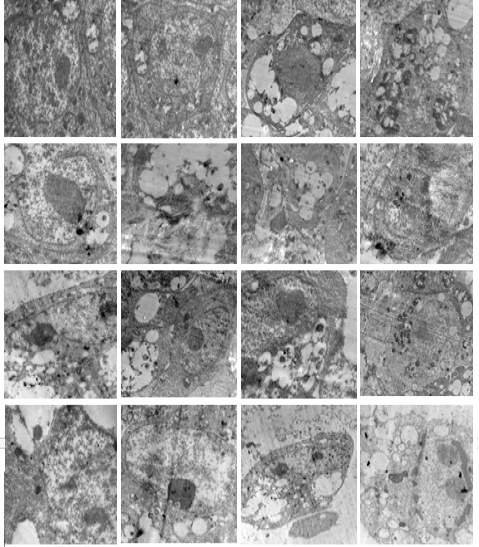
Micrographs for TEM examination by thin and ultrastructure examinations in infected MSCs-CD105 with vNDVs after 24 hours in 15000 nm and 3000nm magnification power.
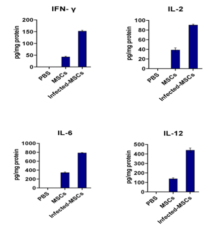
Means of cytokine production levels of IFN-γ, IL-2, IL-6 and IL-12 and evaluated by ELISA in PBS, bone marrow mesenchymal stem cells (BM-MSCs-CD105) and infected BM-MSCs-CD105 by vNDV.


