
Improving the Phytosanitary of Fig Plants Infected with Fig Latent Virus by Nanochitosan and Biomagic

Improving the Phytosanitary of Fig Plants Infected with Fig Latent Virus by Nanochitosan and Biomagic
Hanaa H.A. Gomaa1*, Dalia Y.A. Amin1, Mona A. Ismail1, Basma Hamdy2, Khaled A. El-Dougdoug3

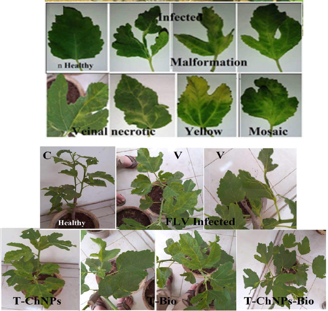
Fig plants showing healthy and infected showed a wide range of foliar discoloration and malformation symptoms, Vein feathering, Vein banding, Leaf deformation, Chlorosis, Mosaic. Fig trees treated with ChNPs (T-ChNPs), fig trees treated with Biomagic (T-bio) and fig trees treated with ChNPs and Biomagic (T-ChNPs and Bio) correction of the effects caused by the Fig latent virus infection.

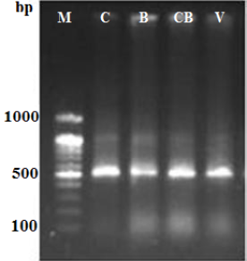
Agarose gel electrograph 1.5% showing PCR products at expected size of 520 bp for CP gene of FLV detected infected fig plant cv. Sultani. C = infected fig plant treated with ChNPs; B= infected fig plant treated with biomagic; CB= infected fig plant treated with ChNPs +Bio magic and V= Infected fig plant.
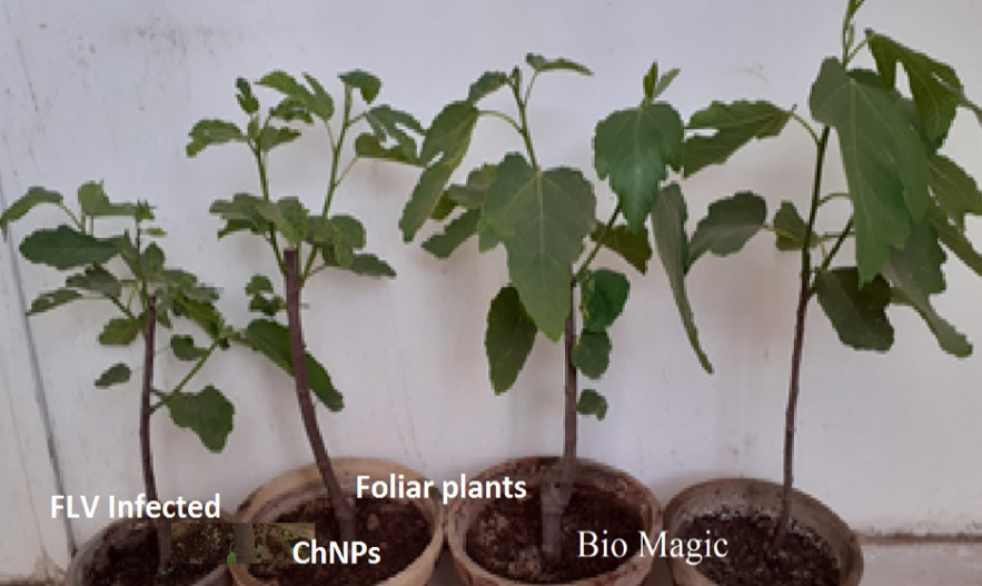
Photograph showing increased leaf area and shoot length of FLV infected fig plants and foliar with ChNPs and biomagic compared with infected fig plants.

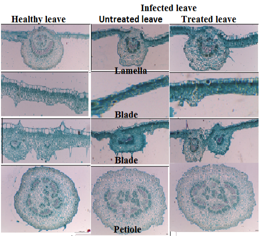
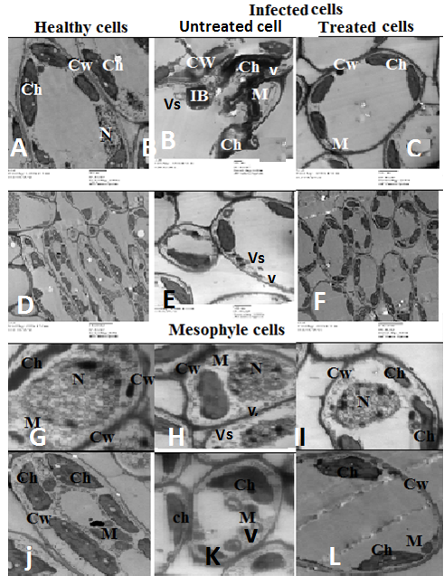
Transverse sections of the middle part of the leaf lamina, blade and petiole of healthy and FLV infected untreated and treated fig plant with ChNPs and or biomagic (X400).

Ultra-micrograph section in mesophyll tissue of healthy infected and treated fig leaf ChNPs and biomagic showing: (A) Impact cells and (B) Magnified nucleus (N) showing compact nucleus in healthy cell and deformed nucleus in infected cell. CW: cell wall; Ch: chloroplast; V: Virus; Vs: vesicles.


