
Histomorphological and Histochemical Study of Ileum and Jujenum of the Camel (Camelus dromedarius)


Histomorphological and Histochemical Study of Ileum and Jujenum of the Camel (Camelus dromedarius)
Khayreia Kadhim Habib*, Masarat S. Al-Mayahi
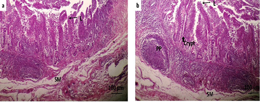
Figure 1
Histological stain: (a) Jejunum, (b) İleum: L: lumen, SM: submucosa, arrow: villus intestinalis, PP: Peyer patches, arrowhead: crypt. Mallory’s trichrome method X 20.
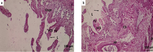
Figure 2
Histological stain: (a) Jejunum, (b) Ileum: TV: Tip of villi, CD: Crypt depth, VCS: Villus-crypt space. PAS stain X 20.
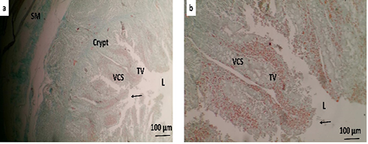
Figure 3
Histological stain: (a) Jejunum, (b) Ileum. PAS/AB stain X 20.
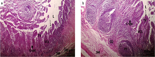
Figure 4
Histological stain: (a) Jejunum, (b) Ileum. Crossman stain X 20.

December 2024
S. Asian J. Life Sci., Vol. 12

