
Epithelial Periostin Expression in Canine Mammary Tumors


Epithelial Periostin Expression in Canine Mammary Tumors
Islam S Alani*, Huda Sadoon Al-Biaty
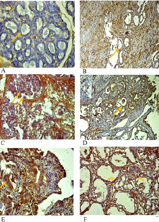
Figure 1:
Immunohistochemical section of the epithelial compartment in canines with mammary tumors shows.
*A: negative expression in the normal mammary gland ; B: low expression in benign tumor ; C: high expression in ductal carcinoma ; D: high expression in invasive Mammary carcinoma ; E: high expression in invasive papillary carcinoma ; F: high expression in intra ductal papillary carcinoma A,C,E,F: 40X, B: 4X, D: 10X.
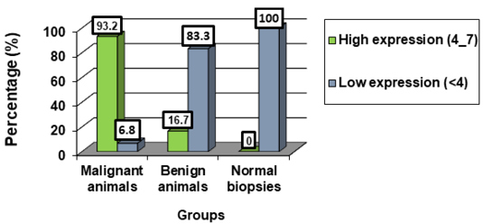
Figure 2:
Expression of periostin in mammary gland tumor tissues and normal biopsies of dogs.
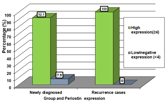
Figure 3:
TissuesExpression of periostin in newly dignosed and recurrent canine mammary carcinoma.
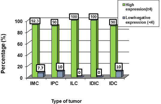
Figure 4:
Expression of periostin in mammary gland cancer in dogs according to type of tumor.
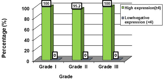
Figure 5:
Expression of periostin in mammary gland cancer in dogs according to Grade of tumor.
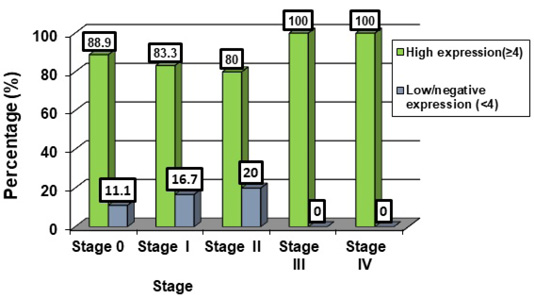
Figure 6:
Expression of periostin in canine mammary gland cancer according to stage.

September 2024
Vol. 12, Iss. 9, pp. 1622-1845
