Effect of Aloe Vera Extract on Some Parameter Complete Blood Count and Liver Functions Induced by Azathioprine in Changes Male Rats
Effect of Aloe Vera Extract on Some Parameter Complete Blood Count and Liver Functions Induced by Azathioprine in Changes Male Rats
Ahmed Al-Hasnawi*, Wafaa Kadhim Jasim
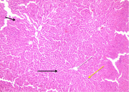
Photomicrograph of rats liver tissue section from Aloe group, showed hepatic tissue with normal liver lobules (black arrow), normal hepatocytes arrangements radiating (yellow arrow), around central vein (white arrow) (H and E, 10X).
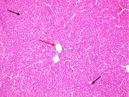
A photomicrograph of a control group rat liver tissue segment revealed hepatic tissue with normal liver lobules (black arrow) and a remarkable normal central vein (red arrow). (10X H and E).
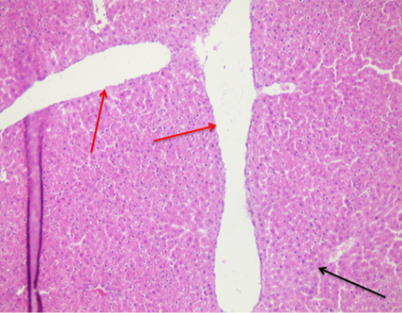
Photomicrograph of rats liver tissue section of AZA group, showed sever central vein dilatation (red arrow), significant hepatic degenerative changes (black arrow). (H and E, 20X).
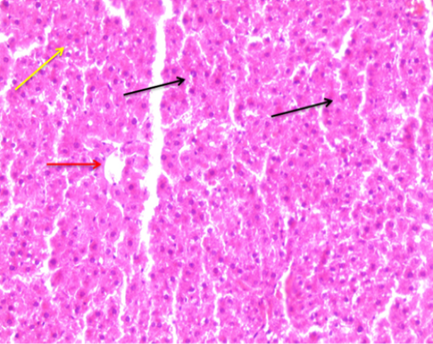
Photomicrograph of AZA + Aloe group rats liver tissue slice showing modest hepatic edema (black arrow), hepatic inflammatory cell infiltration (yellow arrow), and normal central vein (red arrow). 40X (H and E).


