
Determination of prevalence and associated risk factors of Gastrointestinal Nematodes in Cattle at Sylhet Region, Bangladesh


Short Communication
Determination of prevalence and associated risk factors of Gastrointestinal Nematodes in Cattle at Sylhet Region, Bangladesh
Real Datta1*, Md- Tariqul Islam2, Md. Afradul Islam3, Apurbo Kumar Mondal4, Tilak Chandra Nath1, Kazi Mehetazul Islam1, Jamal Uddin Bhuiyan1
1Department of Parasitology, Sylhet Agricultural University, Sylhet, Bangladesh; 2Department of Microbiology & Immunology, Sylhet Agricultural University, Sylhet, Bangladesh; 3Office of the Registrar, Sylhet Agricultural University, Sylhet, Bangladesh; 4Department of Physiology & Pharmacology Bangabandhu Sheikh Mujibur Rahman Agricultural University, Gazipur, Bangladesh.
Abstract | A cross-sectional study was conducted to identify the significant gastrointestinal (GIT) nematodes species affecting cattle and to ascertain the prevalence and associated risk factors of GIT nematodes in cattle in Sylhet district of Bangladesh. The investigation included the identification of nematode species, a questionnaire survey, and fecal testing. This study investigated the connections between various significant risk factors and the prevalence of GIT nematodes in Cattle. A total of 246 fecal samples from cattle grown under various management systems were obtained. A total of 97 of the 246 investigated fecal samples tested positive for GIT nematodes, resulting in a prevalence of 39.43%. The results show that the presence of GIT nematodes is significantly correlated with the type of farm, breed, frequency of deworming, and body condition score of cattle but not significantly with other factors like animal gender and age. Additionally, this study concludes that every farm should employ bio-security measures as well as a regular deworming strategy due to the frequency of GIT nematodes, particularly in indigenous breeds of cattle raised in free range farming systems.
Keywords: GIT nematodes, Prevalence, Risk factors, Cattle
Received | June 24, 2024 Accepted | July 03, 2024; Published | July 25, 2024
*Correspondence | Real Datta, Department of Parasitology, Sylhet Agricultural University, Sylhet, Bangladesh; Email: rdatta.parasitology@sau.ac.bd
Citation: Datta R, Islam MT, Islam MA, Mondal AK, Nath TC, Islam KM, Bhuiyan JU (2024). Determination of prevalence and associated risk factors of gastrointestinal nematodes in cattle at Sylhet Region, Bangladesh S. Asian J. Life Sci. 12: 59-63.
DOI | http://dx.doi.org/10.17582/journal.sajls/2024/12.59.63
ISSN | 2311–0589
INTRODUCTION
An essential component of agriculture, the livestock industry makes a sizable contribution to Bangladesh’s national economy. In addition to contributing 1.43% of the country’s GDP, this subsector also employs 20% of the workforce (BBS 2020; MOFL 2019). Since cattle are domesticated animals that can live and reproduce while being cared for by humans they have a significant positive impact on human livelihoods in emerging economies. Smallholder farmers in Bangladesh own most of the cattle in mixed crop-livestock systems. In this farming system, cattle are essential to the household’s aspirations for a sustainable way of life since they can boost cash income and promote food security. Numerous illnesses, particularly parasite infections of the gastrointestinal tract (GIT), have a negative impact on the growth, development, and productivity of these animals (Thapa et al., 2020). According to Dey et al. (2020), GIT parasites are particularly sneaky and cause economic losses through decreased output and higher death.
Previous research in Bangladesh demonstrated the high prevalence of GIT parasite infections in cattle (Chowdhury et al., 2017). Unfortunately, parasitic infections are commonly ignored in Bangladesh since animals with parasitic infections exhibit minimal or no clinical symptoms (Alim et al., 2012). Several variables that are influenced by parasite host environment interactions define the epidemiology of GIT nematode infections (Maingi et al., 2004). The main risk variables can be broadly divided into three categories: host factors (genetic resistance, age, and physiological status of the animal), parasite factors (anthelmintic resistance of various species), and environmental factors (climate, stocking density, and management). These risk variables have a significant impact on the pathophysiology of GIT parasite infections in animals (Badran et al., 2012).
Only a few epidemiological studies have previously been carried out in Bangladesh to determine the prevalence and potential risk factors of GIT nematode infections. The main obstacle to planning nematode control operations in our study area is the lack of epidemiological data on GIT nematodes causing cattle nematodiasis. To create a baseline for future control and prevention measures, the goals of this study were to assess the prevalence, major species identification, and parameters associated with the epidemiology of GIT nematodes in the area.
Material and methods
Study location and laboratory examination
A cross sectional study was conducted on indigenous and cross breeds of cattle reared and managed under free range and intensive farming systems in two Upazilla of Sylhet district which is in north-east Bangladesh and is one of the four districts in the Sylhet Division. Laboratory examinations were performed in the Laboratory of Department of Parasitology, Sylhet Agricultural University (SAU). Individual research animal feces were collected for coprological analysis using a systematic random sampling technique. As risk variables for the development of GIT nematode infections in cattle, farm type, breed, age, gender, physical condition, and frequency of deworming were taken into consideration. The sample size was estimated using the Thrusfield (2005) calculation with an expected prevalence of 80% with a 5% absolute precision at a 95% confidence range based on prior research on the prevalence of GIT nematodes in cattle at various regions in Bangladesh and throughout the world. Therefore, the sample size was 246, which was utilized as a typical animal to assess the prevalence of GIT nematodes by inserting the values of the variables in the formula.
Fecal samples were collected directly from the rectum of each animal by using rectal gloves and were placed in sampling bottles containing 10% formalin and labeled. For a qualitative analysis of the different types of gastrointestinal nematode eggs, the collected fecal samples were processed and evaluated with Modified McMaster technique as described by Sharma et al (2013).
Data Management and Analysis
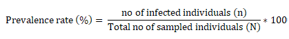
The raw data was entered into Microsoft excel spreadsheet and analyzed using R studio (Version 1.2.1335). Confidence interval was set at 95% and statistically significant association between variables were considered to exist if the computed P-value is less than 0.05. The prevalence rate was calculated for all data by using the following formula:
Microscopic examination of Eggs and identification of nematodes:
To identify nematode eggs, a fecal sample diluted with saturated salt solution was collected. Eggs were initially viewed and then counted using a 10X objective using a sample that was transferred from the beaker’s center level into a two-chambered McMaster chamber (FEC Source, USA). The keys provided by Urquhart (1996) were used to identify the eggs of several parasite species.
Risk factors considered and questionnaires
The study was conducted on cattle with consideration of farm type, breed, age, body condition score, and frequency of deworming of cattle as risk factors. The information was gathered directly from the owners and visual observation.
Results and discussion
Overall Prevalence
Numerous research revealed that gastrointestinal nematodes are the main contributors to productivity declines in Bangladeshi cattle production (Dey et al ., 2020).
An overall prevalence of gastro-intestinal nematode infection of 39.43% of cattle from this area were parasitized by at least one type of GIT nematode, according to the coprological examination carried out for this study. The remaining 52 (21.13%) cattle out of 246 were co-infested (mixed infestation) with several types of GIT endoparasites, as opposed to the 45 (18.29%) cattle out of 246 who were infected with a single helminth parasite. Our results are consistent with those of (Ahmad et al., 2020) who found that buffalo calves in Sylhet, Bangladesh, had a prevalence rate of GIT parasites of approximately 36.47%.
The results of the present study, in contrast, are less significant than those of earlier research by (Dey et al., 2020; Chowdhury et al., 2017; Amran et al., 2018; Rahman et al., 2017) who reported that the prevalence rate of GIT parasites among various species of livestock was 62.1%, 64%, 63.88%, and 63.4%, respectively, in various areas of Bangladesh. These inconsistencies might be explained by variations in the management system, to pography, de-worming procedures, sample size, and climatic conditions that support the survival of the parasite’s infective stage.
We were able to distinguish identify two of the most prevalent species, Trichostrongylus spp. and Nematodirrus spp., based on their physical traits (Figures 1 and 2) (Urquhart et al., 1996).
Risk Factors and Prevalence of Gastrointestinal Nematodes Infection in Cattle
The highest and lowest percentage of GIT nematodes from different potential risk factors of cattle were recorded in Table 1. Chi-square test analysis of the potential risk factors revealed presence of significant difference in the prevalence of GIT nematodes between farm type (P<0.05), breed (P<0.05), body condition (P<0.05) and frequency of deworming (P<0.05) of animals. Conversely, statistically significant difference was not observed for ages and gender of animals (P>0.05).
According to statistical research, there is a connection between the kind of agricultural animals, breed, physical condition, frequency of deworming, and presence of GIT nematodes (P<0.05).As a result, a higher prevalence was seen in cattle raised in a free-range farming system (57%) and cattle of indigenous breeds (57.14%) compared to cattle raised in an intensive farming system (20.83%) and cattle of crossbreeds (20.83%). The fact that nematodes primarily complete their life cycle in grazing pastures may be the cause of the results’ considerable variations. According to studies, the climate at a natural pasture plays a major role in the survival and transmission of parasite eggs and larvae (Pfukenyi et al., 2013).
The prevalence of GIT parasites is significantly influenced by local environmental factors, including humidity, temperature, rainfall, vegetation, and management practices. Ruminants, in particular cattle, sheep, and goats, are vulnerable to GIT parasite infestation in low-lying grazing regions that are frequently water-filled. The general temperature and humidity of the area surrounding the cattle grazing field in the study area may have an impact on the growth of GIT in ruminants (Islam et al., 2015). Animals raised in a free-range agricultural environment are therefore more susceptible to GIT nematodes than those raised in an intensive farming system.
Farm cattle are not allowed to access open fields. The low prevalence of GIT nematode infections in farm animals is a result of these animals being exposed to grass imported from outside the farms. Crossbred cattle are raised in Sylhet in large farms, whilst native cattle are raised in wide-open grazing fields. In this study, the prevalence of GIT nematode infection was substantially correlated with the Body Condition Score (BCS) (P<0.05) and it was shown that poor body condition had a greater prevalence (62.8%) of the infection than excellent body condition (27.3%). This poor physical condition may be caused by hunger, another illness present at the same time, or the ongoing parasitic infection, which results in a weak immune reaction to the parasite’s infective stage. Prior research by (Dey et al., 2020) found a highly significant correlation (P<0.05) between the prevalence of GIT nematodes infection in goats and the animals in poor condition.
In our investigation, we found a substantial correlation between the frequency of deworming and the presence of GIT nematodes in cattle. A higher prevalence of GIT infections was seen in cattle with deworming intervals larger than 6 months. Many of the farmers in this study used a deworming schedule that ranged from three to six months, which contributed to the overall low prevalence of nematodes.
Table 1: Analysis of Risk Factors and Association of Nematodiasis with Different Variable
|
Risk factor
|
Level of risk factor
|
No. of animals examined |
Prevalence (n/N) |
Prevalence rate |
Chi-square (χ2) |
Degrees of freedom (df) |
Significance of difference (p value) |
| Farm type | Intensive farm | 120 | (25/120) | 20.83 |
32.426 |
1 |
0.001
|
| Free range | 126 | (72/126) | 57.14 | ||||
| Total | 246 | 97 | 39.43 | ||||
| Age | 3-12 Month | 36 | (13/36) | 36.11 | 0.065834 | 1 | 0.797 |
| Greater than 12 Month | 210 | (84/210) | 40 | ||||
| Total | 246 | 97 | 39.43 | ||||
| Breed | Cross Breed | 120 | (25/120) | 20.83 | 32.426 | 1 | 0.001 |
| Indigenous | 126 | (72/126) | 57.14 | ||||
| Total | 246 | 97 | 39.43 | ||||
| Gender | Male | 16 | (10/16) | 62.5 | 2.8502 | 1 | 0.091 |
| Female | 230 | 87/230 | 37.82 | ||||
| Total | 246 | 97 | 39.43 | ||||
| Body condition | Good | 166 | 42/166 | 25.30 | 40.871 | 1 | 0.001 |
| Poor | 80 | 55/80 | 68.75 | ||||
| Total | 246 | 97 |
39.40 |
||||
| Frequency of Deworming | 3-6 M | 163 | 37/163 | 22.69 | 54.5 | 1 | 0.001 |
| More than 6 M | 83 | 60/83 | 72.28 | ||||
| Total | 246 | 97 | 39.43 |
P- value less than 0.05 considered significant
Acknowledgements
The authors are cordially acknowledged to the SAURES authority, SAU, Sylhet for providing financial support for successful implementations of the project during the period of research work. Also, we would like to express our heartfelt gratitude to the laboratories of Department of Parasitology, Faculty of Veterinary, Animal and Biomedical Sciences, Sylhet Agricultural University, Sylhet-3100, Bangladesh.
Conflict of interests
No confliction of interests.
novelty statement
This research has determined the prevalence rate of Gastrointestinal nematodes and its associated risk factors of the study location. We also identified two important nematode species in cattle.
Authors’ Contributions
Real Datta; Apurba Kumar Mondal; Md. Afradul Islam; Md. Tariqul Islam; Research, Data Analysis, Writing of Original Draft. Tilak Chandra Nath, Kazi Mehetazul Islam; Jamal Uddin Bhuiyan: Conceptualization Writing, Revision and Editing. All authors tried to edit the final draft of the manuscript.
References
Ahmad S., Chowdhury S. R., Hossain M., Rahman M., Rahman M. (2020). The prevalence of gastrointestinal parasites in buffalo calves in Sylhet district of Bangladesh. Iranian J. Vet. Med., 14(3).
Alim M. A., Das S., Roy K., Sikder S., Mohiuddin M. M., Hossain M. A. (2012). Prevalence of gastrointestinal parasites in cattle of Chittagong division, Bangladesh. Wayamba J. Anim. Sci., 4(7): 1-8.
Amran MA, Yadav SK, Akter F, Sarkar S, Hossain MA, Joy SM, Samrat AAK (2018). Prevalence of gastrointestinal parasitic infections in different existing goat breeds in different districts of bangladesh. J. Adv. Parasitol. 5(1): 11-21.
Badran I., Abuamsha R., Aref R., Alqisi W., Alumor J. (2012). Prevalence and diversity of gastrointestinal parasites in small ruminants under two different rearing systems in Jenin district of Palestine. An-Najah University J. Res.-A (Nat. Sci.)., 26(1): 1-18. https://doi.org/10.35552/anujr.a.26.1.96
Chowdhury R., Sen A., Kar J., Nath S. K. (2017). Prevalence of gastrointestinal parasitism of cattle at Chandaniash Upazilla, Chittagong, Bangladesh. Int. J. Adv. Res. Biol. Sci., 4(6): 144-149.
Dey A. R., Begum N., Alim M. A., Malakar S., Islam M. T., Alam M. Z. (2020). Gastro-intestinal nematodes in goats in Bangladesh: a large-scale epidemiological study on the prevalence and risk factors. Parasit. Epidemiol. Cont., 9: e00146. https://doi.org/10.1016/j.parepi.2020.e00146
Islam M. M., Islam M. S., Howlader M. M. R., Lucky N. S. (2015). Prevalence of gastrointestinal nematodiasis and comparative efficacy of anthelmintics on body weight of cattle in Bangladesh. Int. J. Scient. Res. Agricult. Sci., 2(3): 61-75. https://doi.org/10.12983/ijsras-2015-p0061-0075
Kumsa B., Tolera A., Nurfeta A. (2010). Comparative efficacy of seven brands of albendazole against naturally acquired gastrointestinal nematodes in sheep in Hawassa, southern Ethiopia. Turkish J. Vet. Anim. Sci., 34(5): 417-425. https://doi.org/10.3906/vet-0712-28
Maingi N., Ng’ang’a C. J., Kanyari P. W. N., Munyua W. K. (2004). Development, survival and availability of gastrointestinal nematodes of sheep and pastures in a semi-arid area of Kajiado District of Kenya. Vet. Res. Commun., 28: 491-501. https://doi.org/10.1023/B:VERC.0000040246.22919.cd
Pfukenyi D. M., Mukaratirwa S. (2013). A review of the epidemiology and control of gastrointestinal nematode infections in cattle in Zimbabwe. Onderstepoort J. Vet. Res., 80(1): 1-12. https://doi.org/10.4102/ojvr.v80i1.612
Rahman M. A., Labony S. S., Dey A. R., Alam M. Z. (2017). An epidemiological investigation of gastrointestinal parasites of small ruminants in Tangail, Bangladesh. https://doi.org/10.3329/jbau.v15i2.35071
Sharma S., Iqbal A., Azmi S., Shah H. A. (2013). Study of poultry coccidiosis in organized and backyard farms of Jammu region. Vet. World., 6(8): 467. https://doi.org/10.5455/vetworld.2013.467-469
Thapa Shrestha U., Adhikari N., Kafle S., Shrestha N., Banjara M. R., Steneroden K., Ghimire P. (2020). Effect of deworming on milk production in dairy cattle and buffaloes infected with gastrointestinal parasites in the Kavrepalanchowk district of central Nepal. Vet. Rec. Open., 7(1): e000380. https://doi.org/10.1136/vetreco-2019-000380
Thrusfield M. (2005). Veterinary Epidemiology. (2nd. Ed.). Blackwell Science, UK. pp 180-181.
Urquhart G., Armour J., Duncan J., Dunn A., Jennings F. (1996). Veterinary Parasitology 2nd Edition. University of Oxford.


