
Degenerative Joint Disease in the Skeletal Remains of a Captive Bornean Sun Bear (Helarctos malayanus euryspilus)


Degenerative Joint Disease in the Skeletal Remains of a Captive Bornean Sun Bear (Helarctos malayanus euryspilus)
Awang Hazmi Awang-Junaidi1, Yeoh Boon Nie2, Siew Te Wong2, Nur Nabila Sarkawi3, Wafeq Mu’izzadin Khairul Anuar1, Siti Mariam Zainal Ariffin1*

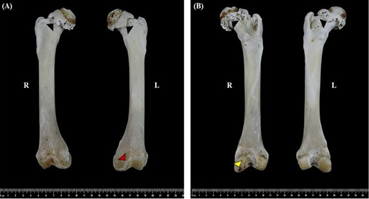
Osteophyte formation along the femoral head-neck junction (black arrowheads) and the trochlear margin of the distal femur (red arrowheads). The right femoral head showed a flattened shape, compared with the rounded appearance observed in the left femoral head. Evidence of subchondral bone degradation was observed on the surface of the right medial femoral condyle (yellow arrowheads). (A) cranial view of right (R) and left (L) femur. (B) caudal view of right (R) and left (L) femur.
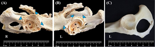
Degenerated acetabulum (A-B) undergoes remodelling, with severe osteophyte formation (blue arrowheads) at the cranial effective acetabular rim and caudal acetabular edge. Evidence of subchondral bone degradation was observed on the entire surface of the right (R) and left (L) acetabulum. A normal acetabulum (C) is characterized by smooth acetabular edge, surface and bone.
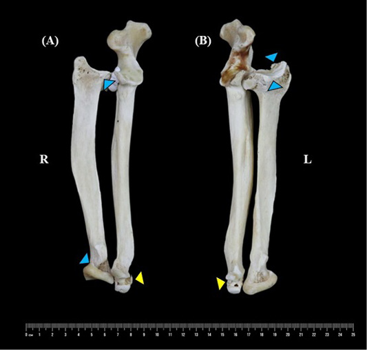
Osteophyte formation on the proximal and distal radius (blue arrowheads) and the distal ulna (yellow arrowheads). (A) Medial view of right radius and ulna. (B) Medial view of the left radius and ulna.
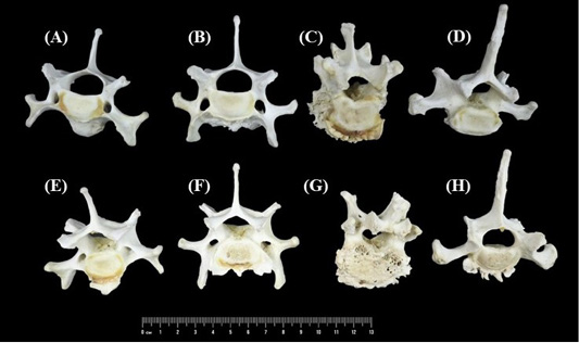
Spondylosis deformans lesion characterized by the osteophyte formation developed at the edge of the ventral vertebral body of C6 (A and E), C7 (B and F), T1 (C and G) and T12 (D and H). Bone degradation, characterized by erosion, roughened and irregular bone texture, was found on both the cranial and caudal intervertebral surfaces of all the affected vertebrae. (A-D) Cranial view; (E-H) Caudal view.

