
Comparative Anatomy, Histology, Histochemistry, and Immunohistochemistry of the Esophagus in Ostrich (Struthio camelus) and Turkey (Meleagris gallopavo)


Comparative Anatomy, Histology, Histochemistry, and Immunohistochemistry of the Esophagus in Ostrich (Struthio camelus) and Turkey (Meleagris gallopavo)
Rabab Abd Alameer Naser1*, Sameer Ahmed Abid Al-Redah2, Enas S. Ahmed3, Fatimah S. Zghair2, Ali Ibrahim Ali Al-Ezzy4
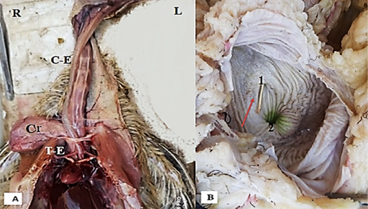
Photograph illustrate: A: The lesophagus in turkey consist from three parts cervical esophagus (C-E), thoracic esophagus (T-E).and crop (Cr), and relative with Trachea (T), B: The crop after fixed by 10% formalin, mucosal fold (brown arrow).
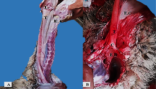
Photograph illustrate, A: The esophagus in ostrich (black arrow).(A):Cervical esophagus(E.C.) longitudinal folds (black arrow), (B):Thoracic Esophagus (E.T), (Trachea (T), Syrinx (C).
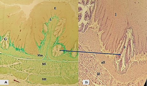
Photomicrograph section illustrated (A): the esophagus male turkey cervical part the epithelium (E),( lamina propria (L.P) ,musculais mucosa (Mm), two layers of tunica muscularis (MI), (ME) , tunica adventitia (brown arrow), Masson’s Trichrome stain 40× ( B)shows tubular acinar gland in turkey H and E) stain100 ×.
Photomicrograph section illustrated the esophagus in ostrich cervical part H and E stain 100×,(B): the gland in ostrich simple tubular gland (G) (mucus gland ) PAS stain 200 ×:B: epithelium (E),( lamina propria (L.P), lamina muscularis (Mm); (Mm), tunica muscularis (MI) and (ME).
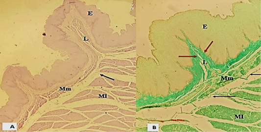
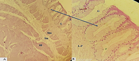
Photomicrograph section showing the mucosal fold in turkey (crop), epithelium (E), lamina propria contain high number of collagen fibers (brown arrow), lamina muscularis (Mm), submucosa (black arrow), inner tunica muscularis (MI), A: (H and E) stain 100 ×, B: Masson’s Trichrome stain 100×.

Photographic section of the thoracic part esophagus in chick turkey, (A): The mucosal folds become, elongated, and unbranched leaving only a narrow lumen, T. serosa (brown arrow) A: Masson’s Trichrome stain A: 100 X, (B) mucus gland in turkey lined by columnar cell with basal nucleus: PAS stain 400 x.
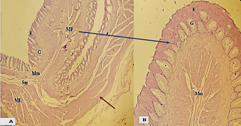
photographic section ostrich showing the mucosal fold (MF) was elongated (H and E) stain 100×, (B): thoracic part of esophagus ostrich B: (H and E) stain 200 x. epithelium gland (G), submucosa (SM ), lamina muscularis (Mm).
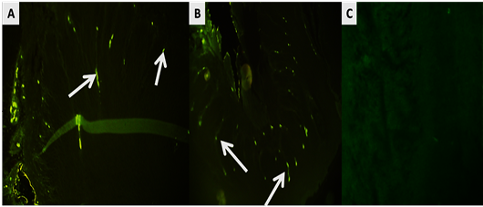
Immunohistochemical image of the esophagus in (A: turkey; B: ostrich, C: negative control) show immunoreactive cells (white arrows) (lable by FITC with the secondary antibody) in the mucosal layer that expression of serotonin cells. And negative control (C) when the omitted of primary antibody and used secondary antibody. (A and B: X200; C: X400).

