Burned Wound Healing Effect of Prepared Pumpkin Seed Oil Nano Phytosome Loaded Lidocaine in Rabbit
Burned Wound Healing Effect of Prepared Pumpkin Seed Oil Nano Phytosome Loaded Lidocaine in Rabbit
Wafa’a Mohammad Abduwhab1*, Waseem Ali Hasan2, Mohanad Abdulsataar AL-Bayati3*
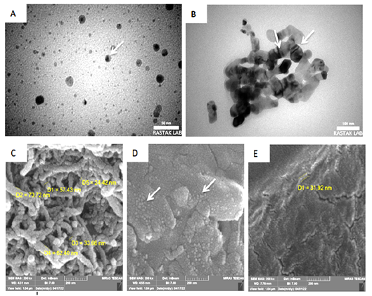
Electron microscope shows scan and transmission of Nano phytosome pumpkin-lidocaine , where (A&B) are the transmission modes, the white arrows indicate multilamellar vesicles, where C, D, and D are the scan mode and the white arrows indicate spherical shapes in different sizes.
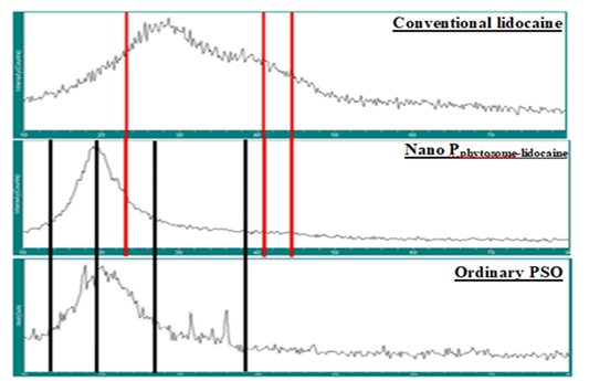
X-ray diffraction of ordinary lidocaine, Nano phytosome pumpkin-lidocaine, and oddinary pumpkin seed oil respectively. Red arrows indicated the diffraction spectrum of lidocaine while black arrows indicated the diffraction spectrum of pumpkin seed oil
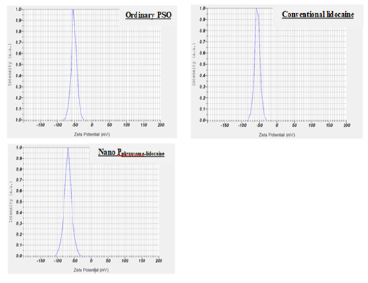
Zeta potential distributions curve of ordinary PSO, ordinary lidocaine , and Nano phytosome pumpkin-lidocaine
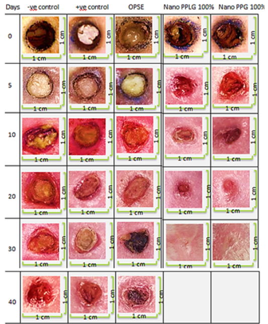
photograph of kinetic profile of burn wound healing (0.8mm) during phases of healing during 40 days and the improvement of closure mentioned in different types of treatment of pumpkin Nano formulated pharmaceutics.
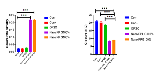
Closure rate and Closure t (½)
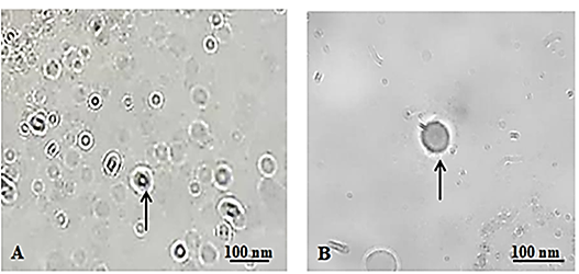
Light microscope of Nano phytosome pumpkin A, Nano phytosome pumpkin-lidocaine B, the arrow denoted to Nano formation and their creation


