
Biology, Histopathology and Treatment Evaluation Against Cryptosporidium meleagirids on Infected Quails


Biology, Histopathology and Treatment Evaluation Against Cryptosporidium meleagirids on Infected Quails
Khitam J. Yahya*, Mohammed T. S. Al-Zubaidi
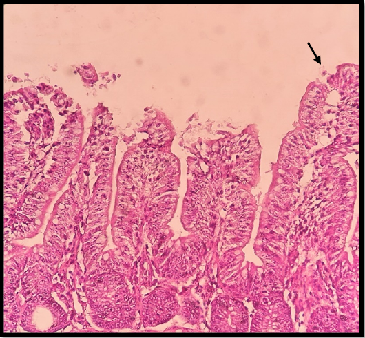
G2 after 3 (DPI). Cryptosporidium oocysts (black arrow) and intact apical microvilli border demonstrated moderate shortening of intestinal villi.
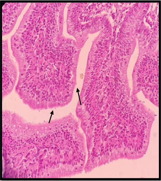
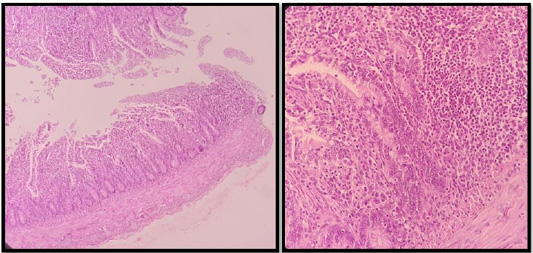
Ileum lesion on G2 (8 DPI), considerable villous shortening, epithelial sloughing acceptance, and multiple established stages of parasites were visible with several Cryptosporidium oocysts attached (black arrow), H & E stain X40.
Ileum lesion on G2 (8 DPI); goblet cell hyperplasia with evident stunting of intestinal villi, (H & E stain X 40).
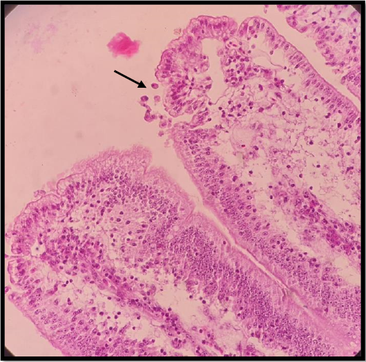
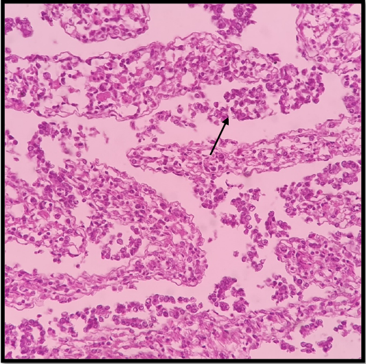
G2 (14 dpi), Cryptosporidium on the epithelial surface lining (oval round ball) (black arrow), with proprial distention tissue with leukocytic infiltration (H & E stain X 40).
ileum examination on G3 (14 DPI), severe infiltration of many cell types with goblet cell hyperplasia, (H& E stain X 40).

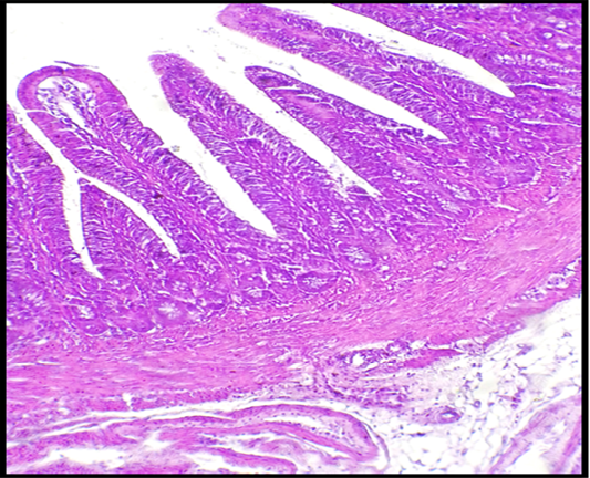
Intestinal segment on G4 (14dpi), expansion of gut-associated lymphoid tissue, and an indication of some villus fusion and shortening, (H & E stain X 40).
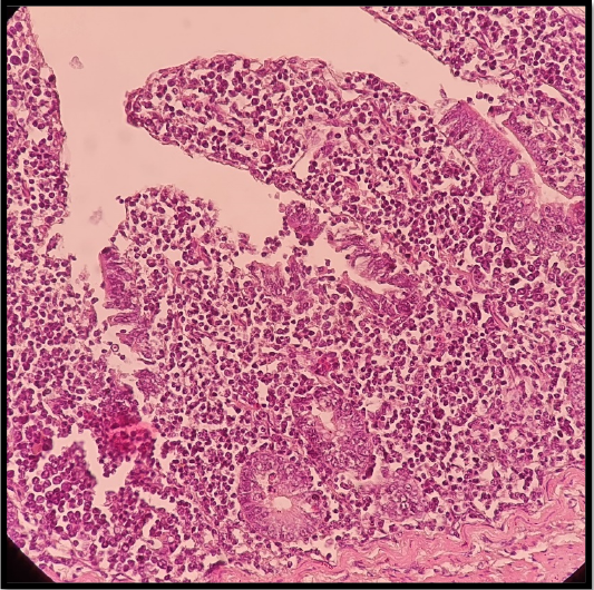
G2 (23 DPI) the mucosa and submucosal tissues were severely disrupted, and there were marked cystic crypt dilations and disorganized villus structures. A higher magnification of the previous section revealed the complete loss of enterocytes along with remnants of proprial tissue that still contained severe inflammatory cells, necrotic debris between villi (black arrow), (H & E stain X 40).
G3 ileum lesion (23 DPI) mild to moderate goblet cell hyperplasia without obvious alterations in the tissue around it, (H & E stain X 40).
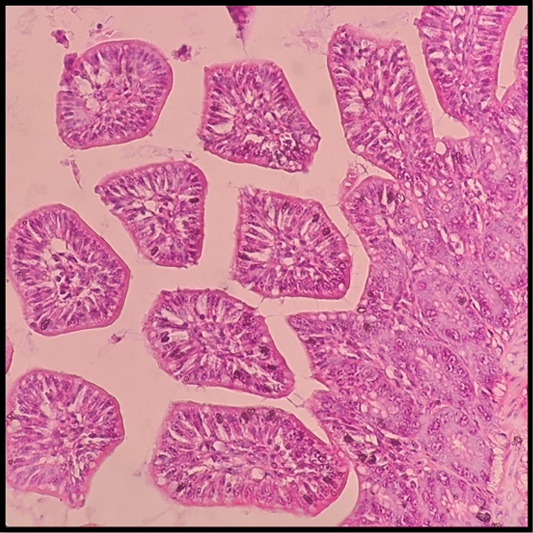
Ileum section G4 (23 DPI) mild mucosal mononuclear cell infiltration with normal crypt tissue, (H & E stain X 40).

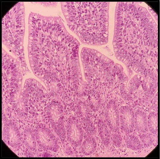
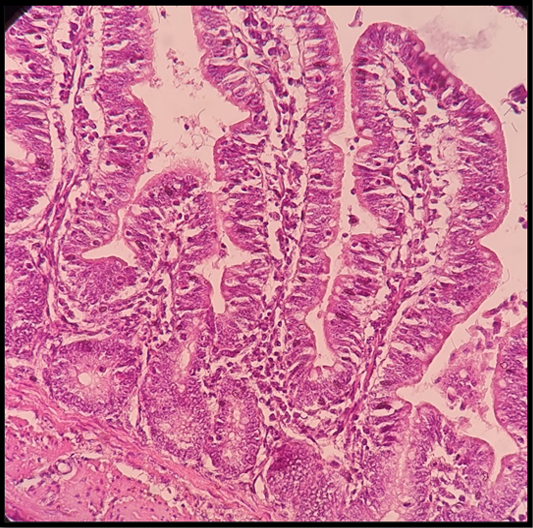
Intestinal section G1 normal intestinal structure, (H & E stain X 40).
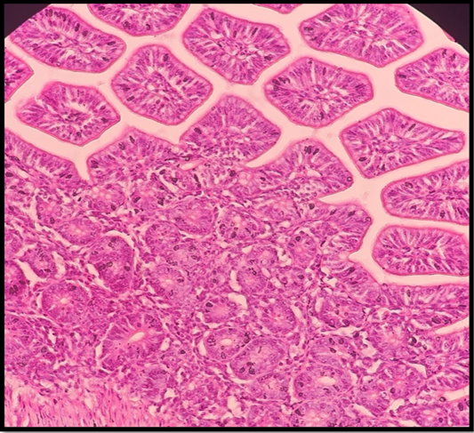
intestinal section of G5 showed normal structural details of epithelial mucosa and lamina propria, (H & E stain X 40).


