
Bee Venom for the Treatment of Rabbit Arthritis Caused by Staphylococcus aureus


Bee Venom for the Treatment of Rabbit Arthritis Caused by Staphylococcus aureus
Heba S.S. Salem1, Hend M. Megahed2, Marwa M. Sarhan2, Maha M. El Alem3, Gehan N. Alagmy3*

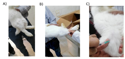
Representative images for A) group 1 normal rabbit, B) group 2 control positive with enlarged joint, and C) group treated with BV

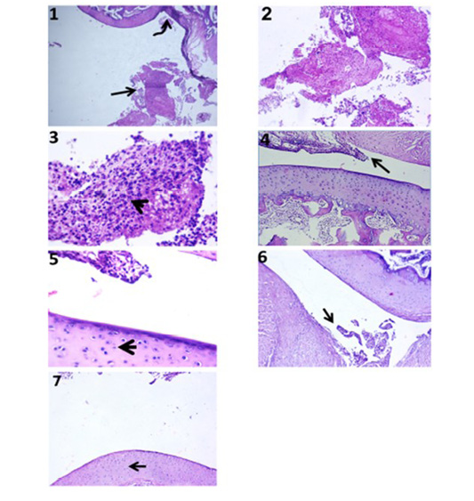
Histopathological findings of S. aureus-inoculated rabbit jointson the7 th day post inoculation
Lane 1 showing group 2 with degenerative changes with fibrillation within the cartilage of articular surfaces (curved arrow) with pannus formation (arrow) (5X), lane 2 showing septic synovitis formed from aggregates of inflammatory exudates (10X), lane 3 showing aggregation of neutrophils (arrowhead) intermixed with fibrin exudates within synovium (40X). Lanes 4 and 5 showing group 4 with restoration of all zones of chondrocytes at articular surfaces with hypertrophied chondrocytes (arrowhead) beside presence of moderate synovitis with cellular proliferation and exudation in the synovium within the joint cavity (arrow) (5, 10X). Lanes 6 and 7 showing group 5 with reduction of synovitis area within the joint cavity (arrow) and hypercellularity of the articular surfaces with reactive chondrocytes (arrowhead) (H&E staining) (10, 40X).
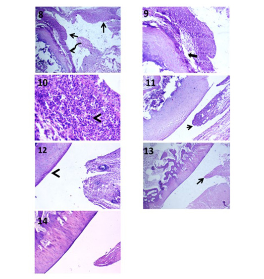
Histopathological findings of S. aureus-inoculated rabbit jointson the7 th day post inoculation
Lane 8 showing group 2 with destructive and septic arthritis beside presence of fibrillation (curved arrow), ostiomalacia and pannus formation (arrows) (5X). Lane 9 showing group 2 with replacement of the superficial and transitional zones of articular cartilage by eosinophilic and basophilic materials (thick arrow) (10X). Lane 10 showing group 2 with cellular proliferation with large areas of inflammatory exudates mainly neutrophils (arrowhead) and fibrin threads in synovial membrane (40X). Lanes 11 and 12 showing group 2 with articular surfaces showing smooth flattened chondrocytes in superficial zone and apparently normal chondrocytes in mid (arrowhead) and deep zones beside mild synovitis (arrow) (10, 40X). Lanes 13 and 14 showing group 5 with reduced area of pannus formation (arrow) and apparently normal histomorphological structures of superficial, mid, and deep zones of chondrocytes at the articular joints (5, 10X)(H&E staining).


