
Aptamer


Aptamer
John G. Bruno and Jeffery C. Sivils

Aptamer Western blot for Shiga-like toxin 2 (SLT-2) from a 12% nonreducing SDS polyacrylamide gel showing a dominant band at ~ 22 kD which correlates well with the reported molecular weight from the manufacturer of the SLT-2
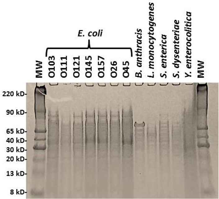
Coomassie blue-stained 12% reducing SDS-polyacrylamide gel electrophoresis (PAGE) of the E. coli serotypes and other bacterial lysates used in the investigation. The molecular weight (MW) standards are shown in the far left lane
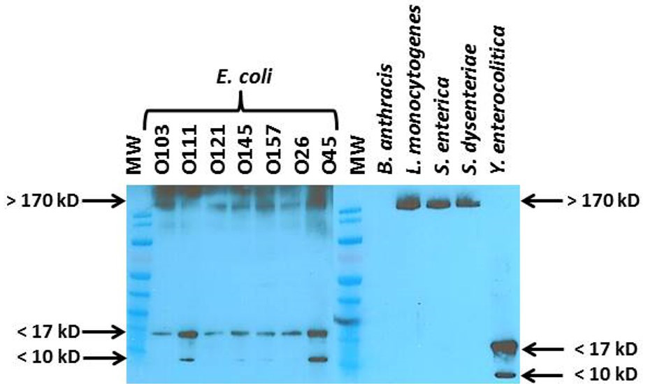
Western blot using the 100 base poly T-EcO 3R-biotinylated aptamer. Bands at ~ 17 kD may represent detection of OmpX

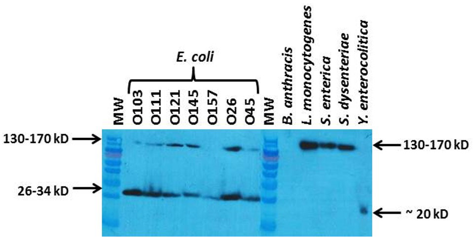
Western blot using the 100 base poly T-EcO 4F-biotinylated aptamer. Bands at ~ 34 kD may represent detection of OmpA

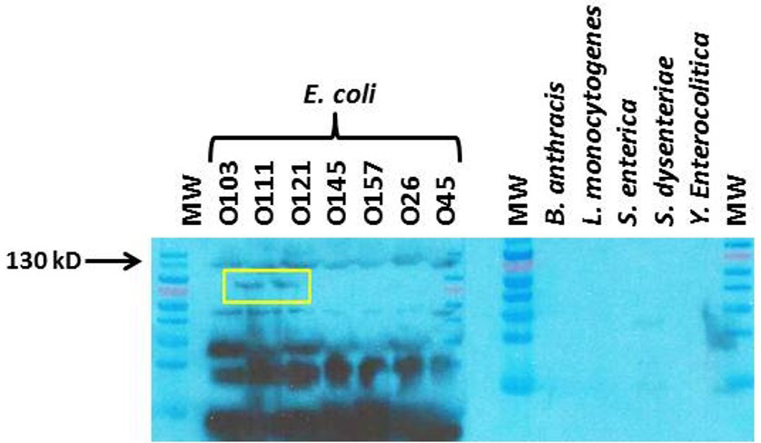
Western blot using an intimin-alpha-biotinylated aptamer. Yellow boxed bands may represent detection of intimin-alpha

Western blots using an intimin-gamma-biotinylated aptamer with exposure times of 10 sec (bottom) and 1 min (top). Yellow boxed bands may represent detection of intimin-gamma
