Special Issue: Emerging and Re-Emerging Animal Health Challenges in Low and Middle-Income Countries · Issue 1, 6 December 2024 · Open Access
Virulence Potential of Aspergillus flavus Isolated from Dogs
·
Affiliations & Notes ⌄
- 1Department of Microbiology, College of Veterinary Medicine, University of Baghdad, Baghdad, Iraq. *Correspondence | Salma Ali Munshed, Department of Microbiology, College of Veterinary Medicine, University of Baghdad, Baghdad, Iraq
- 2Email: Salma.Ali2203m@covm.uobaghdad.edu.iq
Article Info ⌄
- Journal:
- Journal of Animal Health and Production
- Published:
- 6 December 2024

From the article
- The invasiveness of the Aspergillus species may be indicated by the virulence factors found in isolates of the fungus.
- The study determined that the three most significant virulence factors of Aspergillus flavus isolated from dogs are proteinase, hemolysin, and biofilm, with respect to the variations in virulence factors generated by Aspergillus flavus.
Selected from the article's own text.
Abstract
Introduction
The most opportunistic pathogen is Aspergillus flavus, which infect humans and other animal species both superficially and invasively ; . In order to infect the host, hazardous virulence factors are requireds for Aspergillus strains. Aspergillus species produce virulence factors that facilitate the development of an infection and may subsequently lead to invasive or widespread infections. In recent years, the significance of Aspergillus infections has increased (Sales et al., 2013). The most typical form of Aspergillosis in dogs is a localized infection of the nasal region. In dogs systemic Aspergillosis usually affects the thoracic lymph nodes, lung, and renal pelvis . The production of a broad spectrum of hydrolytic enzymes, adhesions, heat resistance, pigment synthesis, and toxic metabolites are thought to be additional virulence characteristics to support Aspergillus species pathogenesis and enable the fungal cells to evade the host’s defense . Aspergillosis pathogenesis is determined by host variables (Immune state) as well as pathogen virulence factors. Extracellular hydrolases are among the principal virulence factors, suggesting that they play an important role in Aspergillus spp. widespread colonization of lung tissue. Enzymes such as α-amylase, proteinase, phospholipase, pectinase, and lipase play a significant role in degrading tissue and erythrocytes lyse (Vila and Rozental, 2016).
Aspergillus species can form biofilms, which are defined with the presence of a mycelium in the extracellular matrix, during different types of chronic Aspergillosis infections. The pathogenicity of Aspergillus is significantly influenced by its biofilm . The development of biofilms, which promotes hyphae adherence to host cells and increases resistance to antifungal treatment, is primarily responsible for fungal virulence . The aim of this study to isolate, identification and detection of virulence factors of A. flavus that increasing the degree of damage of the host as hydrolysis of albumin, hemolytic activity on blood agar and formation of biofilm in dogs.
MATERIALS AND METHODS
Isolation of fungi
In Baghdad province (Al-Rusafa and Al-Karkh) Al-Rusafa district (Zayouna, Al-Mashtel Street, Adamiyah and Al-Karkh district (Baghdad Veterinary Hospital) during November 2023 to January 2024, fifty samples of nasal swab from infected dogs were taken from veterinary clinic and stray dogs from different ages between (2-5years), sexes(male and female) and breeds like (terriers, German shepherds and police dog) by using a sterilizing swab. All of the collected samples were stored in a sterilizer tube containing transport media at 4 °C in an ice container until they cultivated.
Identification of fungi
The samples were cultured on Sabouraud Dextrose Agar (SDA) from (Himedia-India) containing 500 mg/L of Cefalexin, then incubated at 25 oC for 4–7 days for diagnosing macroscopically and microscopically. Furthermore, Aspergillus spp. were cultured on special medium Czapek Dox Agar (Oxoid-England) for appearance of pigmentation on reverse side of plates. The microscopic examination of Aspergillus spp. and other fungi were performed according to Baron and Finegold by putting one drop of lactophenol cotton blue stain on a slide and mixing it with a small part of the fungus by loop, then covering it with a cover slip, and examined by a light microscope under a 40X lens to determine the fungal elements microscopically.
Detection of virulence factors of Aspergillus flavus
Proteinase production medium
This medium was prepared according to . Take 68 mL of a solution containing MgSO4.7H2O 0.04 g, K, HPO 0.5 g, NaCl 1 g, yeast extract 0.2 g, glucose 4 g, and bovine serum albumin medium. The solution was sterilized using a Millipore filter (0.22 µm). After that, it was mixed with 140 ml of agar agar, sterilized in an autoclave at 121 oC under 15 pound/inch for 15 minutes, and put into sterile petri dishes. This medium was used to detect the ability of A. flavus to hydrolyze albumin, which was detected by the well diffusion method carried out one well was made in a proteinase production medium that contained bovine serum albumin in this well culture of A. flavus and incubated at 25 oC for 4–7 days.
Medium for hemolytic activity
Sabouraud Dextrose Agar medium used with 5% glucose and 5% sheep blood added as supplements as mentioned in . Hemolysis in the petri dishes was classified into low, moderate, and high during a maximum of seven days of incubation at 25 °C.
Determination of biofilm formation
Biofilm formation was assessed in sterile 96-well microplates. In a tube containing 2 ml of brain heart infusion broth (BHIB) medium with 0.25% glucose, the culture was placed using a loop, and the tube was then incubated at 37 °C for 24 hours. A final solution was obtained, and 200 μl was added to 96-well polystyrene microliter plates that were sterile. After placing covers on the microplates, they were incubated for 24 hours at 37 °C. Following incubation, the microplate was flipped to a blot and washed three times with PBS. Each well was then filled with 1% crystal violet, and it was incubate for 15 minutes. The microplate was once again washed three times with PBS following incubation. After adding ethanol to each well , (ELISA) reader, the wells’ 450 nm readings were obtained, and the OD was noted for each well.
Statistical analysis
The SAS application, which stands for statistical analysis system, utilized to determine the effect of various variables on the research parameters. To compare means significantly and the least significant difference (LSD). In this study, the chi-square test was utilized to evaluate percentages 0.05 and 0.01 probability (SAS, 2018).
Results and Discussion
Isolation of fungi from respiratory infection samples of dogs
This study found a high percentage of fungal isolates which reached (100%) were isolated from dogs collected from different locations within the Baghdad Governorate. Part of Aspergillus spp., which included A. flavus, A. niger, and A. terreus from these 50 isolates, were identified as significant differences were found between these species at P≤0.05, as shown in (Table 1), and there were also significant differences between about 13 other types of important fungi (2 yeast, 11 mold) as shown in Table 2.
Table 1: The percentage of Aspergillus spp. isolated from dogs respiratory infection.
| Aspergillus spp. | Isolation No. | Percentage % |
| Aspergillus flavus | 9 | 42.8 |
| Aspergillus niger | 9 | 42.8 |
| Aspergillus terreus | 3 | 14.2 |
| Total | 21 | 100% |
| P-value | -- | 0.0476 * |
* (P≤0.05).
Table 2: Distribution of sample study according to fungal isolation from dog’s respiratory infection.
| Fungal spp. | Isolation No. | Percentage % | |
| Absidia | 1 | 2 | |
| Aspergillus flavus | 9 | 18 | |
| Aspergillus niger | 9 | 18 | |
| Aspergillus terreus | 3 | 6 | |
| Candida spp. | 4 | 8 | |
| Chrysonilia sitophilia | 2 | 4 | |
| Cladosporuim spp. | 1 | 2 | |
| Curvularia spp. | 1 | 2 | |
| Geomysis spp. | 2 | 4 | |
| Geotricum | 1 | 2 | |
| Penicillum spp. | 12 | 24 | |
| Phialophora spp. | 1 | 2 | |
| Rhizopus | 2 | 4 | |
| Rodetilla spp. | 1 | 2 | |
| Trichoderm spp. | 1 | 2 | |
| Total | 50 | 100% | |
| P-value | -- | 0.0028 ** | |
Note: There are significant differences at level (P≤0.01). ** (P ≤ 0.01).
Aspergillus flavus causes a variety of superficial and invasive infections in both humans and other animal species and it is a soil borne fungus, which is ubiquitous in distribution. In the present investigation, the clinical and mycological observations conclusively established that dogs were suffering with fungal rhinitis due to A. flavus infection. ; ; ; , 2017). The majority of molds are harmless but many can cause significant infections in humans and dogs. The main Aspergillus species that cause complications for dogs are Aspergillus flavus, Aspergillus niger, and Aspergillus terreus, yet several other species can also cause disease in dogs.
Identification of Aspergillus flavus isolates
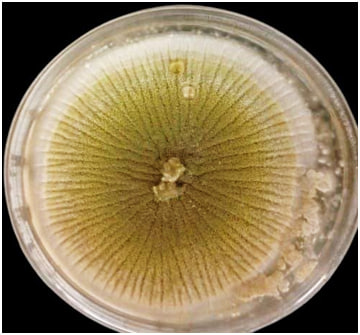
Aspergillus flavus was isolated and identified from dogs by sub culturing on SDA at 25ºC for 5-7 days. The colonies appeared as yellow green in color with fluffy in texture (Figure 1). A white mycelium that spread outward to cover the whole surface of the medium was the beginning of the colony expansion. As sporulation started, the white colony color gradually convert to yellowish green or dark green color of the conidia, which eventually covered the whole surface. This matched the findings of , who observed that the mycelia were white in color and that the reverse of the plates had cream and olive or dark green conidia bordered by a white ring.
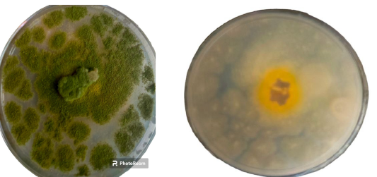
Furthermore, Czapek dox agar colonies initially had a yellowish green appearance before changing to an olive green over time, whereas the reverse side’s color changed from creamy to yellow. The average colony diameter was 4-5 cm (Figure 2). These outcomes were consistent with ; ; ; .
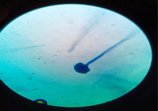
The conidial head appeared complete flower (complete circle phialides) which were arranged as long conidiophores (Figure 3). The microscopic characteristics were in harmony with the A. flavus characteristics previously described ; .
Virulence factors of A. flavus
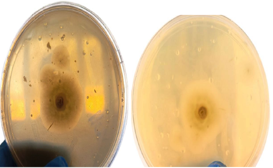
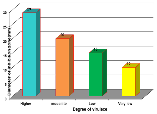
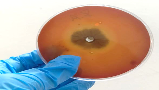
Characteristics of virulence factors that facilitate fungal growth, survival, and spread in tissues of mammals and cause illness . This study measured the diameter of the albumin inhibition zone as shown in (Table 3) and (Figures 4, 5), to determine the degree of virulence (higher, moderate, weak, and very weak) of nine isolates of A. flavus from fifty samples of nasal swab. The findings demonstrated that each isolate of A. flavus was able to degrade albumin protein, with highly significant differences in the degrees of virulence. Similar results were obtained from . The explanation for these observations was the ability of albumin to influence Aspergillus conidial germination and hyphae formation, particularly in A. flavus (Mezher and Bandar, 2016).
Table 3: The results of proteinase action by A. flavus.
| Degree of virulece | Isolated A.flavus | Percentage (%) | Diameter of inhibition zone(mm) |
| Higher | A and B | 11.11 | 29 |
| Moderate | C, D, E, G and I | 27.7 | 20 |
| Weak | H | 0.55 | 15 |
| Very weak | F | 0.55 | 10 |
| P_value | --- | 0.0387 (P≤ 0.05), | 0.0001 (P≤0.01) |
Note: There are high significant differences at level (P≤0.05) and ability to albumin hydrolyze by A. flavus at level (P≤0.01).
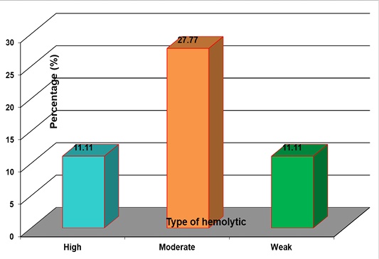
Likewise A. flavus isolates showed positive action of hemolysis on blood agar. The research showed that all isolates of A. flavus had hemolytic action on Blood Agar with significant differences between level of hemolytic (P≤0.05) classified into weak, moderate, and high as shown in (Table 4) and (Figures 6, 7). Hemolytic action is taking into account one of the most significant virulence factors, a transparent or semi-transparent zone surrounding the inoculation site was determined to be positive for hemolytic action. Hemolysin lyses red blood cells (RBCs) by creating pores or holes in their membranes, which releases iron and encourages the growth of microorganisms . The present results are consistent with previous studies by ; ; .
Table 4: The results of Hemolytic activity by A. flavus.
| Isolates of A. flavus | Degree of hemolysis | Percentage (%) |
| Two isolated | High | 11.11 |
| Five isolated | Moderate | 27.77 |
| Two isolated | Weak | 11.11 |
| P value | --* (P≤0.05) | 0.0316 |
Note: the significant differences between level of hemolytic (P≤0.05).
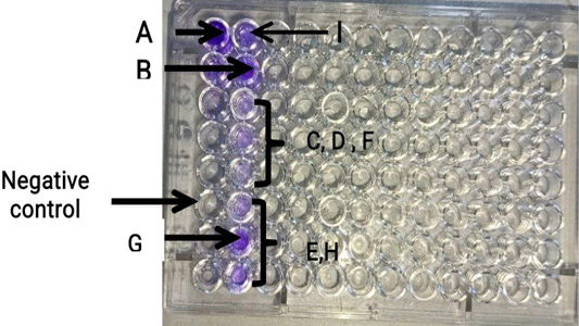
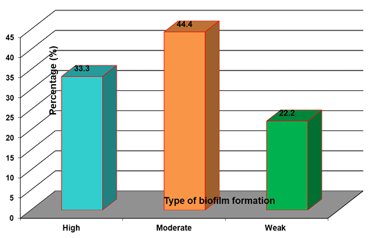
Another virulence factor of A. flavus was biofilm formation. All isolates of A. flavus biofilm formation were classified as weak, moderate, and high. The biofilm mass was higher in (A, B, and C) isolates of this mold with high significant differences (P≤0.01), as shown in (Table 5 and Figure 8), respectively. This result was in agreement with other studies . The reason behind these results may be attributable to the ability of A. flavus to utilize more protein. This result is similar to the biofilm formation by A. flavus reported in a previous study by . The study reported that A. flavus utilized more serum proteins in the production of biofilms. This could suggest that a riskier factor in A. flavus is bioflim, which has been associated with infections of the upper respiratory tract ; .
Table 5: Biofilm formation by A. flavus.
| Isolates of A.flavus | Degree of biofilm formation | Percentage (%) |
| Three isolates | High | 33.3 |
| Four isolates | Moderate | 44.4 |
| Two isolates | Weak | 22.2 |
| P value | --(P ≤0.01) | 0.00839 |
Note: high significant differences between isolation at level (P ≤ 0.01).
Conclusions and Recommendations
Any microorganism’s virulence factors are crucial in distinguishing harmful from non-pathogenic species. The invasiveness of the Aspergillus species may be indicated by the virulence factors found in isolates of the fungus. The study determined that the three most significant virulence factors of Aspergillus flavus isolated from dogs are proteinase, hemolysin, and biofilm, with respect to the variations in virulence factors generated by Aspergillus flavus.
Acknowledgments
The chief veterinarian of the Baghdad governorate, the medical team, and the veterinary clinic are all appreciated by the authors for their help with sample collection. The authors also appreciate the personnel in the microbiology departments of the University of Baghdad, Iraq’s college of veterinary medicine for their assistance and the facilities they provided for the processing of the samples.
Novelty statement
The novelty of the study is focus on isolation and determination the virulence factor of A. flavus (specially biofilm formation) that has capacity to cause disease even in healthy animals without immune suppression, and these are life threatening.
Author’s Contribution
Each of these writers made an equal contribution.
Conflict of interest
The authors have declared no conflict of interest.
Figures

Macroscopic appearance of A. flavus on SDA at 25ºC for 7 days.

Aspergillus flavus on Czapek dox agar at 25 ºC for 4–5 days.

Microscopic appearance of A. flavu using stain lactophenol cotton blue (40x).

Hydrolysis of albumin by A. flavus in proteinase production medium.

Measurement the diameter of inhibition zone in albumin medium.

Hemolytic action by A. flavus on blood agar.

Results of hemolytic activity of A. flavus.

Microplate inoculated with nine A. flavus isolates with negative control for detection of bioflim formation result show high Biofilm formation in sample A, B and G, moderate in sample C, D, F and I and weak biofilm in sample E and H.

Biofilm formation by A. flavus.
References
- Aşkun T (2006). Investigation of fungal species diversity of maize kernels. J. Biol. Sci., 6(2): 275-281. https://doi.org/10.3923/jbs.2006.275.281[CrossRef]
- Baron EJ, Bennion R, Thompson J, Strong C, Summanen P, McTeague M, Finegold SM (1992). A microbiological comparison between acute and complicated appendicitis. Clin. Infect. Dis., 14(1): 227-231. https://doi.org/10.1093/clinids/14.1.227[CrossRef]
- Bavadharani S, Premamalini T, Karthika K, Kindo AJ (2022). In vitro production of virulence factors and antifungal susceptibility pattern of aspergillus isolates from clinical samples in a tertiary care center. J. Lab. Phys., 14(4): 479-484. https://doi.org/10.1055/s-0042-1747680[CrossRef]
- Bonassoli LA, Bertoli M, Svidzinski TI (2005). High frequency of Candida parapsilosis on the hands of healthy hosts. J. Hosp. Infect., 59(2): 159-162. https://doi.org/10.1016/j.jhin.2004.06.033[CrossRef]
- D’Eça Júnior A, Silva AF, Rosa FC, Monteiro SG, Figueiredo PD, Monteiro CD (2011). In vitro differential activity of phospholipases and acid proteinases of clinical isolates of Candida. Rev. Soc. Brasil. Med. Trop., 44: 334-338. https://doi.org/10.1590/S0037-86822011005000036[CrossRef]
- Diba K, Kordbacheh P, Mirhendi SH, Rezaie S, Mahmoudi M (2007). Identification of Aspergillus species using morphological characteristics. Pak. J. Med. Sci., 23(6): 867.
- Gao J, Liu Z, Yu J (2007). Identification of Aspergillus section Flavi in maize in northeastern China. Mycopathologia, 164: 91-95. https://doi.org/10.1007/s11046-007-9029-4[CrossRef]
- Garcia RS, Wheat LJ, Cook AK, Kirsch EJ, Sykes JE (2012). Sensitivity and specificity of a blood and urine galactomannan antigen assay for diagnosis of systemic aspergillosis in dogs. J. Vet. Intern. Med., 26(4): 911-919. https://doi.org/10.1111/j.1939-1676.2012.00935.x[CrossRef]
- Ghorbel D, Hadrich I, Neji S, Trabelsi H, Belaaj H, Sellami H, Cheikhrouhou F, Makni F, Ayadi A (2019). Detection of virulence factors and antifungal susceptibility of human and avian Aspergillus flavus isolates. J. Mycol. Méd., 29(4): 292-302. https://doi.org/10.1016/j.mycmed.2019.100900[CrossRef]
- Hoog GD, Guarro J, Gene JF, Figueras MJ (2000). Atlas of clinical fungi.
- Khodavaisy S, Badali H, Hashemi SJ, Aala F, Nazeri M, Nouripour-Sisakht S, Sorkherizi MS, Amirizad K, Aslani N, Rezaie S (2016). In vitro activities of five antifungal agents against 199 clinical and environmental isolates of Aspergillus flavus, an opportunistic fungal pathogen. J. Mycol. Méd., 26(2): 116-121. https://doi.org/10.1016/j.mycmed.2016.01.002[CrossRef]
- Mahmoud MJ, Jawad AL, Hussain AM, Al-Omari M, Al-Naib A (1989). In vitro antimicrobial activity of Salsola rosmarinus and Adiantum capillus-veneris. Int. J. Crude Drug Res., 27(1): 14-16. https://doi.org/10.3109/13880208909053930[CrossRef]
- Mezher MA, Bandar KI (2015). Identification study some virulence factors of invasive mold infections isolated from patients undergoing chemotherapy in Tikrit teaching Hospital. Egypt. Acad. J. Biol. Sci., G. Microbiol., 7(1): 1-1. https://doi.org/10.21608/eajbsg.2015.16480.[CrossRef]
- Ogundijo OA (2017). Biofilm formation by Aspergillus flavus and Aspergillus niger: Influence of cultural conditions and their controls. Trop. Vet., 35(4): 12-12. Retrieved from https://www.tropivet.com/index.php/tvjournal/article/view/196
- Okayo RO, Andika DO, Dida MM, K’Otuto GO, Gichimu BM (2020). Morphological and molecular characterization of toxigenic Aspergillus flavus from groundnut kernels in Kenya. Int. J. Microbiol., 2020(1): 8854718. https://doi.org/10.1155/2020/8854718[CrossRef]
- Pal M (2007). Veterinary and medical mycology. Directorate of Information and Publications of Agriculture, Indian Council of Agricultural Research ISBN., 8171640710,9788171640713.
- Pal M (2017). Rhinomycosis in a dog caused by Aspergillus flavus Department of Veterinary Public Health, College of Veterinary Science, Anand-388001, Gujarat, India. https://doi.org/10.5455/ijlr.20170216030609[CrossRef]
- Pasqualotto AC (2009). Differences in pathogenicity and clinical syndromes due to Aspergillus fumigatus and Aspergillus flavus. Med. Mycol., 47(Supp. 1): S261-270. https://doi.org/10.1080/13693780802247702[CrossRef]
- Ramage G, Rajendran R, Sherry L, Williams C (2012). Fungal biofilm resistance. Int. J. Microbiol., 2012(1): 528521. https://doi.org/10.1155/2012/528521[CrossRef]
- Rodrigues AG, Araujo R, Pina-Vaz C (2005). Human albumin promotes germination, hyphal growth and antifungal resistance by Aspergillus fumigatus. Med. Mycol., 43(8): 711-717. https://doi.org/10.1080/13693780500129814[CrossRef]
- Rodrigues P, Soares C, Kozakiewicz Z, Paterson R, Lima N, Venâncio A (2007). Identification and characterization of Aspergillus flavus and aflatoxins. Formatex. pp. 527-534. at: https://www.researchgate.net/publication/237377677
- Saleemi MK, Khan MZ, Khan A, Hameed MR, Khatoon A, Abadin ZU, Hassan ZU (2017). Study of fungi and their toxigenic potential isolated from wheat and wheat bran. Toxin Rev., 36(1): 80-88. https://doi.org/10.1080/15569543.2016.1233890[CrossRef]
- Sales-Campos H, Tonani L, Cardoso CR, Kress MR (2013). The immune interplay between the host and the pathogen in Aspergillus fumigatus lung infection. BioMed. Res. Int., 2013(1): 693023. https://doi.org/10.1155/2013/693023[CrossRef]
- SAS (2018). Statistical analysis system, user’s guide. Statistical. Version 9. 6th ed. SAS. Inst. Inst. Cary. N.C. USA.
- Satish HS, Viswanatha B, Manjuladevi M (2013). A clinical study of otomycosis. IOSR J. Dent. Med. Sci., 2: 57-62. https://doi.org/10.9790/0853-0525762[CrossRef]
- Savković ŽD, Stupar MČ, Unković ND, Stančić AZ, Vukojević JB, Ljaljević-Grbić MV (2022). Hemolytic potential of bioaerosol-derived Aspergillus, Penicillium and Talaromyces mould isolates. Zbornik Matice Srpske za Prirodne Nauke, 143: 15-25. https://doi.org/10.2298/ZMSPN2243015S[CrossRef]
- Singh G, Urhekar AD (2017). Virulence factors detection in Aspergillus isolates from clinical and environmental samples. J. Clin. Diagn. Res., 11(7): DC13.
- Steinbach WJ, Marr KA, Anaissie EJ, Azie N, Quan SP, Meier-Kriesche HU, Apewokin S, Horn DL (2012). Clinical epidemiology of 960 patients with invasive aspergillosis from the PATH Alliance registry. J. Infect., 65(5): 453-464. https://doi.org/10.1016/j.jinf.2012.08.003[CrossRef]
- Susan EA, Aiello BS (1998). The merck veterinary manual (8th ed.) Merck and Co. INC., Whitehouse Station, NJ, USA.
- Tell LA (2005). Aspergillosis in mammals and birds: Impact on veterinary medicine. Med. Mycol., 43(Supp. 1): S71-73. https://doi.org/10.1080/13693780400020089[CrossRef]
- Thathana MG, Murage H, Abia AL, Pillay M (2017). Morphological characterization and determination of aflatoxin-production potentials of Aspergillus flavus isolated from maize and soil in Kenya. Agriculture, 7(10): 80. https://doi.org/10.3390/agriculture7100080[CrossRef]
- Vila TV, Rozental S (2017). Biofilm formation as a pathogenicity factor of medically important fungi. Fungal Pathog., 11: 1-24. https://doi.org/10.5772/62768[CrossRef]
- Wuren T, Toyotome T, Yamaguchi M, Takahashi-Nakaguchi A, Muraosa Y, Yahiro M, Wang DN, Watanabe A, Taguchi H, Kamei K (2014). Effect of serum components on biofilm formation by Aspergillus fumigatus and other Aspergillus species. Jpn. J. Infect. Dis., 67(3): 172-179. https://doi.org/10.7883/yoken.67.172[CrossRef]
Services
Article usage statistics
Loading…