Research Article · Volume 10, Issue 2, 2 February 2022 · Open Access
Detection of Intestinal Fungi in Chickens Naturally Infected with Infectious Bursal Disease
· · · · · ·
Affiliations & Notes ⌄
1Poultry Diseases Department, Veterinary Research Institute, National Research Centre, P.O. 12622, Giza, Egypt
2Laboratory of Veterinary Vaccines Technology (VVT), Central Laboratories Network, National Research Centre, P.O. 12622, Giza, Egypt. *Correspondence | Aly M. Ghetas, Poultry Diseases Department, Veterinary Research Institute, National Research Centre, P.O. 12622, Giza, Egypt
This preliminary study was performed to identify intestinal fungal population in immunosuppressed chickens.
Aspergillus isolates were the most identified (42%) fungal isolates from cloacal swabs of IBD infected chickens, followed by Trichosporon (10.5%), Penicillium (10.5%), Fusarium (5%), Candida (1%) and non-identified isolates (26%).
Furthermore, the obtained 8 Aspergillus isolates were further morphologically identified into A. fumigatus which was most frequent species identified (4/19 of total) and (4/8 of Aspergillus species); 2 A. flavus, 1 A. niger and 1 A. terreus.
Furthermore, molecular identification of 5 representative Aspergillus isolates indicated A. fumigatus (2).
Selected from the article's own text.
Abstract
A little is known about intestinal fungal population and its role during health and disease particularly in chickens. Thus, this preliminary study was performed to identify intestinal fungal population in immunosuppressed chickens. Cloacal swabs were collected from freshly dead chickens suffering from infectious bursal disease (IBD) for isolation of fungal species on specific medium. A total of 19 purified fungal isolates have been identified morphologically. Aspergillus isolates were the most identified (42%), followed by Trichosporon (10.5%), Penicillium (10.5%), Fusarium (5%), Candida (1%) and non-identified isolates (26%). Furthermore, the obtained 8 Aspergillus isolates were further morphologically identified into A. fumigatus which was most frequent species identified (4/19 of total) and (4/8 of Aspergillus species); A. flavus (2), A. niger (1) and A. terreus (1). Molecular identification of 5 representative Aspergillus isolates indicated A. fumigatus (2), A. flavus (2), and A. niger (1) were also performed. We recommended further studies to identify the intestinal fungi of chickens in large scale and different ages which help to understand their role.
The gastrointestinal tract (GIT) is occupied by a diverse microbial population (microbiota) consisting of bacteria, fungi, protozoa, and viruses . The microbiota which is essential for gut homeostasis has been extensively characterized. GIT microbiota plays an important role in carbohydrate and lipid metabolism, immunity development, synthesis of amino acids and vitamins, and strengthen the gut barrier by producing short-chain fatty acids .
Dysbiosis, the microbial imbalance, is mostly associated with intestinal inflammation ; ; . Intestinal microbial changes after infection with a wide range of pathogens have been reported. These changes characterized by reduction in the abundance and diversity of the intestinal flora ; ; ; . Dysbiosis has been reported in chickens after infection with Newcastle disease virus , Marek’s disease virus , avian leucosis viruses , or infectious bursal disease virus (IBDV) . IBDV is a non-enveloped virus belongs to the genus Avibirnavirus and the family Birnaviridae ; . It is an immunosuppressive virus which replicates in gut associated lymphoid tissue causing histological lesions, changes in immune cells, and alteration of microbial population ; ; .
Although extensive knowledge is available about GIT microbiota, little is known about intestinal fungal population and its role during health and disease particularly in chickens. The term mycobiome used to differentiate the mycology aspect of the microbiome . A total of 88 different fungal and yeast species were identified from cecal samples taken from commercial broiler and layer flocks, including Aspergillus, Penicillium, and Sporidiobolus species, and 18 unknown genera by using automated repetitive sequence-based PCR . Furthermore, fifty fungal isolates belonged to seven species (Aspergillus fumigatus, Aspergillus niger, Chrysonilia crassa, Mucor circinelloides, Mucor sp, Rhizopus oligosporus and Rhizopus oryzae) have been isolated from different parts of GIT of chickens . Recently, Next-generation sequencing of chicken intestinal mycobiota has shown the presence of four phyla and 125 genera in which 3 genera (Microascus, Trichosporon, and Aspergillus) representing over 80% of the total fungal population . Interestingly, many species belong to these genera (Microascus, Trichosporon, and Aspergillus) are considered opportunistic pathogens particularly in immunocompromised humans ; ; ; . Therefore, it is important to continue monitoring these intestinal fungi which could be a source of contamination in poultry meat in processing plant and increase fungal load inside poultry house to ensure food safety and low personal risk.
The objective of this study was to isolate and identify fungal populations that passed through the GIT of the immunosuppressed chickens during IBDV natural infection.
MATERIALS AND METHODS
Flocks history
Two native chicken flocks suffering from infectious bursal disease were sampled from Menoufia governorate. The first flock was 35 day old chickens (n=5000). While the second flock was 28 day old chickens (n=1200). They only vaccinated with intermediate IBD vaccine at 12 day of age. Chicken flocks showed signs including whitish diarrhea, ruffled feathers, anorexia, depression and mortalities (5%-10%). At postmortem examination enlarged bursa with gelatinous transudate covering its serosal surface was observed. Furthermore, hemorrhagic bursae, hemorrhage on thigh muscle and between gizzard and proventriculus was also observed in dead chickens.
Samples
Cloacal swabs and intestinal specimens
Five cloacal swabs and intestinal specimens per flock were collected from freshly dead birds for isolation and identification of fungi and histopathological study respectively.
Isolation and identification of fungi
Culture media
Sabouraud Dextrose Agar (Oxoid) was used for isolation and primary morphologicl identification of the obtained isolates. Preliminary cultures were established with Sabouraud, and glucose-potato (PDA) agars with the addition of antibiotics, and incubated at 25 and 37 for 48 h up to 7 days. After 72 where fungi mostly showing especial growth.
Morphological identification
Species of fungi including Aspergillus were identified by observing the characteristic conidial head and colony . Taxonomic identification was carried out with the method ; .
Molecular identification of fungi
Four representative isolates from identified were subjected to molecular identification as follows:
DNA extraction
DNA extraction from samples was performed using QIAamp DNeasy Plant Mini kit (Qiagen, Germany, GmbH). Briefly, 100 mg of the sample was added to 400 μl Buffer AP1 and 4 μl RNase. A stock solution (100 mg/ml), tungsten carbide bead were added to the previous mixture in a 2 ml safe-lock tube. Tubes were placed into the adaptor sets, which are fixed into the clamps of the Tissue Lyser. Disruption was performed in two 1–2 minute high-speed (20–30 Hz) shaking steps. The mixture was incubated for 10 min at 65°C and mixed 2 or 3 times during incubation by inverting tube. Then, 130 μl Buffer P3 was added to the lysate, mixed, and incubated for 5 min on ice. The lysate was centrifuged for 5 min at 14,000 rpm, and then pipetted into the QIA shredder Mini spin column (lilac) placed in a 2 ml collection tube, and centrifuged for 2 min at 14,000 rpm. The flow-through fraction from was transferred into a new tube without disturbing the cell-debris pellet and then applied to silica column. The lysate was then washed and centrifuged following the manufacturer’s recommendations. Nucleic acid was eluted with 50 µl of elution buffer provided in the kit.
Oligonucleotide primers
Primers used were supplied from Biobasic Canada and were listed in Table 1 and cycling conditions in Table 2.
PCR amplification
DNA samples: Primers were utilized in a 25- µl reaction containing 12.5 µl of Emerald Amp Max PCR Master Mix (Takara, Japan), 1 µl of each primer of 20 pmol concentration, 4.5 µl of water, and 6 µl of DNA template. The reaction was performed in an applied bio system 2720 thermal cycler.
Table 1: Primers sequences, target gene, and amplification size.
Target gene
Primers sequences
Amplified segment (bp)
Reference
Fungi ITS
ITS1: TCCGTAGGTGAACCTGCGG
Variable
Tarini et al., 2010
ITS4: TCC TCC GCT TAT TGA TAT GC
Table 2: Cycling conditions for conventional PCR.
Target
Primary denaturation
Amplification cycles
Final extension
Secondary denaturation
Annealing
Extension
Fungi ITS
94˚C 5 min.
94˚C 30 sec.
56˚C 40 sec.
72˚C 45 sec.
72˚C 10 min.
Analysis of the PCR Products
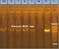
Conventional PCR products: The products of PCR were separated by electrophoresis on 1.5% agarose gel (Applichem, Germany, GmbH) in 1x TBE buffer at room temperature using gradients of 5V/cm. For gel analysis, 15 µl of the products was loaded in each gel slot. A generuler 100 bp DNA Ladder (Fermentas, thermofisher, Germany) was used to determine the fragment sizes. The gel was photographed by a gel documentation system (Alpha Innotech, Biometra) and the data was analyzed through computer software. A. flavus (ATCC® 9643™) was used as positive control (Figure 2).
Gene sequence
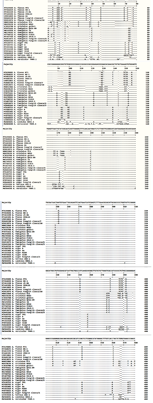
PCR products were purified using QIAquick PCR Product Extraction Kit (Qiagen, Valencia, CA, USA). Big Dye Terminator V3.1 Cycle Sequencing Kit (PerkinElmer, Foster city, CA, USA) was used for the sequence reaction, and then, the product was purified using Centri-Sep™ spin columns. DNA sequences were obtained from Applied Biosystems 3130 genetic: analyzer (Hitachi, Japan). A BLAST® analysis (Basic Local Alignment Search Tool) was initially performed to establish sequence identity to GenBank accessions . Amino acid sequence of the TSI gene of the isolated fungal strains in comparison to published strains were shown (Figure 4). A phylogenetic tree was created using the CLUSTAL W multiple sequence alignment program, MegAlign module of Lasergene DNASTAR version 12.1 (Figure 3).
Phylogenetic analyses
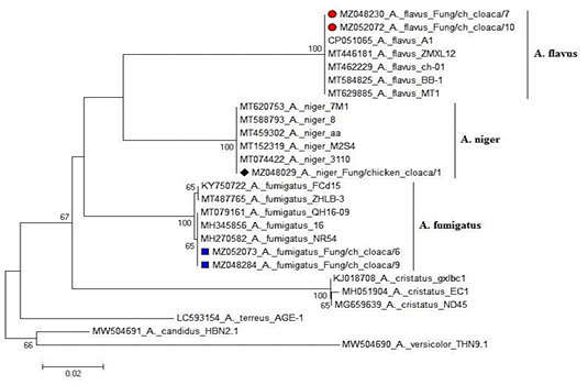
Phylogenetic analyses were performed with published Fungi sequences of ITS gene (Table 3) using the maximum likelihood, neighbor-joining, and maximum parsimony in MEGA6 (Figure 3) .
Histopathological examination
Intestine specimens were collected and fixed in 10% neutral buffered formalin for preparing paraffin tissue sections at 4-6 μm thickness. These sections were stained with hematoxylin and eosin .
RESULTS and DISCUSSION
Pathological findings
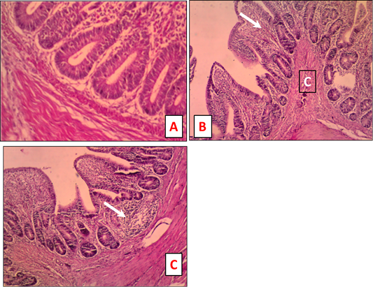
Intestine of chickens naturally infected with IBDV showed inflammatory cells infiltration in the mucosa and congestion of the submucosa (Figure 1).
Isolation and identification of fungi
Morphological examination of purified 19 fungi isolates on Sabouraud agar revealed the identification of 8 Aspergillus species, 2 trichosporon , 2 pencillium, 1 Fusarium, 1 candida and 5 non-identified species. The obtained 8 Aspergillus isolates were further morphologically identified into A. fumigatus which was most frequent species identified (4/19 of total) and (4/8 of Aspergillus species); A. flavus (2), A. niger (1) and A. terreus (1). A representative 5 Aspergillus species those are invasive fungi associated with excessive morbidity and mortality, also of toxigenic importance to chickens including (A. fumigatus (2), A. flavus (2) and A. niger (1)) were taken for further molecular identification.
The PCR product of ITS gens were sequenced and submitted to genebank: A. fumigatus, A. flavus, and A. niger with accession numbers MZ052073 and MZ048284; MZ048230 and MZ052072 as well as MZ048029, respectively.
The intestinal tract of humans and animals has a diverse populations of bacteria, fungi, archaea, protozoa, and viruses ; . Despite large diversity of the fungal community in humans, it is approximately estimated 0.02% of the intestinal mucosa-associated microbiota and 0.03% of the fecal microbiota . A little is known about intestinal fungal population in chickens particularly during immunosuppression. Thus, this study was carried out to identify fungal population passed through intestine of chickens during IBDV infection. IBDV replicates in gut associated lymphoid tissue causing immunosuppression, histological lesions, changes in immune cells, and alteration of microbial population ; ; .
Samples for fungi isolation were collected as cloacal swabs. Small intestine and caecum are the place where the more existences of microbe . Furthermore, Chicken ileum was dominated by lactobacilli, whereas in caecum was found more diverse microbial community . However, the largest number of fungi was found in ileum, and then followed by caecum, jejenum and duodenum .
The morphological characterization was performed as a preliminary step to identify fungi followed by further identification using molecular methods . Based on fungal morphology on sabouraud agar, we identified 19 purified fungal isolates. A total number of 8 out of 19 isolates (42%) were Aspergillus species. Aspergillus species have been reported to be one of the most identified fungal population from cecal swabs obtained from broiler and layer flocks . Furthermore, it was one of the most 3 identified chicken intestinal mycobiota by next-generation sequencing . In this study, the obtained 8 Aspergillus isolates were further morphologically identified into A. fumigatus which was the most identified species (4/19 of total) and (4/8 of Aspergillus species); A. flavus (2), A. niger (1) and A. terreus (1). Noticeably, A. fumigatus is a ubiquitous pathogen in poultry farms causing aspergillosis which reported in different avian species and production types . Additionally, Aspergillus species produce mycotoxins which cause high economic losses in poultry industry represented by mortalities, immunosuppression, and main cause of vaccination failure .
Histological characteristics of fungi in various organs including GIT were associated to systemic form of Aspergillosis in which fungal hyphae from lungs spread by hematogenous route . Besides, pathological lesions in lungs were developed gradually after infection of specific pathogen free chickens with high dose of A. fumigatus conidia. Inflammatory cell infiltration in parabronchi was observed at 1 day post infection (dpi) followed by emergence of typical granulomatous lesions at 3-5 dpi . In our study, the histopathological findings in intestinal sections were inflammatory cells infiltration of the mucosa and congestion of the submucosa (Figure 1). Since fungal populations are a part of GIT ecosystem ; , further experimental studies are required to illustrate and quantify different fungal species during IBDV infection comparing to non-infected chickens which help to further understanding of its role.
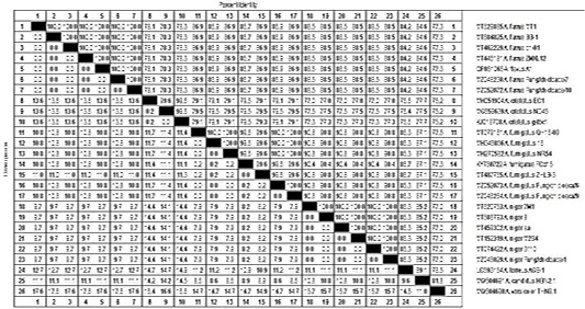
Molecular identification of fungi at the genus or species level by amplification of ITS gene have been used . In this study, a representative 5 Aspergillus species (A. fumigatus (2), A. flavus (2), and A. niger (1)) were taken for further molecular identification. Phylogenetic analysis and nucleotide identity based on ITS sequence (Figure 3 and Table 3) confirmed the morphological identification of these Aspergillus species.
Finally, the opportunistic fungi in intestine may be a source of contamination of poultry meat in processing plant and increasing fungal load inside poultry house. Thus, continuous monitoring of fungal population in chickens during health and disease conditions is required for deep understanding of its role, food safety, and low personal risk.
CONCLUSIONS AND RECOMMENDATIONS
This preliminary study was performed to identify intestinal fungal population in immunosuppressed chickens. A total of 19 purified fungal isolates have been identified morphologically. Aspergillus isolates were the most identified (42%) fungal isolates from cloacal swabs of IBD infected chickens, followed by Trichosporon (10.5%), Penicillium (10.5%), Fusarium (5%), Candida (1%) and non-identified isolates (26%). Furthermore, the obtained 8 Aspergillus isolates were further morphologically identified into A. fumigatus which was most frequent species identified (4/19 of total) and (4/8 of Aspergillus species); 2 A. flavus, 1 A. niger and 1 A. terreus. Furthermore, molecular identification of 5 representative Aspergillus isolates indicated A. fumigatus (2). A. flavus (2), and A. niger (1) were performed.
Novelty Statement
The study of intestinal fungal population and its role during health and disease is essential particularly in chickens. Thus, our study was accomplished to isolate and identify fungal populations that passed through the GIT of the immunosuppressed chickens during IBDV natural infection. Different purified fungal isolates have been identified morphologically in our study. Furthermore, molecular identification of 5 representative Aspergillus isolates were also performed. Our results help to further understand of intestinal fungal population in immunosuppressed chickens.
AUTHOR’S CONTRIBUTION
Mohamed M. Amer and Kh. M. Elbayoumi carried out the research design and revised the manuscript. Aly M. Ghetas, Dalia M. Sedeek, Hanaa S. Fedawy and Hoda M. Mekky isolated and identified fungal isolates. M. A. Bosila performed histopathology for intestinal samples. Aly M Ghetas wrote the manuscript.
Conflict of interest
The authors have declared no conflict of interest.
Figures
Figure 1.
Intestine tissue sections of naturally IBDV infected chicken stained by H and E. A. Normal intestine tissue sections stained (x 200) B: Chicken intestine showing inflammatory cells infiltration of the mucosa (white arrow) and congestion (C) of the submucosa (x 100). C: Chicken intestine showing inflammatory cells infiltration of the mucosa (white arrow) (x 100).
Figure 2.
Amplified ITS gene of isolated fungi. Lane L = 100 bp marker; Lane P = Positive control. Lanes 1 (A. niger), 6 (A. fumigatus), 7 (A. flavus), 9 (A. fumigatus) and 10 (A. flavus); Lane N = Negative control.
Figure 3.
Phylogenetic tree 5 Aspergillus strains named MZ052072, MZ052073, MZ048029, MZ048030 and MZ052073 are compared with 21 sequences of ITS gen published in GenBank. Branched distances correspond to sequence divergence.
Table 3.
Percentage of nucleotide identities for the ITS genes of 5 Aspergillus strains named MZ052072, MZ052073, MZ048029, MZ048030 and MZ052073as compared with 21 sequences published in GenBank.
Figure 4.
Deduced amino acid sequence of the ITS gene of of 5 Aspergillus strains named MZ052072, MZ052073, MZ048029, MZ048030 and MZ052073 as compared with 21 sequences published in GenBank.
References
Altschul SF, Gish W, Miller W, Myers EW, Lipman DJ (1990). Basic local alignment search tool. J. Mol. Biol., 215(3): 403-410. https://doi.org/10.1016/S0022-2836(05)80360-2[CrossRef]
Arné P, Thierry S, Wang D, Deville M, LeLoch G, Desoutter A, Féménia F, Nieguitsila A, Huang W, Chermette R, Guillot J (2011). Aspergillus fumigatus in poultry. Int. J. Microbiol., 2011: 746356. https://doi.org/10.1155/2011/746356[CrossRef]
Bancfort JD, Stevens A (1996). Theory and practice of histological technique. 4th Ed., New York: Churchill Livingstone.
Beytut E, Özcan K, Erginsoy, S (2004). Immunohistochemical detection of fungal elements in the tissues of goslings with pulmonary and systemic aspergillosis. Acta Vet. Hungar., 52(1): 71–84. https://doi.org/10.1556/avet.52.2004.1.8[CrossRef]
Bjerrum L, Engberg RM, Leser TD, Jensen BB, Finster K, Pedersen K (2006). Microbial community composition of the ileum and cecum of broiler chickens as revealed by molecular and culture-based techniques. Poult. Sci., 85: 1151-1164. https://doi.org/10.1093/ps/85.7.1151[CrossRef]
Byrd JA, Caldwell DY, Nisbet DJ (2017). The identification of fungi collected from the ceca of commercial poultry. Poult. Sci., 96: 2360–2365. https://doi.org/10.3382/ps/pew486[CrossRef]
Chang C, Lin H (2016). Dysbiosis in gastrointestinal disorders. Best Pract. Res. Clin. Gastroenterol., 30: 3–15. https://doi.org/10.1016/j.bpg.2016.02.001[CrossRef]
Cheng Z, Li M, Wang Y, Chai T, Cai Y and Li N (2020). Pathogenicity and immune responses of aspergillus fumigatus infection in chickens. Front. Vet. Sci., 7: 143. https://doi.org/10.3389/fvets.2020.00143[CrossRef]
Cisek AA, Binek M (2014). Chicken intestinal microbiota function with a special emphasis on the role of probiotic bacteria. Pol. J. Vet. Sci., 17(2): 385–394. https://doi.org/10.2478/pjvs-2014-0057[CrossRef]
Colombo AL, Padovan AC, Chaves GM (2011). Current knowledge of Trichosporon spp. and Trichosporonosis. Clin. Microbiol. Rev., 24: 682–700. https://doi.org/10.1128/CMR.00003-11[CrossRef]
Cui N, Huang X, Kong Z, Huang Y, Huang Q, Yang S, Zhang L, Xu C, Zhang X, Cui Y (2018). Newcastle disease virus infection interferes with the formation of intestinal microflora in newly hatched specific-pathogen-free chicks. Front. Microbiol., 9: 900. https://doi.org/10.3389/fmicb.2018.00900[CrossRef]
Day JM, Oakley BB, Seal BS, Zsak L (2015). Comparative analysis of the intestinal bacterial and RNA viral communities from sentinel birds placed on selected broiler chicken farms. PLoS One, 10(1): 15. https://doi.org/10.1371/journal.pone.0117210[CrossRef]
Delmas B, Kibenge FSB, Leon JC, Mundt E, Vakaharia VN, Wu JL (2005). Birnaviridae. In: Virus Virus Taxonomy. 8th Report ICTV; Fauquet, C.M., Mayo, M.A., Maniloff, J., Desselberger, U., Ball, L.A., Eds.; Elsevier Academic Press: San Diego, CA, USA, 2005; pp. 561–569.
Fan Y, Pedersen O (2021). Gut microbiota in human metabolic health and disease. Nat. Rev. Microbiol., 19(1): 55–71. https://doi.org/10.1038/s41579-020-0433-9[CrossRef]
Frank DN, Zhu W, Sartor RB, Li E (2011). Investigating the biological and clinical significance of human dysbioses. Trends Microbiol., 19(9): 427–434. https://doi.org/10.1016/j.tim.2011.06.005[CrossRef]
Gaskell GJ, Carter DA, Britton WJ, Tovey ER, Benyon, FH, Løvborg U (1997). Analysis of the internal transcribed spacer regions of ribosomal DNA in common airborne allergenic fungi. Electrophoresis, 18(9): 1567-1569. https://doi.org/10.1002/elps.1150180914[CrossRef]
Gillevet PM, Sikaroodi M, Torzilli AP (2009). Analyzing salt-marsh fungal diversity: Comparing arisa fingerprinting with clone sequencing and pyrosequencing. Fungal Ecol., 2: 160–167. https://doi.org/10.1016/j.funeco.2009.04.001[CrossRef]
Girma G, Abebaw M, Zemene M, Mamuye Y (2016). A review on aspergillosis in poultry. J. Vet. Sci. Technol., 7: 6. https://doi.org/10.4172/2157-7579.1000382[CrossRef]
Hoerr FJ (2010). Clinical aspects of immunosuppression in poultry. Avian Dis., 54; 2–15. https://doi.org/10.1637/8909-043009-Review.1[CrossRef]
Hughes ER, Winter MG, Duerkop BA, Spiga L, de Carvalho TF, Zhu WH, Gillis CC, Buttner L, Smoot MP, Behrendt CL, Cherry S, Santos RL, Hooper LV, Winter SE (2017). Microbial respiration and formate oxidation as metabolic signatures of inflammation-associated dysbiosis. Cell Host Microbe., 21: 208–219. https://doi.org/10.1016/j.chom.2017.01.005[CrossRef]
Iwen PC, Schutte SD, Florescu DF, Noel-Hurst RK, Sigler L (2012). Invasive Scopulariopsis brevicaulis infection in an immunocompromised patient and review of prior cases caused by Scopulariopsis and Microascus species. Med. Mycol., 50: 561–569. https://doi.org/10.3109/13693786.2012.675629[CrossRef]
Jackwood DJ (2017). Advances in vaccine research against economically important viral diseases of food animals: Infectious bursal disease virus. Vet. Microbiol., 206: 121–125. https://doi.org/10.1016/j.vetmic.2016.11.022[CrossRef]
Kurtzman CP, Fell JN, Bekhout T (2011). The yeasts. A taxonomic study. Elsevier, Tokyo, 2011, pp. 2080.
Li L, Kubasová T, Rychlik I, Hoerr FJ, Rautenschlein S (2018). Infectious bursal disease virus infection leads to changes in the gut associated lymphoid tissue and the microbiota composition. PLoS One, 13: e0192066. https://doi.org/10.1371/journal.pone.0192066[CrossRef]
Liu LY, Lin LL, Zheng LN, Tang H, Fan XZ, Xue NG, Li M, Liu M, Li XY (2018). Cecal microbiome profile altered by salmonella enterica, serovar Enteritidis inoculation in chicken. Gut Pathog., 10: 14. https://doi.org/10.1186/s13099-018-0261-x[CrossRef]
Ma X, Wang Q, Li H, Xu C, Cui N, Zhao X (2017). 16S rRNA genes Illumina sequencing revealed differential cecal microbiome in specific pathogen free chickens infected with different subgroup of avian leukosis viruses. Vet. Microbiol., 207: 195–204. https://doi.org/10.1016/j.vetmic.2017.05.016[CrossRef]
Macdonald SE, Nolan MJ, Harman K, Boulton K, Hume DA, Tomley FM, Stabler RA, Blake DP (2017). Effects of Eimeria tenella infection on chicken caecal microbiome diversity, exploring variation associated with severity of pathology. PLoS One, 12(9): 17. https://doi.org/10.1371/journal.pone.0184890[CrossRef]
Müller H, Islam M.R, Raue R (2003). Research on infectious bursal disease the past, the present and the future. Vet. Microbiol., 97: 153–165. https://doi.org/10.1016/j.vetmic.2003.08.005[CrossRef]
Ott SJ, Kuhbacher T, Musfeldt M, Rosenstiel P, Hellmig S, Rehman A, Drews O, Weichert W, Timmis KN, Schreiber S (2008). Fungi and inflammatory bowel diseases: Alterations of composition and diversity. Scand J. Gastroenterol., 43: 831-841. https://doi.org/10.1080/00365520801935434[CrossRef]
Peixoto RS, Harkins DM, Nelson KE (2021). Advances in microbiome research for animal health. Annu. Rev. Anim. Biosci., 9(1): 289–311. https://doi.org/10.1146/annurev-animal-091020-075907[CrossRef]
Perumbakkam S, Hunt HD, Cheng HH (2014). Marek’s disease virus influences the core gut microbiome of the chicken during the early and late phases of viral replication. FEMS Microbiol. Ecol., 90(1): 300–312. https://doi.org/10.1111/1574-6941.12392[CrossRef]
Raper KB, Fennel DI (1965). The genus Aspergillus. Williams Wilkins, Baltimore, pp. 685.
Robinson K, Xiao Y, Johnson TJ, Chen B, Yang Q, Lyu W, Wang J, Fansler N, Becker S, Liu J, Yang H, Zhang G (2020). Chicken intestinal mycobiome: Initial characterization and its response to bacitracin methylene disalicylate. Appl. Environ. Microbiol., 86: e00304-20. https://doi.org/10.1128/AEM.00304-20[CrossRef]
Romach EA, Sklan D, Uni Z (2004). Microflora ecology of the chicken intestine using 16S ribosomal DNA primers. Poult. Sci., 83: 1093-1098. https://doi.org/10.1093/ps/83.7.1093[CrossRef]
Roto SM, Rubinelli PM, Ricke SC (2015). An introduction to the avian gut microbiota and the effects of yeast-based prebiotic-type compounds as potential feed additives. Front. Vet. Sci., 2: 28. https://doi.org/10.3389/fvets.2015.00028[CrossRef]
Sandoval-Denis M, Sutton DA, Fothergill AW, Cano-Lira J, Gené J, Decock CA, de Hoog GS, Guarro J (2013). Scopulariopsis, a poorly known opportunistic fungus: spectrum of species in clinical samples and in vitro responses to antifungal drugs. J. Clin. Microbiol., 51: 3937–3943. https://doi.org/10.1128/JCM.01927-13[CrossRef]
Sugui JA, Kwon-Chung KJ, Juvvadi PR, Latgé J-P, Steinbach WJ (2014). Aspergillus fumigatus and related species. Cold Spring Harb. Perspect. Med., 5: a019786. https://doi.org/10.1101/cshperspect.a019786[CrossRef]
Tamura K, Stecher G, Peterson D, Filipski A, Kumar S (2013). MEGA6: Molecular evolutionary genetics analysis version 6.0. Mol. Biol. Evol., 30: 2725–2729. https://doi.org/10.1093/molbev/mst197[CrossRef]
Thompson JD, Higgins DG, Gibson TJ (1994). CLUSTAL W: Improving the sensitivity of progressive multiple sequence alignment through sequence weighting position-specific gap penalties and weight matrix choice. Nucl. Acids Res., 22(22): 4673-4680. https://doi.org/10.1093/nar/22.22.4673[CrossRef]
Yudiarti T., VD Yunianto BI, Murwani R and Kusdiyantini E (2012). Isolation of fungi from the gastrointestinal tract of indigenous chicken. J. Indones. Trop. Anim. Agric., 37(2): 2012. http://www.jppt.undip.ac.id/pdf/37(2)2012p115-120.pdf, https://doi.org/10.14710/jitaa.37.2.115-120[CrossRef]
Zulkifli NA, Zakaria L (2017). Morphological and molecular diversity of Aspergillus from corn grain used as livestock feed. Hayati J. Biosci., 24(1): 26-34. https://doi.org/10.1016/j.hjb.2017.05.002[CrossRef]