Special Issue: Emerging and Re-Emerging Animal Health Challenges in Low and Middle-Income Countries · Volume 12, Issue 1, 6 December 2024 · Open Access
Clinical Isolation and Genetic Established and Saprophytic Bacteria During Normal Estrus Cycle of Cow
· · ·
Affiliations & Notes ⌄
1Department of Surgery and Obstetrics, College of Veterinary Medicine, University of Baghdad, Iraq. *Correspondence | Hayder A.H. Al-Mutar, Department of Surgery and Obstetrics, College of Veterinary Medicine, University of Baghdad, Iraq
In case of Escherichia coli present in vagina of cows, the percentage of proliferation was highest in proestrus, diestrus and metestrus phases (45.45%, 41.66% and 40%, respectively) while there was no bacterial proliferation of Escherichia coli estrus phase.
Opposite of that, the result shows no proliferation of Streptococcus lutetiensis bacteria in bovine vaginal mucosa in proestrus and estrus phase while there was low proliferation (20%) of this bacteria in metestrus phase and (25%) in diestrus and phase.
The study concluded that the highest percentage of bacterial normal flora present in pro-estrus, estrus, metestrus and dieasrus were (Escherichia coli 45%, Klebsiella pneumoniae 50%, Escherichia coli 40% and Escherichia coli 41%, respectively).
The study concluded that there are many types of Saprophytic bacterial proliferation during estrus cycle inside vagina and uterus of cows with a variation of different countries, bread and age.
Selected from the article's own text.
Abstract
There are many cases of vaginitis and metritis occurring in animals farms specially in cows In majority of these cases, the infection remained unnoticed caused by proliferation of saprophytic bacteria that present normally in mucosal membrane of vaginal. These infections predispose cow more susceptible to infection and bacterial proliferation during the stages of estrus periods (proestrus, estrus, diestrus and metestrus). Here, we investigated the presence of saprophytic bacteria in vaginal smears, and characterize them using PCR. All vaginal swabs collected from 43 sexually matured cows were subjected to Stuart transport media, before planted in blood agar media to identify the number of colonies counted. The bacteria was confirmed by Gram’s staining and biochemical tests. The PCR products from the bacterial samples were genetically analyzed (Al-Mutar et al., 2018). The result show that the most bacterial present in bovine vagina were Streptococcus thermophilus and Streptococcus uberis (92.65%) while the lowest percent of bacteria present in bovine vagina was Corynebacterium frankenforstense (78.8%). The other saprophytic bacteria were Streptococcus lutetiensis, Klebsiells pneumoniae, Escherichia coli and Staphylococcus aurreus were present in a percentage from 94.61%, 93.25%, 80.5% and 78.69%. respectively. The study concluded that there are many types of Saprophytic bacterial proliferation during estrus cycle inside vagina and uterus of cows with a variation of different countries, bread and age.
During the normal estrus cycle of cows, the presence of saprophytic bacteria in the reproductive tract does not significantly affect reproductive efficiency . Studies have shown that the uterine microbiota composition differs between cows exhibiting estrus and non-estrual cows ; . Additionally, shifts in the intestinal microbiota have been observed during estrus synchronization in Simmental cows, influenced by reproductive hormones . Abnormal cervical mucus discharge (A-CMD) in cows and heifers has been associated with specific pathogens affecting fertility, emphasizing the importance of evaluating cervical mucus discharge for infertility issues . Overall, while the reproductive tract microbiota may vary during the estrus cycle, the presence of saprophytic bacteria does not seem to hinder the reproductive performance of cows. Saprophytic bacteria have been isolated from the vaginal flora of cows in various studies. The vaginal microbiota of Criollo Limonero cows revealed the presence of Gram-positive bacteria like Staphylococcus aureus and Arcanobacterium pyogenes . Additionally, research on crossbred cows identified Bacillus, Corynebacterium, and Staphylococcus as predominant vaginal bacteria during different physiological states . Furthermore, studies on postpartum cows showed the presence of Bacillus, Streptococcus, and Escherichia coli in the vaginal flora, with varying antimicrobial resistance profiles . These findings highlight the diverse bacterial populations present in the vaginal environment of cows, including saprophytic species, which play a role in the overall reproductive health and microbial balance in these animals .
The normal flora of the cow vagina consists predominantly of Gram-positive bacteria, including Arcanobacterium pyogenes, Staphylococcus aureus, coagulase-negative Staphylococcus, Erysipelothrix rhusiopathiae, Bacteroides spp., and Peptostreptococcus spp. ; ; . Additionally, Streptococci and Staphylococci are the dominant bacteria in the cow’s vaginal microbiota, with low levels of Enterobacteriaceae and lactobacilli present . The vaginal microbiota of cows and ewes exhibit differences in terms of α-diversity, with both being mainly colonized by Bacteroidetes, Fusobacteria, and Proteobacteria, along with the presence of Archaea, mainly Desulfurococcales . The microbial community in the cow’s vaginal tract is influenced by factors such as hormonal changes, pregnancy status, and potential interactions with the gastrointestinal microbes.
Genetic detection of saprophytic bacteria from the vaginal tract of cows can be achieved through molecular techniques like PCR . Studies have successfully utilized PCR assays for specific identification of bacteria like Staphylococcus saprophyticus in different contexts ; . Similarly, in the context of bovine genital campylobacteriosis, a real-time PCR assay coupled with high-resolution melting analysis was developed for the identification of Campylobacter fetus subspecies in bull preputial samples . Furthermore, research on Leptospira colonization in cows utilized PCR for detecting leptospiral DNA in vaginal fluid samples ; . These studies highlight the effectiveness of genetic detection methods, such as PCR, in identifying specific bacteria in the vaginal tract of cows, showcasing the potential for accurate and sensitive detection of Saprophytic bacteria.
Factors effecting growth of saprophytic bacteria
The growth of saprophytic bacteria in the vagina of cows is influenced by various factors. Research indicates that the normal microbial flora of a cow’s vagina is predominantly composed of Streptococci and Staphylococci ; . Additionally, lactobacilli levels increase during the cow’s growth, potentially due to hormonal influences . Factors such as age, stage of the sexual cycle, and pH value do not seem to directly correlate with the bacterial flora in the cow’s vagina . Moreover, the presence of specific microbes in the reproductive organs of cows, including the uterus and vagina, can impact reproductive success . Understanding these microbial communities and their dynamics is crucial for managing reproductive inefficiencies in cattle operations and ensuring overall productivity. The death of saprophytic bacteria in the cow genital system can be influenced by various factors. For instance, Staphylococcus saprophyticus, a common bacterium found in ticks, has been identified as a causative agent of pathological symptoms in fully engorged female ticks, leading to their death . Additionally, bacterio-spermia caused by bacteria like S. saprophyticus and Escherichia coli can impact sperm parameters and clinical outcomes in semen samples, potentially affecting assisted reproductive techniques and reducing the likelihood of clinical pregnancy . Moreover, the presence of bacteria like E. coli and Staphylococcus coagulase-negative in semen samples can lead to infections and impact fertility outcomes . Therefore, the interaction of saprophytic bacteria with the reproductive system can have significant implications for the health and fertility of cows.
Materials and Methods
Vaginal swabs
A vaginal swab collected from 43 sexually matured cows randomly. These samples consist of 11 swabs collected from cows in proestrus phase, 10 in estrus phase, 10 metestrus phase and 12 in diestrus phase. Then the swabs samples are flood in Stuart transport media, after that planted in blood agar media to identify the number of colonies counted, followed by gram staining and biochemical tests then the results were presented descriptively.
Blood samples
A 3-5 ml of blood samples were collected form jugular vein and stored in EDTA collecting tube in very sterile conditions.
Template preparation and DNA extraction
In compliance with the Manufacturer’s instructions, the DNA of cow used in the study were extracted using a kit for extraction DNA (kit no. 17045). The augmented product of the 16s gene was exposed to (2%) agarose gel electrophoresis.
Polymerase chain reaction (PCR) reaction
Through amplification of the 16s gene, the prolactin receptor (PRLR) was identified by PCR. forward primer (5’-AGAGTTTGA TCCTGGCTCAG-3’) and reverse primer (5’-GGTTACCTTGTTACGACTT-3’) (1250 bp), (each 10 Picomoles/μL) and the annealing (56°C for 35 sec.) .
DNA sequences
All the samples products of PCR from these primers (1250bp) obtained from the Iraqi cows were used. The PCR product was sequencing using online BLASTNCBI and BioEdit.
Statistical analysis
By using the statistical SPSS-USA-v.23 software, statistical analysis of the data was performed. These data were track using (HWE) and Fisher’s-exact test .
RESULTS AND DISCUSSION
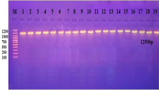
Results of conventional PCR for detection of extracted DNA of 16s RNA gene: Gel electrophoresis revealed bands of extracted nucleic acid of 16s gene (Figure 1).
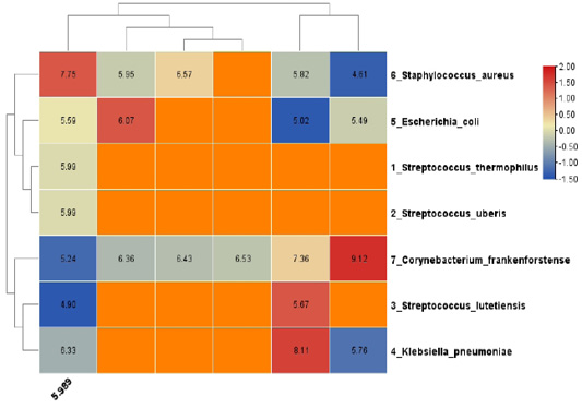
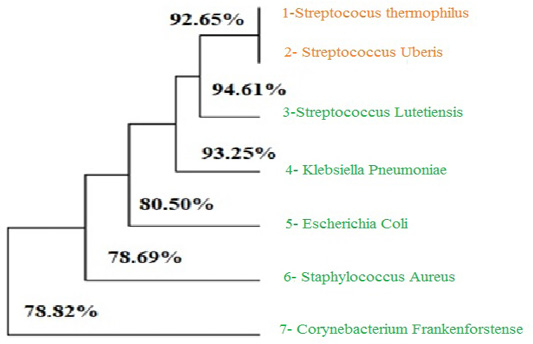
The result show as present in Figures 2 and 3 that the most more bacterial present in bovine vagina were Streptococcus thermophilus and Streptococcus uberis in percent 92.65% while the lowest percent of bacteria present in bovine vagina was Sorynebacterium frankenforstense (78.8%). The other saprophytic bacteria were Streptococcus lutetiensis, Klebsiells pneumoniae, Escherichia coli and Staphylococcus aurreus were present in a percent of 94.61%, 93.25%, 80.5 % and 78.69%, respectively.
According to Table 1, the results show high bacterial proliferation Klebsiella pneumoniae (50 %) in estrus, less of these in metestrus (30 %) and diestrus (33.33 %) phasein comparison with proestrus phase (9.09). In case of Escherichia coli present in vagina of cows, the percentage of proliferation was highest in proestrus, diestrus and metestrus phases (45.45%, 41.66% and 40%, respectively) while there was no bacterial proliferation of Escherichia coli estrus phase. The result either show proliferation of Streptococcus thermophiles (18.18% and 20%, respectively), Streptococcus uberis (9.09% and 20%, respectively) and Streptococcus pneumoniae (18.18% and 20%, respectively) in proestrus and estrus phases while there were no proliferation of these bacteria in metestrus and diestrus. Opposite of that, the result shows no proliferation of Streptococcus lutetiensis bacteria in bovine vaginal mucosa in proestrus and estrus phase while there was low proliferation (20%) of this bacteria in metestrus phase and (25%) in diestrus and phase. Finally, the Corynebacterium frankenforstense found in vaginal mucosa in percentage of 10% in metestrus only while not presented in the other phases of estrus cycle (proestrus, estrus and diestrus). The study concluded that the highest percentage of bacterial normal flora present in pro-estrus, estrus, metestrus and dieasrus were (Escherichia coli 45%, Klebsiella pneumoniae 50%, Escherichia coli 40% and Escherichia coli 41%, respectively).
Table 1: Percentage of bacteria present in vagina of cow during phases of estrus cycle.
Diestrus phase (%)
Metestrus phase (%)
Estrus phase (%)
Proestrus phase (%)
Bacteria
33.33
30
50
9.09
Klebsiella pneumoniae
41.66
40
0
45.45
Escherichia coli
0
0
20
18.18
Streptococcus thermophilus
0
0
20
9.09
Streptococcus uberis
0
0
10
18.18
Streptococcus pneumoniae
25
20
0
0
Streptococcus lutetiensis
0
10
0
0
Corynebacterium frankenforstense
It was long believed that the healthy uterus and vagina are a sterile environment. However, the microbial population in the uterus and vagina of cows has been characterized thanks to recent advancements in sequencing technology. The result of bacterial present in vaginal mucosa agreed with ; they found that there are many of saprophytic bacteria in vaginal mucosa during estrus such as Escherichia, Streptococcus, Corynebacterium and Staphyococcus bacteria. While reported that Firmicutes, Clostridia, Clostridiales, Lachnospiraceae, Ruminococcaceae, Peptostreptococcaceae, Ruminiclostridium, Oscillibacter, Romboutsia, Lachnoclostridium, Coprococcus, Ruminococcus, Clostridium leptum, and Dorea spp. were significantly increased, while Bacteroidetes, Bacteroidia, and Bacteroidales were significantly decreased in vaginal mucosa. The result also agreed with they found K. pneumonia, A. pyogenes, S. aureus, present in vaginal mucosa in low percentage ranged from (3%-17%). Bacillus spp. were the most frequently isolated bacteria during the follicular phase of the estrous cycle, followed by Corynebacterium spp., Staphylococcus spp., and Streptococcus spp. In contrast, Staphylococcus spp. were the most frequently isolated bacteria during the luteal phase, followed by Corynebacterium spp., Bacillus spp., E. coli, and Streptococcus spp. In descending order, the most common vaginal bacterial isolates found during pregnancy were Bacillus, Streptococcus, E. Coli, Klebsiella, and pseudomonas. While the main isolates in endometritis were Staphylococcus spp., Bacillus spp., Streptococcus spp., E. coli, Salmonella, and Corynebacterium, the vaginal aspirates of acyclic cows contained Streptococcus spp., Bacillus spp., and Staphylococcus spp., Corynebacterium spp., and Micrococcus. Rich bacterial diversity was found in the vagina of crossbred calves with a range of physio-pathological disorders .
Conclusions and Recommendations
The study concluded that there are many types of Saprophytic bacterial proliferation during estrus cycle inside vagina and uterus of cows with a variation of different countries, bread and age.
Acknowledgment
This study supported by College of Veterinary Medicine, University of Baghdad.
NOVELTY STATEMENT
This study introduces an innovative approach to obtaining clinical and molecular bacterial isolation from pregnant and non-pregnant in cow
AUTHOR’S CONTRIBUTION
This work was carried out in collaboration between all authors. HAHA-M: Designed the study, performed the statistical analysis. HAHA-M, BJH, KAH, and SOH: Wrote the protocol, and wrote the first draft of the manuscript managed the analyses of the study and the literature searches.
Ethical approval
The research started in Jan 2022, lasting for one year, samples were taken from Baghdad government fields. All samples were examined at the laboratory of Bacteriology in the College of Veterinary Medicine, University of Bagdad.
Conflict of interest
The authors have declared no conflict of interest.
Figures
Figure 1:.
Product of PCR, the size of band was 1250 bp. The product electrophoresis on 2% agarose for 1:20 hrs. M: DNA ladder (100).
Figure 2:.
The heat map of the differentially expressed genes.
Figure 3:.
The phylogenetic tree of the type of Bacteria.
References
Al-Mutar HA (2017). Investigation the polymorphism of gonadotropin releasing hormone receptor gene in Iraq sheep. Iraqi J. Vet. Med., 41(1): 138-144. https://doi.org/10.30539/iraqijvm.v41i1.96[CrossRef]
Al-Mutar HAK, Younis HL, Khawla H (2018). Effect of the point mutation in growth differentiation factor 9 gene in Awassi sheep oocytes on sterility and fertility. J. Pure Appl. Microbiol., 12(4): 2095-2102. https://doi.org/10.22207/JPAM.12.4.46[CrossRef]
Ayhan A, Hulya T, Mehmet K, Mehmet ŞG, Faruk P (2010). Microbial flora of normal and abnormal cervical mucous discharge associated with reproductive performance of cows and heifers in Estrus. Asian-Austral. J. Anim. Sci., 23(8): 1007-1012. https://doi.org/10.5713/ajas.2010.90645[CrossRef]
Biao D, Haiyan Y, Hua W, Hu J, Jigang Y, Shen Y, Wang Y, Weibo W (2012). Saprophyte culturing medium for oil field sewage treatment system.
Brooke AC, Sydney TR, Felipe GD, Franco, Timothy PL, Ishola A, Ky GP, Phillip RM (2017). Vaginal and uterine bacterial communities in postpartum lactating cows. Front. Microbiol., 8: 1047-1047. https://doi.org/10.3389/fmicb.2017.01047[CrossRef]
Diana SÁ, Sonia AN, Jorge LB, Pablo RR (2020). Incidencia de vaginosis bacterianay factores asociados, en pacientes con secreciones vaginales anormales. FACSALUD-UNEMI. 4(7): 61-66. https://doi.org/10.29076/issn.2602-8360vol4iss7.2020pp61-66p[CrossRef]
Dong-Yan W, Chunjie W, Huasai S, Bo L, Zhimeng C, Pengfei Z (2022). Reproductive hormones mediate intestinal microbiota shifts during estrus synchronization in grazing simmental cows. Animals, 12(14): 1751-1751. https://doi.org/10.3390/ani12141751[CrossRef]
Estefhan M, Raquel C, Ma DR, Quezada-Delgado B, Sachman-Ruiz ER (2010). Staphylococcus saprophyticus is a pathogen of the cattle tick Rhipicephalus (Boophilus) microplus. Biocontr. Sci. Technol., 20(10): 1055-1067. https://doi.org/10.1080/09583157.2010.505325[CrossRef]
Fernanda S, Oliveira D, de Oliveira, Emanoel F, Martins F, Federico C, Paula R, Melissa HP, Walter L (2016). Avaliação histológica e imuno-histoquímica da colonização vaginal por Leptospira em vacas com fluido vaginal positivo à PCR. 38: 163-167.
Francisco MCJ, Ariza GM, Alberto HMF, Aragón GC (2018). Saprophyticus bacteria: Cause of fulminant and lethal sepsis? https://www.ijcriinfectiousdiseases.com/archive/article-full-text/100002Z16JC2018
Friedrich T (2010). Die Bakterienflora der Vagina des gesunden Rindes unter Berücksichtigung des Geschlechtszyklus, der Trächtigkeitsphase, des Alters und des pH wertes. J. Vet. Med. Ser. Binfect. Dis. Vet. Publ. Health, 5(4): 373-390. https://doi.org/10.1111/j.1439-0442.1958.tb00525.x[CrossRef]
Gloria I, Morales P (2015). Aspectos clínicosy diagnóstico de laboratorio de la vaginosis bacteriana. 14(5): 611-623.
Ibraheem RS, Abbas BA (2015). Study on Candida species isolated from cows with molecular detection of some virulence gene. Bas. J. Vet. Res., 14(2): 31-43. https://doi.org/10.23975/bjvetr.2022.174022[CrossRef]
James RH (2005). Bacterial and fungal organisms in the vagina of normal cows and cows with vaginitis. https://oaktrust.library.tamu.edu/handle/1969.1/1310
Jeffrey D, Swartz, MM, Lachman KW, Thomas J, O’Neill TW (2014). Characterization of the vaginal microbiota of Ewes and cows reveals a unique microbiota with low levels of Lactobacilli and near-neutral pH. Front. Vet. Sci., 1: 19-19. https://doi.org/10.3389/fvets.2014.00019[CrossRef]
Julio BO, Sunny ZN, Jexenia N, Germán PM (2010). Perfil de la flora bacteriana vaginal. Un riesgo potencial para la reproducción de vacas criollo limonero profile of vaginal bacterial flora. A potential risk for reproduction in criollo limonero cows.
Laith SY, Al-Mutar HAK, Ali AA (2018). Effect of leptin gene polymorphism on reproductive efficiency in Awassi Ewes. Adv. Anim. Vet. Sci., 7(1): 18-23. https://doi.org/10.17582/journal.aavs/2019/7.1.17.23[Full text][CrossRef]
Luis O, Alba G, Enrique A, Silveira P (2005). Perdurabilidad de la efectividad de la combinación penicilina- estreptomicina en la reducción de la carga bacteriana del semen de toro congelado en pastillas (Durability of the effectiveness of the penicillin-streptomycin combination in the reduction of the bacterial content of frozen bulls semen in concentrated pellet form).
MacKichan J, Kato-Maeda M, Miller S (2015). Use of 16S rRNA gene for identification of a broad range of clinically relevant bacterial pathogens. PLoS One, 10(2): e0117617. https://doi.org/10.1371/journal.pone.0117617[CrossRef]
Madison BW, Matthew PH, Jeferson M, Pedro LPF (2022). 299 differences in microbial community composition between uterine horns ipsilateral and contralateral to the corpus luteum in beef cows on day 15 of the estrous cycle. J. Anim. Sci., 100(Suppl. 3): 142-143. https://doi.org/10.1093/jas/skac247.263[CrossRef]
Marta S, Sabine K, Gonçalo P, Luísa M, Luís L, Elisabete S (2022). Molecular diagnosis of bovine genital campylobacteriosis using high-resolution melting analysis. Front. Microbiol., 13. https://doi.org/10.3389/fmicb.2022.969825[CrossRef]
Maryam R, Ezzat AG (2023). Detection of virulence genes among Staphylococcus saprophyticus isolated from women with urinary tract infections: First report from Iran.
Mateus L, Kelly M, Grillo R, Branco M, Ribeiro G, Silvia G, Laura RL (2015). Vaginal microbiome characterization of Nellore cattle using metagenomic analysis. PLoS One, 10(11). https://doi.org/10.1371/journal.pone.0143294[CrossRef]
Otero C, Lucila SC, Silva ROR, Wilde A, Pesce DR, Holgado M, Elena N (2000). Vaginal bacterial microflora modifications during the growth of healthy cows. Lett. Appl. Microbiol., 31(3): 251-254. https://doi.org/10.1046/j.1365-2672.2000.00809.x[CrossRef]
Patel CI, Panchal MT, Dhami AJ, Bhanderi BB, Mathakiya RA (2019). Isolation of bacteria from the vaginal aspirates of cyclic, acyclic, endometritic and pregnant crossbred cows. Int. J. Curr. Microbiol. Appl. Sci., 8(3): 536-542. https://doi.org/10.20546/ijcmas.2019.803.065[CrossRef]
Qodri WNL, Sri M, Sri PM, Rimayanti R, Suherni S, Wiwiek T (2020). Efisiensi reproduksi pada sapi perah yang teridentifikasi Bakteri non spesifik dalam saluran reproduksi. 9(2): 41-47. https://doi.org/10.20473/ovz.v9i2.2020.41-47[CrossRef]
Rahawy MA, Al-Mutar HAK (2021). Association of the KiSS1 gene with litter size in Cyprus and Iraqi black goats. Vet. World, 14(8). https://doi.org/10.14202/vetworld.2021.1995-2001[CrossRef]
Raymond M, Rousset F (1995). Population genetics software for exact tests and ecumenisms. J. Hered., 86(3): 248-249. https://doi.org/10.1093/oxfordjournals.jhered.a111573[CrossRef]
Sunny AZ, Julio B, Jexenia N (2011). Normal bacterial flora from vaginas of Criollo Limonero cows. Trop. Anim. Health Prod., 43(2): 291-294. https://doi.org/10.1007/s11250-010-9701-4[CrossRef]
Yvonne W, Burim NA, Divakar JA, Michael GG (2013). Characterisation of the bacterial microbiota of the vagina of dairy cows and isolation of pediocin-producing Pediococcus acidilactici. BMC Microbiol., 13(1): 19-19. https://doi.org/10.1186/1471-2180-13-19[CrossRef]