
Antibiotic susceptibility testing is the most suitable strategy for antibiotic resistance detection in bacteria. This project was an initial step for the production of β-lactamase for clinical diagnostics in Pakistan. The current work deals with the optimization of conditions for higher level production of recombinant β-lactamase from Bacillus subtilis R5. Supplementation of LB medium with various carbon sources including wheat bran, rice bran and molasses could increase the enzyme production from 25 to 43.7, 44.3 and 49 µmole/min whereas the nitrogen sources including tryptone, peptone and yeast extract could enhance the enzyme production from 25 to 40.21, 41 and 43.76 µmole/min, respectively. The highest β-lactamase production was recorded when the LB medium was supplemented with 4% wheat bran and 2% yeast extract. The locally produced recombinant β-lactamase exhibited strong potential regarding the hydrolysis of β-lactam ring containing antibiotics and showed comparable results to β-lactamase from Hi-media kit available in the market. The β-lactamase from current study was found suitable for its use as positive control in antibiotic susceptibility testing and this enzyme will be utilized for the development of antibiotic susceptibility testing kit at domestic level in near future.
Short Communication · Volume 53, Issue 6, 17 October 2021 · Open Access
Production and Utilization of Locally Characterized Recombinant β-Lactamase as Positive Control in the Antibiotic Susceptibility Testing
· · · · · · ·
Affiliations & Notes ⌄
- 1Institute of Biochemistry and Biotechnology, University of Veterinary and Animal Sciences, Abdul Qadir Jillani Road, Lahore 2Riphah International University, Raiwind Road Campus, Lahore 3Department of Biotechnology, Virtual University, Lahore , * Corresponding author: tayyab_pakistan@yahoo.com
- 2muhammad.tayyab@uvas.edu.pk
Article Info ⌄
- Journal:
- Pakistan Journal of Zoology
- Published:
- 17 October 2021

From the article
- The LB medium could produce 25 µmole/min of β-lactamase whereas this production was increased to 43.7, 44.3 and 49 µmole/min when the LB medium was supplemented with molasses (3%), rice bran (3%) and wheat bran (4%), respectively (Table I).
- Comparative analysis for the production of β-lactamase showed maximal enzyme production after 5 h of post induction incubation at 37°C (Fig. 1A, B) while further incubation decreased the enzyme production.
- The locally characterized β-lactamase showed comparable results to the β-lactamase from Hi-Media kit that was used as positive control.
- The β-lactamase from present study was found suitable for its utilization in the antibiotic susceptibility testing and this enzyme will be produced and utilized for the development of antibacterial susceptibility testing kit at domestic level.
Selected from the article's own text.
Abstract
Materials and methods
The chemicals utilized in this study were of high purity and were procured from Sigma Aldrich, USA and β-lactamase used as control was from Hi-Media (Mumbai, India).
Recently coding region for β-lactamase from Bacillus subtilis R5 was cloned in pET21a and was expressed in BL21CodonPlus (DE3) cells. This β-lactamase has been characterized (unpublished data). Expression vector (pET-21a) harboring the β-lactamase gene from Bacillus subtilis R5 (pET-LAC) was available in the lab and was utilized in the current study for its enhanced production and its efficacy was evaluated against various β-lactam ring containing antibiotics.
For optimization of conditions for the higher level production of recombinant β-lactamase the recombinant BL21 CodonPlus (DE3) cells with pET-LAC were grown on LB medium supplemented with 1-6% carbon sources (Wheat bran, Rice bran and Molasses) and 1-3% nitrogenous sources (yeast extract, Tryptone and peptone) before sterilization. The sterilized medium was inoculated with the overnight grown recombinant cells at 37°C under shaking conditions. The cells were induced with 0.6 mM Isopropyl β-D-1-thiogalactopyranoside (IPTG) when the optimal density reached to 0.4 followed by further incubation under same conditions. The samples were drawn after every hour, up to 6 h and were utilized to examine production of β-lactamase after lysing of cells by sonication ; ; .
For evaluation of efficacy of β-lactamase in antibiotic susceptibility testing antibiotic susceptibility assay was performed in collaboration with Chughtai Labs, Lahore. The petri plates were prepared using sterilized Hinton agar medium (Merck) under sterilized conditions. Clinical isolates of pathogenic strains of Staphylococcus aureus and Escherichia coli were spread homogenously on the above said petri plates. These plates were utilized for exploring the effect of antibiotics on the clinical isolates in the presence or absence of β-lactamase.
Five antibiotics containing β-lactam ring including tetracycline (30 µg); erythromycin (15 µg); ampicillin (10 µg); penicillin G (6 µg) and lincomycin (2 µg) were utilized to examine their activity against the selected microbial strains. The antibiotics concentrations were selected as per guidance by the Clinical and Laboratory standard institute (CLSI), USA and European society of clinical Microbiology and infectious diseases EUCAST, Sweden, Europe.
Five sterilized paper discs (1 to 5) were placed on each petri plate having pathogenic culture. In each plate, a separate antibiotic was applied on all the discs. In addition to antibiotic on one of the discs (disc 1), β-lactamase from Hi-Media kit was applied as control whereas on the second disc (disc 2) the locally characterized recombinant β-lactamase was applied to explore the efficacy of locally characterized β-lactamase to degrade the antibiotics. The results were recorded in term of formation of zone due to availability of antibiotic and absence of zone due to presence of β lactamase as this enzyme is involved in the degradation of β lactam containing antibiotics.
The data was analyzed using univariate analysis of variance tool using SPSS 16 software .
Table I.- Optimization of conditions for maximal production of β-lactamase.
| Medium | Activity (µmole/min) |
| Lauria Bertani (LB) medium | 25 |
| Supplementation of LB medium with carbon sources | |
| Wheat bran (4%) | 49 |
| Rice bran (3%) | 44.3 |
| Molasses (3%) | 43.7 |
| Supplementation of LB medium with nitrogen sources | |
| Yeast extract (2%) | 43.76 |
| Peptone (1.5%) | 41 |
| Tryptone (1.5%) | 40.21 |
Results and discussion
The LB medium could produce 25 µmole/min of β-lactamase whereas this production was increased to 43.7, 44.3 and 49 µmole/min when the LB medium was supplemented with molasses (3%), rice bran (3%) and wheat bran (4%), respectively (Table I). The immense production of β-LACBS by wheat bran might be due to its richness in macro and micro nutrients, minerals, vitamins and bioactive compounds which resulted in enhanced microbial growth and β-lactamase production ; ; . It had been reported from the literature that wheat bran is most suitable substrate for production of enzymes from the Bacillus subtilis strains . Less fat contents, dry matter, crude proteins, more ash contents in case of rice bran and viscosity of molasses might be the possible reason for lower production of β-lactamase .
Similarly the supplementation of LB medium with Tryptone (1.5%), Peptone (1.5%) and Yeast extract (2%) could enhance the production of β-lactamase from 25 to 40.21, 41 and 43.76 µmole/min, respectively (Table I). These results are in agreement with the previous report for the production of β-lactamase from Bacillus licheniformis . This is might be due to the composition of yeast extract as it is rich in nucleic acids, amino acids, peptides, vitamins, trace elements and growth factors that could possibly elicit more microbial growth and enzyme production than peptone and tryptone ; . Comparative analysis for the production of β-lactamase showed maximal enzyme production after 5 h of post induction incubation at 37°C (Fig. 1A, B) while further incubation decreased the enzyme production.
The development of zone of inhibition around all the antibiotics containing discs showed the intactness and working of antibiotics. Presence of ampicillin, tetracycline, lincomycin, erythromycin and penicillin G showed 23.16, 17.66, 21.33, 22.83 and 23.16 mm zone of inhibition against the E. coli, respectively (Supplementary Fig. S1) and 14.16, 26.16, 15.33, 23.66 and 23.5 mm zone of inhibition against the S. aureus, respectively (Supplementary Fig. S2). The application of locally characterized β-lactamase on the disc 2 containing antibiotic in each petri plate resulted in the degradation of antibiotics and this antibiotic degrading ability was confirmed with no appearance of zone of inhibition around the disc on each plate (Supplementary Figs. S1, S2). The locally characterized β-lactamase showed comparable results to the β-lactamase from Hi-Media kit that was used as positive control. The β-lactamase from present study was found suitable for its utilization in the antibiotic susceptibility testing and this enzyme will be produced and utilized for the development of antibacterial susceptibility testing kit at domestic level.
Acknowledgements
The authors are thankful to the Higher Education Commission Pakistan for providing the funds for the completion of this work.
Supplementary material
There is supplementary material associated with this article. Access the material online at: https://dx.doi.org/10.17582/journal.pjz/20210718150740
Statement of conflict of interest
The authors have declared no conflict of interest
Figures
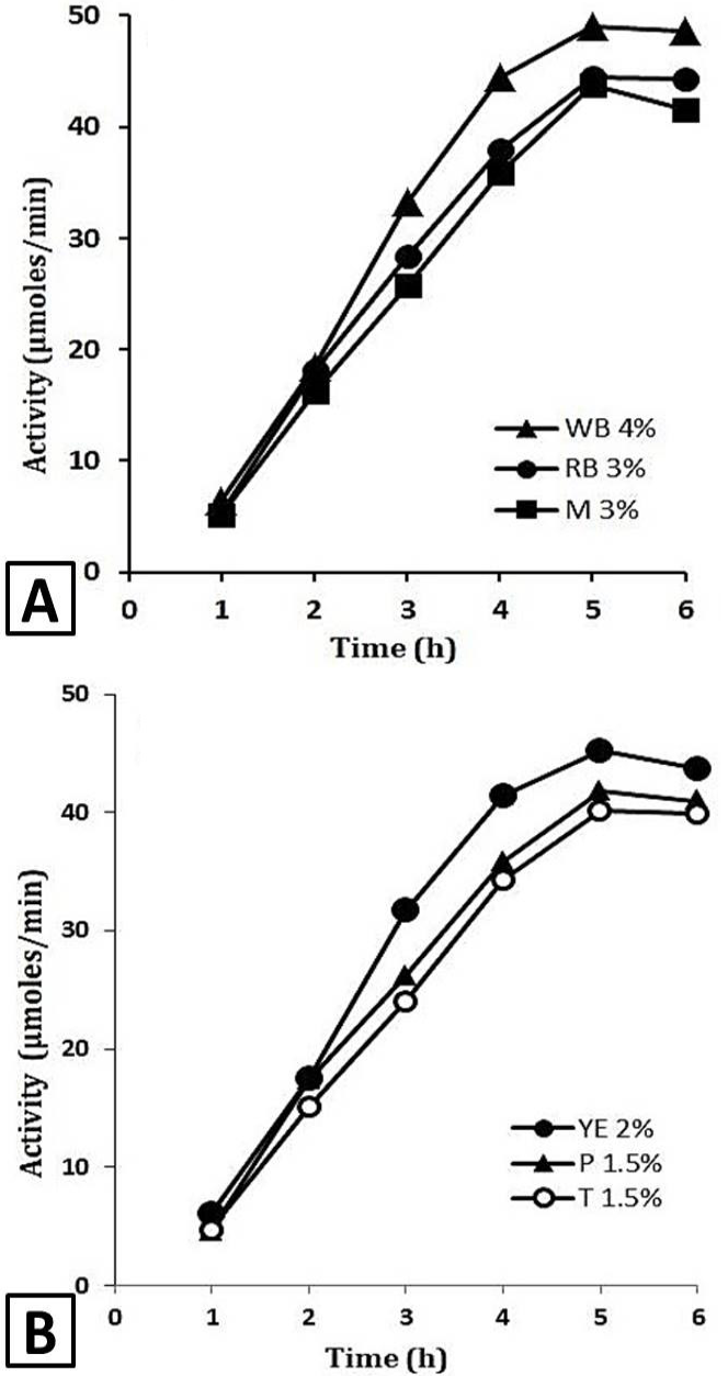
Comparative analysis of impact of carbon and nitrogen sources for the production of β-Lactamase. A, comparative analysis of carbon sources on the production of β-Lactamase. Molasses, rice bran and wheat bran were used as carbon sources for the supplementation of LB medium and for the enhanced production of β-Lactamase. B, comparative analysis of nitrogen sources on the production of β-Lactamase. Tryptone, peptone and yeast extract were used as nitrogen sources for the supplementation of LB medium and for the enhanced production of β-Lactamase. X-axis shows the time of incubation (h) while Y-axis shows the β-lactamase activity in µmoles/min.
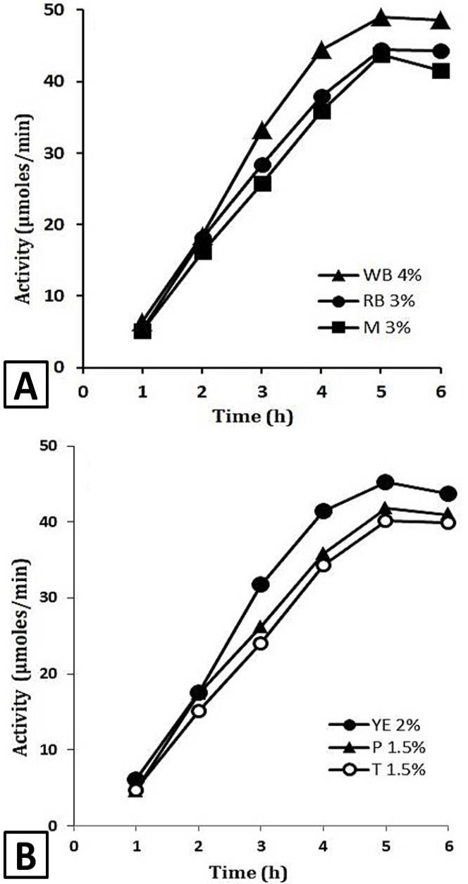
Comparative analysis of impact of carbon and nitrogen sources for the production of β-Lactamase. A, comparative analysis of carbon sources on the production of β-Lactamase. Molasses, rice bran and wheat bran were used as carbon sources for the supplementation of LB medium and for the enhanced production of β-Lactamase. B, comparative analysis of nitrogen sources on the production of β-Lactamase. Tryptone, peptone and yeast extract were used as nitrogen sources for the supplementation of LB medium and for the enhanced production of β-Lactamase. X-axis shows the time of incubation (h) while Y-axis shows the β-lactamase activity in µmoles/min.
References
- Abinaya, R., Ramya, P., Sivakami, V., Ponnusami, V. and Sugumaran, V., 2017. J. chem. Pharmaceut., 10: 488–491.
- Aslam, B., Wang, W., Arshad, M.I., Khurshid, M., Muzammil, S., Rasool, M.H., Nisar, M.A., Alvi, R.F., Aslam, M.A., Qamar, M.U. and Salamat, M.K., 2018. Infect. Drug Resist., 11: 1645. https://doi.org/10.2147/IDR.S173867[CrossRef]
- Biswas, P., Bharti, A.K., Kadam, A. and Dutt, D., 2019. Bioresources, 14: 5788-5806.
- Boucher, H.W., Talbot, G.H., Benjamin Jr, D.K., Bradley, J., Guidos, R.J., Jones, R.N., Murray, B.E., Bonomo, R.A. and Gilbert, D., 2013. Clin. Infect. Dis., 56: 1685-1694. https://doi.org/10.1093/cid/cit152[CrossRef]
- Bush, K., 2018. Antimicrob. Agents Chemother., 62: e01076-18. https://doi.org/10.1128/AAC.01076-18[CrossRef]
- Butt, M.S., Qamar, M.I., Anjum, F.M., Aziz, A. and Randhawa, M.A., 2004. Nutr. Fd. Sci., 34: 161-165. https://doi.org/10.1108/00346650410544855[CrossRef]
- Celik, E., 2003. Bioprocess design parameters for beta-lactamase production by Bacillus species. PhD thesis, The Middle East Technical University, Ankara, Turkey.
- Chen, T., Cheng, G., Ahmed, S., Wang, Y., Wang, X., Hao, H. and Yuan, Z., 2017. Talanta, 175:435-442. https://doi.org/10.1016/j.talanta.2017.07.044[CrossRef]
- Danilova, I.V. and Sharipova, M.R., 2020. Front. Microbiol., 11: 1-7. https://doi.org/10.3389/fmicb.2020.01782[CrossRef]
- Dirar, M., Bilal, N., Ibrahim, M.E. and Hamid, M., 2020. Cureus, 12: e7260.
- Djembi, L.F., Hodille, E., Jaboulay, C.S., Coudrais, S., Santis, D.N., Gardes, S., Mauranne, C.C., Mourey, N., Fredenucci, I. and Girard, R., 2017. J. Infect. Publ. Hlth., 10: 185-190. https://doi.org/10.1016/j.jiph.2016.04.010[CrossRef]
- Essack, S.Y., 2001. Pharm. Res., 18: 1391-1399. https://doi.org/10.1023/A:1012272403776[CrossRef]
- Falgenhauer, L., Imirzalioglu, C., Oppong, K., Akenten, C.W., Hogan, B., Krumkamp, R., Poppert, S., Levermann, V., Schwengers, O., Sarpong, N. and Dabo, O.E., 2019. Front. Microbiol., 9: 3358. https://doi.org/10.3389/fmicb.2018.03358[CrossRef]
- Garcia, S.B., Galan, F., Iglesias, R.M.A. and Gracia, P.M.T., 2019. Vector Borne Zoonot. Dis., 19: 115-120. https://doi.org/10.1089/vbz.2018.2333[CrossRef]
- Jalal, A., Rashid, N., Rasool, N. and Akhtar, M., 2009. J. Biosci. Bioengin., 107: 360-365. https://doi.org/10.1016/j.jbiosc.2008.12.005[CrossRef]
- Javed, M.M., Zahoor, S., Shafaat, S., Mehmooda, I., Gul, A., Rasheed, H., Bukhari, S.A.I. and Aftab, M.N., 2012. Afr. J. Microbiol. Res., 6: 724-733.
- Jubeh, B., Breijyeh, Z. and Karaman, R., 2020. Molecules, 25: 1543. https://doi.org/10.3390/molecules25071543[CrossRef]
- Khalid, A., Tayyab, M., Hashmi, A.S., Yaqub, T., Awan, A.R., Wasim, M., Saeed, S., Firyal, S. and Shakoori, A.R., 2019. Pakistan J. Zool., 51: 1371. https://doi.org/10.17582/journal.pjz/2019.51.3.925.934[Full text][CrossRef]
- Kraemer, S.A., Ramachandran, A. and Perron, G.G., 2019. Microorganisms, 7: 180. https://doi.org/10.3390/microorganisms7060180[CrossRef]
- Krasauskas, R., Labeikyte, D., Markuckas, A., Povilonis, J., Armalyte, J., Planciuniene, R., Kavaliauskas, P. and Suziedeliene, E., 2015. Annls. clin. Microbiol. Antimicrob., 14: 1-8. https://doi.org/10.1186/s12941-015-0113-1[CrossRef]
- Krishna, C., 2005. Crit. Rev. Biotechnol., 25: 1-30. https://doi.org/10.1080/07388550590925383[CrossRef]
- Kummerer, K., 2009. Chemosphere, 75: 417-434. https://doi.org/10.1016/j.chemosphere.2008.11.086[CrossRef]
- Kusuma, S.A.F., Hargono, Y.C. and Winarno, H., 2018. J. Pharm. Sci. Res., 10: 1036-1040.
- Latorre, J.D., Hernandez, V.X., Wolfenden, R.E., Vicente, J.L., Wolfenden, A.D., Menconi, A., Bielke, L.R., Hargis, B.M. and Tellez, G., 2016. Front. Vet. Sci., 3: 95-103. https://doi.org/10.3389/fvets.2016.00095[CrossRef]
- Loh, M.C., Mamphweli, S., Meyer, E. and Okoh, A., 2018. Molecules, 23: 795. https://doi.org/10.3390/molecules23040795[CrossRef]
- Park, K. and Reardon, K.F., 1996. Biotechnol. Lett., 18: 737-740. https://doi.org/10.1007/BF00130775[CrossRef]
- Pereckaite, L., Tatarunas, V. and Giedraitiene, A., 2018. Methods, 152: 154-164. https://doi.org/10.1016/j.mimet.2018.07.014[CrossRef]
- Ramirez, D.M. and Bentley, W.E., 1995. Biotechnol. Bioengin., 47: 596-608. https://doi.org/10.1002/bit.260470512[CrossRef]
- Sawai, T., Takahashi, I. and Yamagishi, S., 1978. Antimicrob. Agents Chemother., 13: 910-913. https://doi.org/10.1128/AAC.13.6.910[CrossRef]
- Senter, P.D. and Springer, C.J., 2001. Adv. Drug Deliv. Rev., 53: 247-264. https://doi.org/10.1016/S0169-409X(01)00206-X[CrossRef]
- Schallmey, M., Singh, A. and Ward, O.P., 2004. Can. J. Microbiol., 50: 1-17. https://doi.org/10.1139/w03-076[CrossRef]
- Singh, Y., Palombo, M. and Sinko, P.J., 2008. Curr. med. Chem., 15: 1802-1826. https://doi.org/10.2174/092986708785132997[CrossRef]
- Smyth, T.P.O, Donnell, M.E., Connor, O.M.J. and Ledger, S.J.O., 2000. Tetrahedron, 56: 5699-5707. https://doi.org/10.1016/S0040-4020(00)00419-1[CrossRef]
- Tham, J., Walder, M., Melander, E. and Odenholt, I., 2012. Infect. Drug Resist., 5: 143.
- Tooke, C.L., Hinchliffe, P., Bragginton, E.C., Colenso, C.K., Hirvonen, V.H., Takebayashi, Y. and Spencer, J., 2019. J. mol. Biol., 431: 3472-500. https://doi.org/10.1016/j.jmb.2019.04.002[CrossRef]
- Ventola, C.L., 2015. Pharm. Ther., 40: 277.
Supplementary material
Services
Article usage statistics
Loading…