
In this work, sub-lethal (1/3rd, 1/5th and 1/6th of LC50) effects of chlorpyrifos (CPF) on biochemical markers such as glutathione S-transferase (GST) activity and total protein contents in various tissues (gills, hepatic, renal, brain, muscle and cardiac) of Labeo rohita was determined. Fish was exposed for 60-day and sampling was done after 7-day. Results showed that the GST activity was considerably increased in L. rohita as compared to control. The GST activity was enhanced in various tissues (hepatic, brain, cardiac, gills, renal and muscle) of CPF treated fish as compared to control group. Comparison among different concentrations indicated that 1/3rd of LC50 caused greater increase in GST activity followed by that of 1/5th and 1/6th. The GST activity in selected tissues of fish varied with duration of exposure as 28>21>35>42>14>7>49>56. Total protein contents in all selected tissues of CPF exposed fish were decreased with the passage of time. Among all the concentrations, 1/3rd of LC50 cause greater decrease in protein contents. The total protein contents in various tissues of L. rohita followed the order: muscle>hepatic>brain>gills>renal>cardiac.
Short Communication · Volume 54, Issue 3, 5 March 2022 · Open Access
Sub-lethal Effects of Chlorpyrifos on Glutathione S-Transferase Activity and Total Protein Contents of Fish, Labeo rohita
· · · · ·
Affiliations & Notes ⌄
- 1Department of Zoology, Wildlife and Fisheries, University of Agriculture, Faisalabad 2Department of Zoology, Cholistan University of Veterinary and Animal Sciences, Bahawalpur, Pakistan 3College of Animal Sciences and Technology, Guangxi University, China * Corresponding author: qaisrasiddique711@gmail.com, dr.humanaz98@gmail.com
Article Info ⌄
- Journal:
- Pakistan Journal of Zoology
- Published:
- 5 March 2022

From the article
- The present study showed that selected tissues of CPF treated L. rohita had increased GST activity compared to control group.
- The GST activity in tissues of treated fish followed the order: hepatic>gills>renal>brain>cardiac>muscle.
- Results showed that total protein contents of kidney, heart, gills, liver, brain and muscle of L. rohita was considerably decreased after exposure to sub-lethal concentrations (1/3rd, 1/5th and 1/6th of LC50) of CPF in contrast to untreated group.
- It is concluded that chlorpyrifos significantly enhance the GST activity and reduce the protein contents in different tissues of fish.
Selected from the article's own text.
Abstract
Materials and methods
The experimental fish (Labeo rohita) were procured from Fish Seed Hatchery, Faisalabad and transferred to Fisheries Research Farm, University of Agriculture, Faisalabad. After that juveniles of experimental fish were kept in cemented tank and adapted to laboratory conditions for 2-week. The experiment was carried out in triplicates in aquarium, which is made up of glass and have a capacity of 100 L water. The 10 juveniles were placed in each aquarium. The LC50 value (96h) of CPF for L. rohita was calculated as 16.53 mgL-1 . L. rohita were exposed to different sub-lethal (1/3rd, 1/5th and 1/6th of LC50) concentration of technical grade CPF for 2-month and sampling was done after 7 days. The physico-chemical parameters of water such as total hardness (250 mgL-1), temperature (30ºC), and pH (7.00) were kept constant throughout the experimental period. However, CO2, Na, Ca, K, Mg, NH3 and electrical conductivity (EC) were also measured . Air pump with capillary system is used to supply continuous air to all the test and control mediums. After each sampling, GST activity and total protein contents were measured in different tissues viz., cardiac, hepatic, renal, gills, brain, and muscle of both control and treated fish. The GST activity was calculated by observing obsorbance of the conjugated molecules of GSH with 1-chloro, 2, 4-dinitrobenzene (CDNB) at 340 nm . One unit of enzyme make conjugated molecule with GSH with 10.0 nmol of CDNB per minute at 25oC. Total protein content was measured by Biuret method (Gornall et al., 1994).
The obtained data was analyzed by suitable methods of statistics (Steel et al., 1997). ANOVA was applied to compare the variables of both treated and control fish.
Results
The present study showed that selected tissues of CPF treated L. rohita had increased GST activity compared to control group. The GST activity in tissues of treated fish followed the order: hepatic>gills>renal>brain>cardiac>muscle. During the first 28-days GST activity was enhanced and then declined up to 56-day (Fig. 1). Comparison among different concentrations showed that 1/3rd of LC50 had greater impact on GST activity followed by 1/5th and 1/6th.
Results showed that total protein contents of kidney, heart, gills, liver, brain and muscle of L. rohita was considerably decreased after exposure to sub-lethal concentrations (1/3rd, 1/5th and 1/6th of LC50) of CPF in contrast to untreated group. Total protein contents in tissues of L. rohita followed the order: muscle>hepatic>brain>gills>renal>cardiac (Fig. 2).
Discussion
CPF has broadly and successfully been used all over the world. In fish, oxidative stress has been caused by pesticide exposure and other pollutants ; . In fishes, oxidative stress takes place by two ways, firstly due to unnecessary buildup of the ROS. Secondly, when the critical ratio between anti-oxidants and oxidants is disturbed, due to decline in the ration of antioxidants. Both of these mechanisms ultimately cause damage . In aquatic species, ROS causes alterations in antioxidant enzyme systems which play
important role in scavenging of these free radicals (Livingstone, 2001). GSTs play protective role against these free radicals ; . Various studies reported the mechanism of enzyme action and detoxification of free radicals in fish ; .
In this study the GST activity was stimulated in various tissues of CPF treated fish when compared to that of control group. GST enzyme is concerned with the detoxification of various xenobiotics, hence the activation of GST enzyme has been considered as a pollutant indicator. It has been reported by various studies that hepatic tissue showed enzymatic induction under pesticide exposure in fish. Therefore, GSTs are vital in detoxification and elimination of electropositive compounds from the body ; .
Hepatic and renal tissues showed greater response towards pesticide toxicity because they are vital organs in detoxification and elimination pathways . The greater sensitivity of these tissues is vital for controlling the balance between oxidants and antioxidants under pesticide exposure ; . CPF is processed by microsomal enzymes having oxidative activity at active oxons which ultimately causes oxidative damage ; . According to Keramati et al. (2010) exposure of CPF and methyl parathione altered the GST activity in all the chosen tissues of Oreochromis niloticus. Similarly, O. niloticus showed enhanced GST activity under CPF exposure .
L. rohita exposed to CPF showed significantly higher GST activity in all selected tissues when compare with control. Yonar (2013) reported that treatment with OP pesticide (malathion) showed variations in GST activity in gills, hepatic and renal tissues of carp. Sharbidre et al. (2011) described alterations in GST activity in gills, hepatic and muscle tissues of P. reticulate when exposed to OP pesticide (methyl-parathion and CPF) concentrations (1/4th, 1/8th and 1/10th of LC50).
Xing et al. (2012) observed significant changes in renal and brain GST activity of Cyprinus carpio under the long term administration of CPF and atrazine. Oruc (2010) reported that sub-lethal concentrations (5, 10 and 15 ppb) of CPF showed significant increase in GST activity after 30 days in O. niloticus. Similarly, Karmakar et al. (2016) investigated the enhanced GST activity in gills, hepatic and renal tissues of L. rohita under sub-lethal (18.12 mgL-1 to 105.2 mgL-1) exposure of malathion.
In current study, protein contents in different tissues of L. rohita declined under sub-lethal concentrations (1/3rd, 1/5th, 1/6th) of CPF. CPF is found to be more toxic for fish . In the presence of OP pesticide protein content is reduced either due to the inhibition of protein synthesis or increased degradation/oxidation of proteins by ROS ; . Glucose is synthesized by the metabolic consumption of keto acids which ultimately result in the depletion of protein contents ; ; ; ; Chezhian et al., 2010) and related alterations were also investigated in C. punctatus under exposure of technical grade malathion (Agrhari et al., 2006).
Conclusion
It is concluded that chlorpyrifos significantly enhance the GST activity and reduce the protein contents in different tissues of fish. Furthermore, these parameters of fish can be used as a valuable biomarker of insecticides toxicity in water bodies.
Statement of conflict of interest
The authors have declared no conflict of interest.
Figures
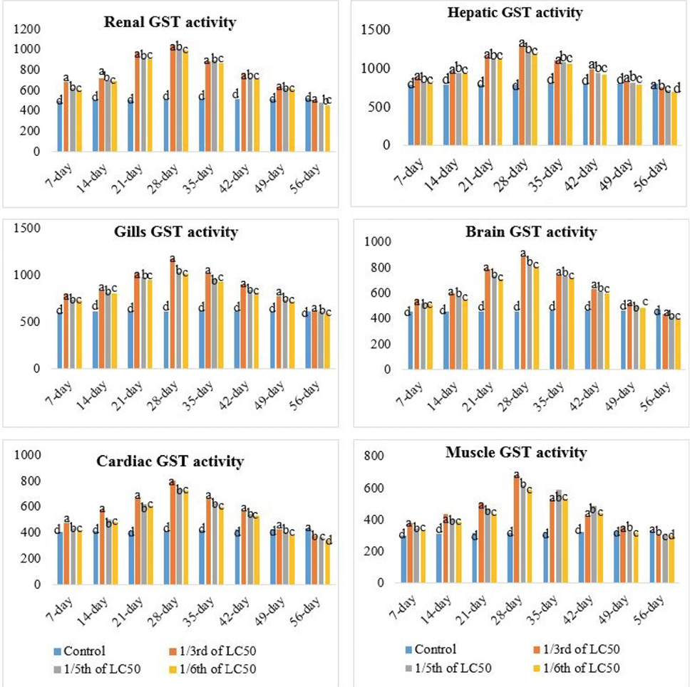
GST activities in gills, heart, cardiac, renal, brain and muscle of Labeo rohita under sub-lethal exposure of chlorpyrifos for different time intervals.
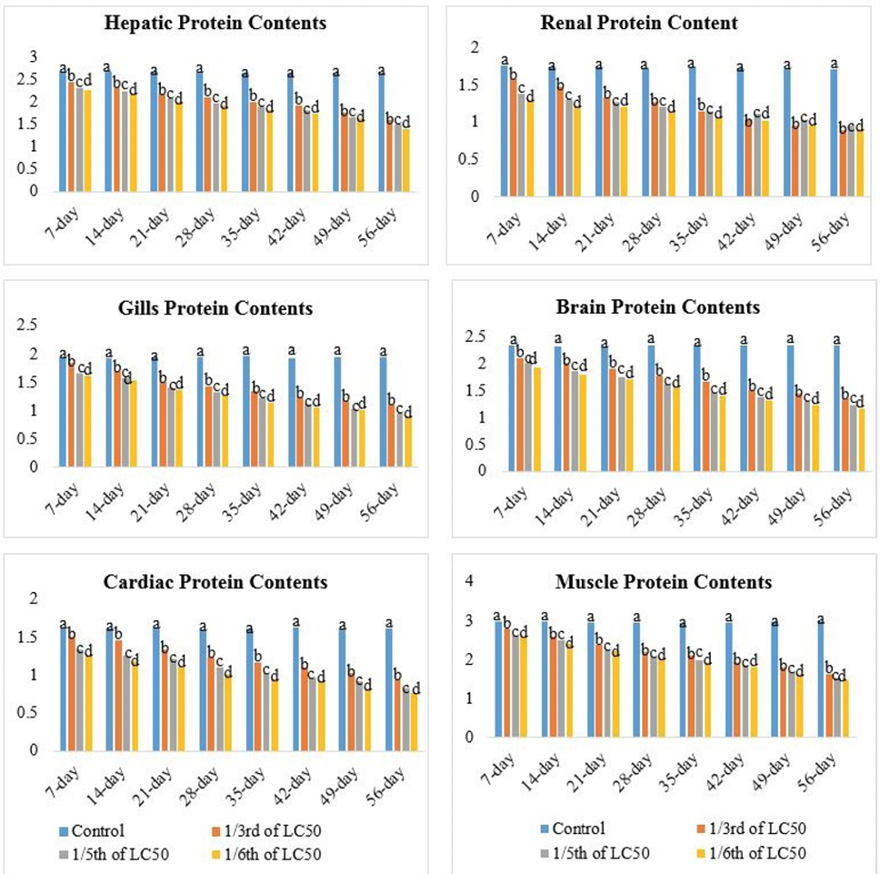
Total protein contents in gills, cardiac, liver, renal, brain and muscle of Labeo rohita under sub-lethal exposure of chlorpyrifos for different time intervals.
References
- Abdollahi, M., Ranjbar, A., Shadnia, S., Nikfar S. and Rezaie, O.E., 2004. Med. Sci. Monit., 10: 141-147.
- Agrahari, S., Gopal, K. and Pandey, K.C., 2006. J. environ. Biol., 27: 453-457.
- Albores, A., Ortega-Mantilla, G., Sierra-Santoyo, A., Cebrian, M.E., MunozSanchez, J.L., Calderon-Salinas, J.V. and Manno, M., 2001. Toxicol. Lett., 124: 1-10. https://doi.org/10.1016/S0378-4274(01)00382-4[CrossRef]
- Ali, D., Nagpure, N.S., Kumar, S., Kumar, R., Kushwaha, B. and Lakra, W.S., 2009. Fd. Chem. Toxicol., 47: 650-656. https://doi.org/10.1016/j.fct.2008.12.021[CrossRef]
- APHA, 2005. Standard methods of the examination of water and waste water. New York, pp. 1193.
- Blanchette, B., Feng, X. and Singh, B.R., 2007. Mar. Biotechnol., 23: 513-542. https://doi.org/10.1007/s10126-007-9034-0[CrossRef]
- Cicchetti, R. and Argentin, R.G., 2003. Mutagenesis, 18: 127-32. https://doi.org/10.1093/mutage/18.2.127[CrossRef]
- Egaas, E., Sandvik, M., Fleld, E., Kallqvist, T., Goksoyr, A. and Svensen, A., 1999. Comp. Biochem. Physiol., 122: 337-344. https://doi.org/10.1016/S0742-8413(98)10133-0[CrossRef]
- Frova, C., 2006. Biomol. Eng., 23: 149-169. https://doi.org/10.1016/j.bioeng.2006.05.020[CrossRef]
- Gornall, A.G., Bardawill, C.J. and David, M.M., 1949. J. biol. Chem., 177: 751-766. https://doi.org/10.1016/S0021-9258(18)57021-6[CrossRef]
- Illyas, R., 2015. Effect of selected pesticides on growth and DNA damage in peripheral blood erythrocytes of fish. Ph.D. thesis, University of Agriculture, Faisalabad. pp. 1-329.
- Karmakar, S., Patra, K., Jana, S., Mandal, D.P. and Bhattacharjee, S., 2016. Pestic. Biochem. Physiol., 126: 49-57. https://doi.org/10.1016/j.pestbp.2015.07.006[CrossRef]
- Keramati, V., Jamili, S. and Ramin, M., 2010. Fish. aquat. Sci., 5: 368-376. https://doi.org/10.3923/jfas.2010.368.376[CrossRef]
- Kumari, M., 2007. Environ. Ecol., 225: 1164-1166.
- Livingstone, D.R., 2001. Mar. Pollut. Bull., 42: 656-666. https://doi.org/10.1016/S0025-326X(01)00060-1[CrossRef]
- Mannervik, B., 1985. Adv. Enzymol. Relat. Areas mol. Biol., 57: 357-417.
- Mates, J.M., 2000. Toxicology, 153: 83-104. https://doi.org/10.1016/S0300-483X(00)00306-1[CrossRef]
- Mehta, A., Verma, R.S. and Srivastava, N., 2008. Environ. mol. Mutagen., 49: 426-433. https://doi.org/10.1002/em.20397[CrossRef]
- Monferran, M.V., Pesce, S.F., Cazenave, J. and Wunderlin, D.A., 2008. Environ. Toxicol., 23: 184-192. https://doi.org/10.1002/tox.20326[CrossRef]
- Mortensen, A.S. and Arukwe, A., 2007. Aquat. Toxicol., 85: 113-123. https://doi.org/10.1016/j.aquatox.2007.08.004[CrossRef]
- Muley, D., Karanjkar, D. and Maske, S., 2007. J. environ. Biol., 28: 245-249.
- Ojha, A., Yaduvanshi, K.S. and Srivastava, N., 2011. Pest. Biochem. Physiol., 99: 148-156. https://doi.org/10.1016/j.pestbp.2010.11.011[CrossRef]
- Oruc, E.O., 2010. Pestic. Biochem. Physiol., 96: 160-166. https://doi.org/10.1016/j.pestbp.2009.11.005[CrossRef]
- Peebua, P., Kosiyachinda, P., Pokethitiyook, P. and Kruatrachue, M., 2007. Bull. environ. Contam. Toxicol., 78: 138-141. https://doi.org/10.1007/s00128-007-9027-8[CrossRef]
- Poet, T.S., Wu, H., Kousba, A.A. and Timchalk, C., 2003. Toxicol. Sci., 72: 193-200.
- Rao, J.V., 2006. Pestic. Biochem. Physiol., 86: 8-84. https://doi.org/10.2307/25161863[CrossRef]
- Rusyniak, D.E. and Nanagas, K.A., 2004. Semin. Neurol., 24: 197-204. https://doi.org/10.1055/s-2004-830907[CrossRef]
- Scandalios, J.G., 2005. Braz. J. Med. biol. Res., 38: 995-1014. https://doi.org/10.1590/S0100-879X2005000700003[CrossRef]
- Sharbidre, A.A., Metkari, V. and Patode, P., 2011. Pestic. Biochem. Physiol., 10: 132-141. https://doi.org/10.1016/j.pestbp.2011.09.002[CrossRef]
- Sharma, Y., Bashir, S., Irshad, M., Gupta, S.D. and Dogra, T.D., 2005. Toxicology, 206: 49-57. https://doi.org/10.1016/j.tox.2004.06.062[CrossRef]
- Sinhorin, V.D., Sinhorin, A.P., Teixeira, J.M., Mileski, K.M., Hansen, P.C., Moreira, P.S., Kawashita, N.H., Baviera, A.M. and Loro, V.L., 2014. Ecotoxicol. environ. Safe., 106: 181-187. https://doi.org/10.1016/j.ecoenv.2014.04.040[CrossRef]
- Slotkin, T.A., Olivier, C.A. and Seidler, F.J., 2005. Brain Res. Dev., 157: 172-180. https://doi.org/10.1016/j.devbrainres.2005.04.001[CrossRef]
- Steel, R.G.D., Torrie, J.H. and Dickey, D., 1997. Principles and procedure of statistics: A biometrical approach, 3rd Ed. McGraw Hill Book Co. Inc., New York, pp. 352-358.
- Taju, G., Abdul-Majeed, S., Nambi, K.S.N. and Sahul-Hameed, A.S., 2014. Toxicol. Pharmacol., 161: 41-52. https://doi.org/10.1016/j.cbpc.2014.01.007[CrossRef]
- Tang, J., Cao, Y., Rose, R.L., Brimfield, A.A., Dai, D., Goldstein, J.A. and Hodgson, E., 2001. Drug Metabol. Disposit., 29: 1201-1204.
- Tilak, K.S., Veeraiah, K. and Ramanakumari, G.V., 2001. Neurol. Res., 20: 438-445.
- Tilak, K.S., Veeraiah, K. and Rao., D.K., 2005. J. Environ. Biol., 26: 341-347.
- Tripathi, G. and Shasmal, J., 2010. Ecotoxicol. environ. Safe., 73: 1397-1401. https://doi.org/10.1016/j.ecoenv.2010.07.022[CrossRef]
- Van der-Oost, R.J. and Beyer, N.P., 2003. Environ. Toxicol. Pharmacol., 13: 57-149. https://doi.org/10.1016/S1382-6689(02)00126-6[CrossRef]
- Venktramana, G.V., Sadhya-Rani, P.N. and Murthy, P.S., 2006. J. environ. Biol., 27: 119-122.
- Verma, R.S., Mehta, A. and Srivastava, N., 2009. Pestic. Biochem. Physiol., 95: 152-158. https://doi.org/10.1016/j.pestbp.2009.08.004[CrossRef]
- Vutukuru, S.S., 2005. Int. J. environ. Res. Publ. Hlth., 2: 456-462. https://doi.org/10.3390/ijerph2005030010[CrossRef]
- Wu, J.Q. and Laird, D.A., 2003. Environ. Toxicol. Chem., 22: 261-264. https://doi.org/10.1002/etc.5620220204[CrossRef]
- Xing, H., Li, S., Wang, Z., Gao, X., Xu, S. and Wang, X., 2012. Chemosphere, 88: 377-383. https://doi.org/10.1016/j.chemosphere.2012.02.049[CrossRef]
- Yonar, S.M., 2013. Ecotoxicol. environ. Safe., 97: 223-229. https://doi.org/10.1016/j.ecoenv.2013.07.020[CrossRef]
Cited by 1 article
In journals published by ResearchersLinks.
Services
Article usage statistics
Loading…