
Fifty rats were randomly divided into sham group, model group, propofol group, vitexin group and propofol + vitexin group,with 10 rats each group. Rat model of whole liver ischemia-reperfusion injury was established according to Pringle’s method. Results showed that compared with propofol group and vitexin group, levels of serum ALT, AST and LDH in propofol + vitexin group were reduced (P < 0.05), apoptosis index was reduced (P < 0.05), Bcl-2 protein expression was increased (P < 0.05), Bax protein and caspase-3 protein expression was decreased (P < 0.05). In conclusion, propofol and vitexin could up-regulate expression of Bcl-2 protein, down-regulate expression of Bax and caspase-3 protein, inhibit cell apoptosis, reduce liver damage indexes, and have certain protective effects on liver ischemia-reperfusion injury. The effect of propofol combined with vitexin is better than that of propofol and vitexin alone.
Short Communication · Volume 54, Issue 3, 5 March 2022 · Open Access
Effects of Propofol and Vitexin on Apoptosis in Rat Liver Ischemia Reperfusion Injury
·
Affiliations & Notes ⌄
- 1Department of Anesthesiology, Zhuji People’s Hospital of Zhejiang Province, No. 9, Jianmin road, Taozhu street, Zhuji, Zhejiang province, 311800, China * Corresponding author: v5rkhy@163.com
Article Info ⌄
- Journal:
- Pakistan Journal of Zoology
- Published:
- 5 March 2022

From the article
- The results showed that propofol combined with vitexin treatment could better reduce liver injury and have more obvious protective effect on hepatocyte necrosis than propofol and vitexin treatment alone.
- Six hours after reperfusion, compared with sham group, apoptosis index (AI) value of liver tissue cells in propofol group, vitexin group and propofol + vitexin group was significantly increased (P < 0.05, Fig. 2).
- This suggested that propofol and vitexin could inhibit apoptosis during liver ischemia-reperfusion injury and has protective effect on liver ischemia-reperfusion injury.
- The expression of Bcl-2 protein in liver tissues of propofol group and vitexin group was lower than that of propofol + vitexin group (P < 0.05, Fig. 3).
Selected from the article's own text.
Abstract
Materials and methods
Fifty healthy adult male Sprague-Dawley (SD) rats (body mass 230-280 g) were used as experimental animals. Rats were offered routine mixed feed anad drinking water ad libitum and fasted for 12 h before operation.
Intraperitoneal injection of 10% chloral hydrate (0.4 g/kg) was used as anesthesia. The rat model of whole liver ischemia-reperfusion injury was established according to Pringle’s method . Liver artery, portal vein and bile duct were clamped together with non-invasive vascular clip. After 30 min of liver portal occlusion, the non-invasive vascular clip was removed, the blood flow into the liver was restored, and the abdomen was closed layer by layer. Rats were fasted after awakening from anesthesia but water was given freely.
Fifty rats were randomly divided into 5 groups, each of 10 rats, namely sham group, model group, propofol group, vitexin group and propofol + vitexin group. Sham group in which only the liver portal was dissociated without liver portal occlusion was treated after laparotomy with normal saline (2 ml/kg) infused through femoral vein by micro pump. Model group was treated with 2 ml/kg normal saline infused through femoral vein by micropump 10 min before liver portal occlusion. Propofol group was treated with propofol which was continuously infused through femoral vein at a rate of 15 mg· kg -1·h -1 for 10 min before liver portal occlusion. Vitexin group was treated with vitexin solution (15 mg/kg) which was infused through femoral vein by micropump 10 min before portal liver occlusion. Propofol + vitexin group was treated 10 min before liver portal occlusion, with vitexin solution (15 mg/kg) whcih was infused through femoral vein by micropump, followed by continuous infusion of propofol at a rate of 15 mg·kg -1 ·h -1.
Six h after reperfusion, 1 ml blood was collected from inferior vena cava, centrifuged at 4 oC at 3 000 rpm for 15 min, and then serum was collected for biochemical analysis. All animals in each group were killed by cervical dislocation and liver tissue specimens were taken for later use.
Levels of alanine aminotransferase (ALT), aspartate aminotransferase (AST), lactate dehydrogenase (LDH) in serum of rats in each group were determined by automatic biochemical analyzer. TUNEL method was used to detect apoptosis of liver tissue cells. Apoptotic cells were observed under light microscope, and the nucleus was brown yellow as positive cells. Apoptosis index (AI) was calculated as follows: AI (%) = number of apoptotic cells/total number of cells × 100.
The contents of apoptosis-related proteins (Bcl-2, Bax and caspase-3) were detected in liver tissue by immunohistochemistry. Under optical microscope, the cytoplasm showed uniform yellow staining or brownish yellow particle-like protein positive expression, while those without staining were negative. The average optical density (OD) value represented the expression of positive products for semi-quantitative analysis.
SPSS 25.0 (SPSS Inc., Chicago, IL, USA) software package was used for statistical analysis. One-factor analysis of variance with LSD-t test was used to compare the deference for multiple groups. The P < 0.05 was significant difference.
Results and discussion
The ALT, AST and LDH levels do not only reflect the degree of hepatocyte injury, but also indirectly reflect the microcirculation of liver reperfusion. Enzymatic indices increase when cell permeability increases and/or necrosis does not reflect apoptosis ; . At 6 h after reperfusion, serum levels of ALT, AST and LDH in model group were significantly higher than those in sham group (P < 0.05, Fig. 1); compared with model group, levels of ALT, AST and LDH in propofol group, vitexin group and propofol + vitexin group decreased significantly (P < 0.05, Fig. 1); compared with propofol group and vitexin group, levels of serum ALT, AST and LDH in propofol + vitexin group were significantly reduced (P < 0.05, Fig. 1). The results showed that propofol combined with vitexin treatment could better reduce liver injury and have more obvious protective effect on hepatocyte necrosis than propofol and vitexin treatment alone.
Six hours after reperfusion, compared with sham group, apoptosis index (AI) value of liver tissue cells in propofol group, vitexin group and propofol + vitexin group was significantly increased (P < 0.05, Fig. 2). This is because liver tissue is injured after ischemia reperfusion which leads to increase in AI. AI values of propofol group, vitexin group and propofol + vitexin group were lower than those of model group (P < 0.05, Fig. 2). AI values of propofol group and vitexin group were higher than those of propofol + vitexin group (P < 0.05, Fig. 2). This suggested that propofol and vitexin could inhibit apoptosis during liver ischemia-reperfusion injury and has protective effect on liver ischemia-reperfusion injury. The results of this study were basically consistent with previous study .
The expression of Bcl-2 protein in each group was higher than that in sham group after reperfusion 6 h (P < 0.05, Fig. 3). The expression of Bcl-2 protein in liver tissues of propofol group, vitexin group and propofol + vitexin group was higher than that of model group (P < 0.05, Fig. 3). The expression of Bcl-2 protein in liver tissues of propofol group and vitexin group was lower than that of propofol + vitexin group (P < 0.05, Fig. 3).
The expression of Bax and caspase-3 proteins in each group was higher than thart in sham group (P < 0.05, Fig. 3). The expression of Bax and caspase-3 proteins in liver tissue of propofol group, vitexin group and propofol + vitexin group was lower than those of model group (P < 0.05, Fig. 3). The expression of Bax and caspase-3 proteins in liver tissues of propofol group and vitexin group was higher than that of propofol + vitexin group (P < 0.05, Fig. 3). This was an indication that propofol and vitexin could inhibit apoptosis during liver ischemia-reperfusion injury and has protective effect on liver ischemia-reperfusion injury. The possible reason was that propofol could block the apoptosis process, promote the expression of Bcl-2 protein and inhibit the expression of Bax protein by reducing the generation of free radicals and intracellular calcium overload, and has anti-apoptosis effect . Vitexin could block the process of apoptosis, inhibit the expression of Bax protein and promote the expression of Bcl-2 protein by reducing the generation of free radicals, protecting mitochondria and inhibiting the release of cytokines, and has the effect of anti-apoptosis ; . Meanwhile, it was suggested that propofol + vitexin group had better protective effect on liver ischemia reperfusion injury than propofol and vitexin alone. However, whether the interaction between propofol and vitexin is synergistic remains to be further studied.
In conclusion, propofol and vitexin could up-regulate expression of Bcl-2 protein, down-regulate expression of Bax and caspase-3 protein, inhibit cell apoptosis, reduce liver damage indices, and have certain protective effect on liver ischemia-reperfusion injury. The effect of propofol combined with vitexin is better than that of propofol and vitexin alone.
Statement of conflict of interest
The authors have declared no conflict of interest.
Figures
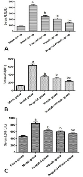
Effect of propofol and vitexin on serum ALT(A), AST(B), LDH(C) levels in each group (n=10). compared with sham group (aP<0.05), model group (bP<0.05), propofol group and vitexin group (cP<0.05).
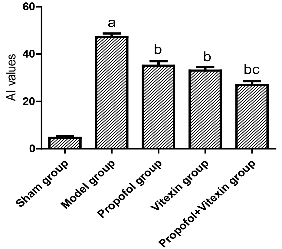
Effect of propofol and vitexin on changes in apoptoxic index (AI) of liver tissue 6 h after reperfusion (n=10). aP<0.05 vs sham group; bP<0.05 vs model group; cP<0.05 vs propofol group and vitexin group.
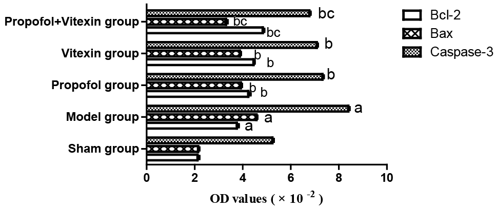
Effect of propofol and vitexin on Bcl-2, Bax and caspase-3 proteins expression level in liver tissue 6 h after reperfusion (n=10). aP<0.05 vs sham group; bP<0.05 vs model group; cP<0.05 vs propofol group and vitexin group.
References
- Abdel-Gaber, S.A., Geddawy, A. and Moussa, R.A., 2019. Pharmacol. Rep., 71:1044-1049. https://doi.org/10.1016/j.pharep.2019.06.006[CrossRef]
- Che, X., Wang, X., Zhang, J., Peng, C., Zhen, Y., Shao, X., Zhang, G. and Dong, L., 2016. Am. J. Transl. Res., 8: 3319-3328.
- Conzen, P.F., Fischer, S., Detter, C. and Peter, K., 2003. Anesthesiology, 99: 826-833. https://doi.org/10.1097/00000542-200310000-00013[CrossRef]
- Czabotar, P.E., Lessene, G., Strasser, A. and Adams, J.M., 2014. Nat. Rev. Mol. Cell Biol., 15: 49-63. https://doi.org/10.1038/nrm3722[CrossRef]
- Deng, W.S., Xu, Q., Liu, Y.E., Jiang, C.H., Zhou, H. and Gu, L., 2016. Exp. Ther. Med., 11: 1955-1960. https://doi.org/10.3892/etm.2016.3160[CrossRef]
- Dong, L., Fan, Y., Shao, X. and Chen, Z., 2011. Fd. Chem. Toxicol., 49: 3211-3216. https://doi.org/10.1016/j.fct.2011.09.040[CrossRef]
- Dong, L.Y., Li, S., Zhen, Y.L., Wang, Y.N., Shao, X. and Luo, Z.G., 2013. Am. J. Chin. Med., 41: 1251-1266. https://doi.org/10.1142/S0192415X13500845[CrossRef]
- He, M., Min, J.W., Kong, W.L., He, X.H., Li, J.X. and Peng, B.W., 2016. Fitoterapia, 74-85. https://doi.org/10.1016/j.fitote.2016.09.011[CrossRef]
- Herr, D.L., Sum-Ping, S.T. and England, M., 2003. J. Cardiothorac. Vasc. Anesth., 17: 576-584. https://doi.org/10.1016/S1053-0770(03)00200-3[CrossRef]
- Ibrahim, S.G., El-Emam, S.Z., Mohamed, E.A. and Abd-Ellah, M.F., 2020. Int. Immunopharmacol., 80: 106131. https://doi.org/10.1016/j.intimp.2019.106131[CrossRef]
- Motayagheni, N., Phan, S., Eshraghi, C., Nozari, A. and Atala, A., 2017. Am. J. Nephrol., 46: 380-389. https://doi.org/10.1159/000482014[CrossRef]
- Murata, R., Hamada, N., Nakamura, N., Kobayashi, A., Fukueda, M., Taira, A. and Sakata, R., 2003. In Vivo, 17: 567-572.
- Ruan, J., Gao, S., Yang, J., Li, H., Huang, H. and Zheng, X., 2018. Leuk. Lymphoma., 59: 162-170. https://doi.org/10.1080/10428194.2017.1312368[CrossRef]
- Sahin, M., Avsar, F.M., Ozel, H., Topaloglu, S., Yilmaz, B., Pasaoglu, H., Avunduk, M.C., Erikoglu, M. and Hengirmen, S., 2004. Transplant. Proc., 36: 2590-2592. https://doi.org/10.1016/j.transproceed.2004.09.057[CrossRef]
- Xi, H.J., Zhang, T.H., Tao, T., Song, C.Y., Lu, S.J., Cui, X.G. and Yue, Z.Y., 2011. Brain Res., 1410: 24-32. https://doi.org/10.1016/j.brainres.2011.06.060[CrossRef]
- Yao, C., Cao, X., Fu, Z., Tian, J., Dong, W., Xu, J., An, K., Zhai, L. and Yu, J., 2017. Med. Sci. Monit., 23: 2059-2064. https://doi.org/10.12659/MSM.901381[CrossRef]
- Zhao, G., Ma, H., Shen, X., Xu, G.F., Zhu, Y.L., Chen, B., Tie, R., Qu, P., Lv, Y., Zhang, H. and Yu, J., 2013. J. Surg. Res., 185: 388-398. https://doi.org/10.1016/j.jss.2013.05.004[CrossRef]
Services
Article usage statistics
Loading…