
Silk is harvested from spiders and silkworms throughout the world. Spider silk varies in types and functions according to the gland producing them. The silk in mulberry and non-mulberrys silkworms also varies from each other. Spidroin is the protein found in spider silk while silkworm silk is made up of an inner core of fibroin and outer layer of sericin protein. After the removal of sericin, silk is non-immunogenic and non-allergic. It is a renowned biomaterial due to its biocompatible nature. Silk proteins have been found to possess antibacterial properties and application in culturing tissues including skin, bone, cartilage and nerves. Sericin has been hailed to have antitumor activity and has also been widely used in different cosmetics. The major hindrance in using spider silk for different purposes is due to its small obtainable amount. Efforts are being made to overcome this problem by modern biotechnological techniques including of making transgenic organisms.
Review · Volume 34, Issue 2, 30 December 2019 · Open Access
Silk of Spider and Bombyx mori: An Overview
· · ·
Affiliations & Notes ⌄
- 1Department of Zoology, GC University, Lahore, Pakistan , Corresponding author: Hafiz Muhammad Tahir, hafiztahirpk1@yahoo.com
Article Info ⌄
- Journal:
- Punjab University Journal of Zoology
- Published:
- 30 December 2019
From the article
- Abstract | Silk is harvested from spiders and silkworms throughout the world.
- Spidroin is the protein found in spider silk while silkworm silk is made up of an inner core of fibroin and outer layer of sericin protein.
- The former one is present in spider silk while the later constitutes the silk of silkworm.
- Spider and mulberry silkworm silks as compatible biomaterials.
Selected from the article's own text.
Abstract
Introduction
The extracorporeal, protein-based, structural material made by the arthropods is known as silk. Although spiders and silkworms are the most well-known silk-producers but many of the insects, arachnids and myriapods also produce silk . There are many types of silkworms around the world that synthesize natural silk as indicated in the Figure 1. Spiders seem to have made the most substantial use of silk, but the most diverse use of it is made by the orb-weavers. The hind end of the spider’s abdomen possesses different silk glands and spinnerets that work together to produce and spin seven different types of silk . The different types of silk produced by spider have been described briefly in Figure 2. Differences in the properties of spider silk and silk of mulberry silkworm are given in Table 1. However, differences between the properties of spidroin and fibroin have been depicted in Table 2. Silk spidroin and silk fibroin are the two prime silk protein families. The former one is present in spider silk while the later constitutes the silk of silkworm. The fibroin in silkworm silk is then further coated with a protein named sericin . Table 3 enlists the major differences between them.
Biomaterial is an extensive term and it covers a range of materials with many uses including implants, scaffolds, conduits and other for cell culture systems. These applications require materials with different and occasionally with intricate properties. Fortunately, hundreds of biomaterials are in use and also undergoing the process of
Table 1: Differences in the properties of spider silk and silk of mulberry silkworm.
| Sr. No. | Properties | Spider silk | Mulberry silkworm silk |
| 1 | Functions | Capture and wrap prey, egg-case or cocoon formation, support web, dragline formation | Cocoon formation |
| 2 | Structural protein | Spidroin | Fibroin |
| 3 | Types produced per specie | Up to eight | One type |
| 4 | Outer covering | Mixture of glycoproteins | Sericin |
| 5 | Effect of water | Dragline silk shows super contraction | No change in length or shape |
| 6 | Age of production | Throughout the life but significant production in mature spiders. | Produced only by the caterpillar form. |
| 7 | Glands involved | Upto seven different types of glands | A pair of gland |
| 8 | Origin of glands | From epidermal invaginations of opisthosoma | From salivary glands |
| 9 | Position of spigots | Caudal ventral position in opisthosoma | Lower lip of silkworms’ mouth |
| 10 | Thickness of silk fiber | 3-6 µm (dragline silk of Naphila clavipes) | 10-16 µm |
Note: This information was taken from ; ; Pérez Rigueiro et al., 2001; ; Asakaura et al., 2007; and .
Table 2: Difference between the properties of spidroin and fibroin.
| Sr No. | Characters | Spidroin | Fibroin |
| 1 | Size | 275-320 kDa | ~350 kDa |
| 2 | Percentage of major amino acids | 37.1% Gly, 21.1% Ala, 9.2% Glu, 7.6% Arg, 4.5% Ser | 46% Gly, 29% Ala, 12% Ser |
| 3 | Repeating motifs | GPGG , GPGQQ, (A)n, (GA)n, and GGX | GAGAGS and GAGAGY |
| 4 | β-sheet content | 35% | 40-50% |
| 5 | Content of crystalline regions | Almost 4-5 units of Alanine joined together, occasionally in the presence of a Serine unit | Frequently the repeating units |
| 6 | Content of non-crystalline regions | They have different composition and length, and are rich in Glycine | Aromatic residues those are negatively charged, polar and bulky in nature. |
| 7 | Heavy chain (N-Terminal) | 130 amino acids, alpha helical at both high and low pH | 151 amino acids, random coil at high pH and beeta sheet rich dimer at low pH |
Source: ; ; and .
development ; ; . Silk is a wonderful biomaterial due to its two of the many exceptional properties: Biodegradability and Biocompatibility. Virgin silkworm silk (unprocessed) presents problems related to biocompatibility when used as sutures by causing a range of skin reactions. But the absence of sericin coating in processed silk (degummed), results in successful biocompatibility. Thus, the silk-based biomaterials exhibit processing-dependent biocompatibility . Cells belonging to different tissues like bone, cartilage, nerves, hippocampus and skin exhibit normal morphology and viability when cultured on scaffolds or in medium containing silk proteins. Silk is a biodegradable protein. The constituent amino acids of its enzymatic breakdown are naturally resolved in the animal’s body without releasing any harmful or cytotoxic byproducts .
Table 3: Percentage mole amino acid composition of spidroin, fibroin and sericin.
| Amino Acids | Spidroin | Fibroin | Sericin |
| Glycine | 37.1 | 42.8 | 8.8 |
| Alanine | 21.1 | 32.4 | 4.0 |
| Valine | 1.8 | 3.0 | 3.1 |
| Leucine | 3.8 | 0.7 | 0.9 |
| Isoleucine | 0.9 | 0.9 | 0.6 |
| Serine | 4.5 | 14.7 | 30.1 |
| Threonine | 1.7 | 1.2 | 8.5 |
| Aspartic acid | 2.5 | 1.9 | 16.8 |
| Glutamic acid | 9.2 | 1.7 | 10.1 |
| Phenyalanine | 0.7 | 1.2 | 0.6 |
| Tyrosine | --- | 11.8 | 4.9 |
| Lysine | 0.5 | 0.5 | 5.5 |
| Histidine | 0.5 | 0.3 | 1.4 |
| Arginine | 7.6 | 0.9 | 4.2 |
| Proline | 4.3 | 0.6 | 0.5 |
| Tryptophan | 2.9 | 0.5 | 0.5 |
| Cysteine | 0.3 | 0.1 | 0.3 |
| Methionine | 0.4 | 0.2 | 0.1 |
Source: Ude et al. (2014) and Saravanan et al. (2006).
Silk proteins
Silk spidroin and silk fibroin are the two prime silk protein families. The former one is present in spider silk while the later constitutes the silk of silkworm73.
Silk fibroin
Silk fibroin being a biopolymer of semi-crystalline nature consists of both crystalline and amorphous regions. Silk I and silk II makes up the crystalline part . Silk І is obtained from spining dope and is water soluble ; . It is unstable but can be stabilized by spining it into crystalline silk II, comprising of β-sheets. Random coils comprise the amorphous parts of the fibroin ; ; . These β-sheets are asymmetrical and form hydrophobic domains, with methyl side chain of alanine projecting from one side, while hydrogen side chain of glycine projecting from the other side of the chain. A thermodynamically stable structure is generated by stacking the sheets together . This is done via strong hydrogen and Vander Waal forces between opposite hydrogen and methyl groups. This crystalline structure is responsible for most of the prominent characteristics of silk including its high tensile strength, resistance to microorganisms and chemicals, and low extensibility and elasticity .
The diameter of silk fiber derived from B. mori is about 10-25 mm. The structure of this silk fibroin consists of three subunits; a heavy chain of ~390 kDa; a light chain of ~26 kDa, and a small glycoprotein of ~30 kDa named P25. Light and heavy chains are linked together via disulphide binds, while non-covalent hydrophobic bonds help in attachment of P25 ; ; . The heavy chain of silk fibroin possesses both the hydrophobic and hydrophilic regions, and thus is amphiphilic in nature. A repeating sequence, Gly-Ala-Gly-Ala-Gly-Ser, makes up the hydrophobic region, and is responsible for folding of silk fibroin chain into β-sheets and thus makes its crystalline structure. On the other hand, the hydrophilic segment is non-repetitive and short, comparatively ; ; . Fibroin is not soluble in water, and is thus, primarily a glycoprotein with hydrophobic nature .
Sericin
Sericin, a protein of 20-310 kDa is the second component of silk of B. mori. it has two subunits. The α-sericin is found in external layer, while β-sericin is present in the inner layer of cocoon . Sericin being water soluble, can be easily removed from fibroin via degumming . It is amorphous and has a form resembling glue. Hence, it maintains the cocoon’s structure by binding together the fibers of fibroin protein .
Silk spidroin
As a contrast, the silk derived from spider do not possess the sericin layer and it can be used in its natural in the form of spidroin solution. Both the inner core and upper coat of the spider silk is primarily composed of the spidroin proteins . These proteins have a high concentration of Gly, Glu and Ala, comparatively large number of Tyr and Leu units, and have a limited number of amino acids in common ; Shen et al., 2004). The Ala rich crystalline regions form compact β-sheets. The hydrophilicity of spider silk is due to the presence of Glu rich amorphous regions ; Bini et al., 2004).
Amongst all the silk of all spiders, the dragline silk of Nephila clavipes is the most frequently studied . This silk is made up of two proteins i.e., MaSp 1 (Major ampullate Spidroin 1) and MaSp 2 (Major ampullate Spidroin 2) and it is produced from the major ampullate gland . Many hydrophilic GGX (X usually being tyrosine, leucine or glutamine) motifs and a single hydrophobic polyalanine block constitutes the MaSp 1 protein. GPGXX replaces the GGX motif in the MaSp 2 . The crystalline β-sheets are formed by the cross linking of several polyalanaine blocks that are further stabilized by the hydrogen bonds and hence result in the elevated tensile strength of the silk. The less organized hydrophilic blocks are present between the crystalline β-sheets. Probably, 310 – helices are formed by the GGX blocks of MaSp 1. While β-turn spirals are formed by the GPGXX motifs found in MaSp 2 that makes the proteins flexible .
Properties and Applications of Silk
Silk protein has various other characteristics that make it an excellent biomaterial for use in biomedical applications:
Biocompatibility
Silk is reported to have no immunogenic response when used for tissue engineering purposes in various animal models including rats, mice, dogs and pigs. Padol et al. (2011) performed skin sensitization tests on mice for silk. The results for acute dermal toxicity came out to be negative as the rats dermis treated with silk films did not exhibited any clinical anomaly and abnormal body weight. The dermal sensitivity tests gave negative results for edema, erythma and eschar. While in case of skin sensitization, no skin reaction was observed 24 and 48 hours after the removal of silk film . Spider silk did not cause hemolysis or direct cytotoxicity during in vitro studies . RGD-recombinat (Arg-Gly-Asp) spider silk protein based bilayer vascular scaffold was prepared and its biosafety has been studied by different tests, involving skin sensitization test, single gel electrophoresis, pyrogenous test, micronucleus and chromosomal abberation test. The scaffold was dense, highly porous and non- adhesive .
Antimicrobial
Spider silk possesses both antibacterial and antifungal properties . Spider silk possess natural antimicrobial properties. Wright and Goodacre (2012) gave evidence for the antibacterial properties of Tegenaria domestica, common house spider . In another study the inhibitory effect of silk of spider Pholcus Phalangioides was observed against two bacterial species, Listeria monocytogenes and Escherichia coli . A research has been carried where spider silk and silver nanoparticle composites were prepared using green synthesis method and the resultant product had great potential as an antimicrobial and biofilm-disrupting agent . Silk taken from Neoscona theisi demonstrated antifungal properties, as it inhibited the growth of fungi on bread . The silkworm silk fibroin-chitosan blend films that were laden with plant extract demonstrated antibacterial property . Spider silk is antimicrobial due to the presence of 12-methyltetradecanic acid and 14-methylhexadecanoic acid .
The effect of sericin concentration from eri silkworms was evaluated on the growth of gram positive (Staphylococcus aureus) and gram negative (E. coli) via disc diffusion and critical-dilution microdilution methods. Lesser the degumming time, more the sericin left on fibroin and hence more the silk has antibacterial activity against the microbes . Whereas silk fibroin blend films were prepared and it was found that they have a pronounced antibacterial effect on Staphylococuss epidermidis .
Vitamin k
Silk proteins react with the blood clotting cascade and aid in the formation of thrombin. Spider silk is a rich source of vitamin K that promotes the process of wound healing. The ancient Greeks and Romans used the cobwebs of spiders as bandages on their wounds, and fortunately it did help in healing . The presence of vitamin k enhances blood clotting and hence prevents bleeding of wounds .
The chemical composition of silk proteins in spider silk has been of immense importance for imparting various properties to it. Table 3 shows percentage composition various amino acids in spider silk proteins. Four motifs are present in all types of silk of all the spiders, which include Poly (Ala) or Poly (Gly-Ala), GPGGX/GPGQQ, GGX, and spacer sequences. These sequences help in the formation and structural alignment of silk fibers, and also provide surface for various interactions. The hydrophilicity of spider silk is due to the presence of two amino acids i.e. glutamine and glycine in the amorphous regions. The presence of glycoproteins, Sulphur containing compounds, amino acids, ionic forms of imines, and inorganic salts imparts the properties to silk that allows the retention and check of water content in it, protects it from microorganism, and allows the identification spider species (Saravanan, 2006).
Antitumor
Sericin present outside the fibroin in silkworm silk has been studied to possess antitumor activity ; . In humans, it has shown to decrease the effect of colon tumor by increasing apoptosis in characteristic cells i.e. SW480 and FHC by increased activity of caspase-3 and decreased Bcl-2 expression . Similarly, silk fibroin irradiated with gamma-rays has also demonstrated antitumor effect in mouse model .
Wound healer
Silk from silkworm has been used as sutures since a long time . Membranes made from silk sericin act as good wound dressings due to their adhesiveness and biocompatible nature . They increase the regeneration of collagenous tissues and decrease inflammation by reducing lymphocyte infiltration . All these properties make it a good wound healing material.
Cosmetic applications
Sericin obtained from silk has been used in different cosmetic products due to its moisturizing and antiaging effects . They enhance the hydration of epidermal structures by increasing the amount of hydroxyproline in them .
Tissue culture
Silk proteins from both the spider and silkworm can be used as a biomaterial for engineering bone, nerve, cartilage and skin ; Gellynk et al., 2008; ; ; ; . Silk fibers support cell attachment and proliferation of cells. Injectable silk-based hydrogel when studied promoted bone healing in rabbits’ femur having critical defects, and 21 days later the regrown bone appeared similar to the normal one . Artificial nerve grafts were prepared using spider silk, accelularized veins and Schwann cells mixed with metrigel. The cells adhered quickly to the fibers, with normal survival and proliferation rate. As a result the cell completely ensheathed the fiber and the formed graft could be cultivated up to one week in vitro . Artificial nerve constructs containing spider silk promote regeneration of the peripheral nerves. Nerve conduits prepared from silkworm silk gave results that increased the possibility of their use as an alternative in nerve autografts . In a study, 3D aqueous-derived silkworm silk protein based scaffolds were used for generating cartilage tissue using adult human chondrocytes and mesenchymal stem cells (MSCs), separately. The cultured hCHs (adult human chondrocytes) attached and proliferated within the scaffold in a medium free of any serum. The constructs derived from hCHs were quite different from those derived from MSCs, thus diversifying the cell sources for cartilage tissue engineering . Mesenchymal cells proliferate rapidly with fusiform morphology when cultured on RGD-recombinant spider silk bilayer scaffold . Chondrocytes respond to silk hydrogel in an excellent manner and exceptional cartilage constructs were obtained after 42 days of culturing of on silk hydrogel .
Limitations and how to overcome these limitations
Different organisms naturally produce silk but the most commonly it is harvested from silkworm and spiders. It is impractical to breed spiders due to their territorial and cannibalistic nature, and as a result, enough amounts of silk can never be obtained for fabricating different materials. Furthermore, almost seven different types of silk with varying physical properties and composition can be produced by some spider species (Volrath, 2000). Consequently, any material made from such a silk would add inconsistencies between different batches of the material, due to the presence of various types of silk proteins in them. While the dragline silk can be obtained in pure form but the method used to do so is comparatively inefficient, and factors such as diet given to spider and the rearing temperature and humidity effects the quality of silk produced .
One more loom for obtaining silk is via the processing of cocoons belonging to silkworm. Two fibroin proteins coated with an adhesive sericin protein makes the structure of these cocoons. Sericins impart stability to the structure of cocoon . Degumming is the thermochemical process through which sericin can be removed . Sericin in combination with fibroin elicit immunological response and that is why its removal is important . Even though the production of silk via regeneration is efficient and cheap, but still the materials fabricated form such a silk need additional changes and processing to make them appropriate for use as biomaterials ; . The tools of biotechnology can be used to surmount several of these drawbacks related to the use of silk. These tools allow one to produce synthetic genes that code for bioengineered silk. The hitch of silk accessibility can be overcome by such bioengineered silk. Additionally, silk biomaterials can be functionalized or modified via genetic engineering. This can be done by adding a fragment that codes for a particular function or by managing the amino acid composition. Such alterations can enlarge the existing brilliant properties of silk and offer a prospect to further modify silk materials for more customized uses.
Future Prospects
Several biotechnological techniques are developing for producing recombinant-spider silk proteins, for providing sufficient spider silk; and for combining spider silk proteins with other molecules, thus resulting in enhanced chemical, physical and biological features . Various strategies have been developed to produce recombinant and chimeric spider silk in various heterologous hosts systems including mice and goat (in milk), transgenic silk worms, and transgenic plants ; .
Statement of conflict of interset
The Authors declare there is no conflict of interset.
Figures
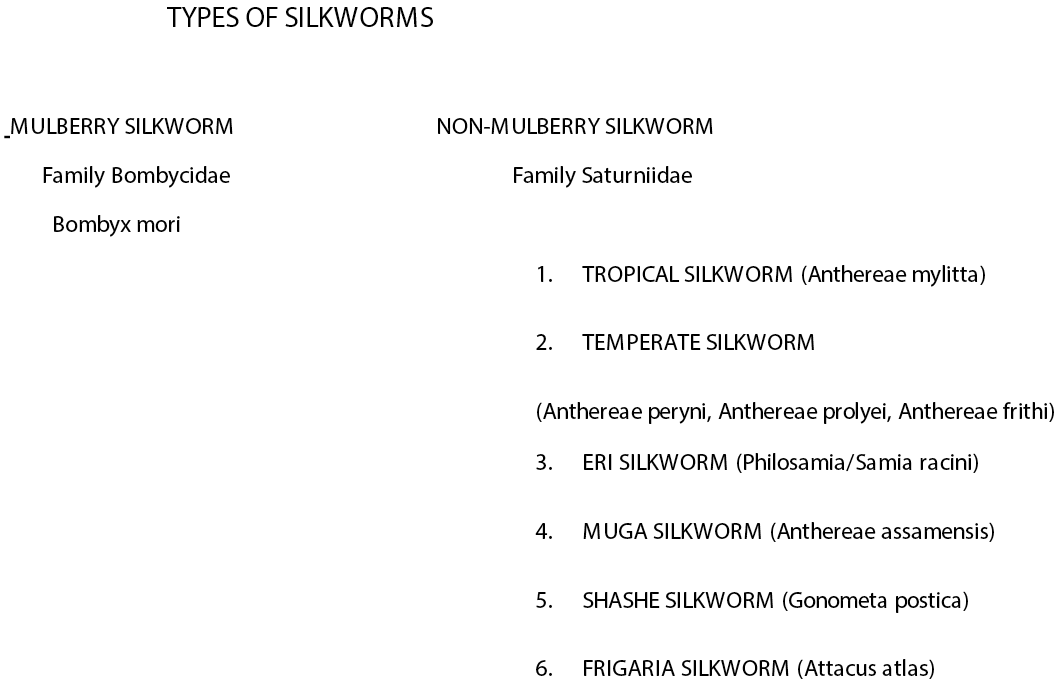
Types of silkworm.
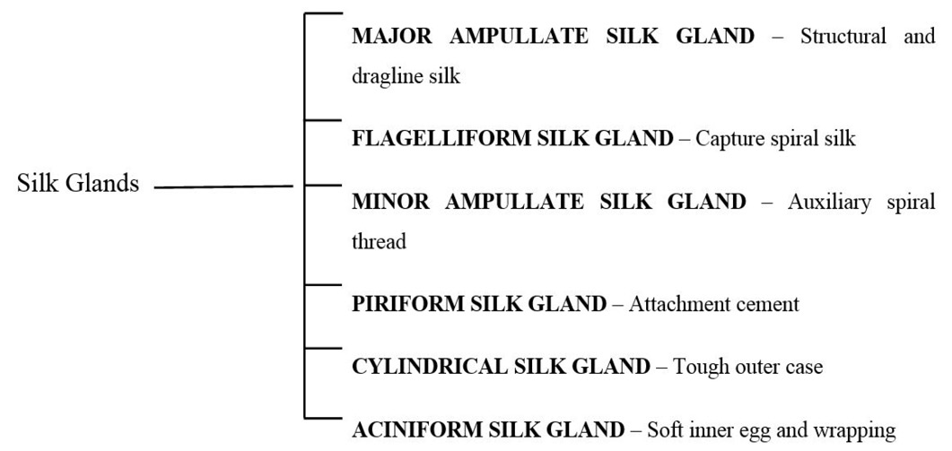
Types of spider silk glands.
References
- Acharya, C., Ghosh, S.K. and Kundu, S.C., 2008. Silk fibroin protein from mulberry and non-mulberry silkworms: cytotoxicity, biocompatibility and kinetics of L929 murine fibroblast adhesion. J. Mater. Sci. Mater. Med., 19: 2827-2836. https://doi.org/10.1007/s10856-008-3408-3[CrossRef]
- Allmeling, C., Jokuszies, A., Reimers, K., Kall, S., Choi, C.Y., Brandes, G. and Vogt, P.M., 2008. Spider silk fibres in artificial nerve constructs promote peripheral nerve regeneration. Cell Prolif., 41: 408-420. https://doi.org/10.1111/j.1365-2184.2008.00534.x[CrossRef]
- Allmeling, C., Jokuszies, A., Reimers, K., Kall, S. and Vogt, P.M., 2006. Use of spider silk fibres as an innovative material in a biocompatible artificial nerve conduit. J. Cell. Mol. Med., 10: 770-777. https://doi.org/10.1111/j.1582-4934.2006.tb00436.x[CrossRef]
- Altman, G.H., Diaz, F., Jakuba, C., Calabro, T., Horan, R.L., Chen, J., Lu, H., Richmond, J. and Kaplan, D.L., 2003. Silk-based biomaterials. Biomaterials, 24: 401-416. https://doi.org/10.1016/S0142-9612(02)00353-8[CrossRef]
- Anderson, J.P., 1998. Morphology and crystal structure of a recombinant silk-like molecule, SLP4. Biopolymers: Original Res. Biomol., 45: 307-332. https://doi.org/10.1002/(SICI)1097-0282(19980405)45:4<307::AID-BIP5>3.0.CO;2-P[CrossRef]
- Aramwit, P., Kanokpanont, S., De-Eknamkul, W. and Srichana, T., 2009. Monitoring of inflammatory mediators induced by silk sericin. J. Biosci. Bioeng., 107: 556-561. https://doi.org/10.1016/j.jbiosc.2008.12.012[CrossRef]
- Asakura, T., Ashida, J., Yamane, T., Kameda, T., Nakazawa, Y., Ohgo, K. and Komatsu, K., 2001. A repeated β-turn structure in poly (Ala-Gly) as a model for silk I of Bombyx mori silk fibroin studied with two-dimensional spin-diffusion NMR under off magic angle spinning and rotational echo double resonance1. J. Mol. Biol., l306: 291-305. https://doi.org/10.1006/jmbi.2000.4394[CrossRef]
- Asakura, T., Umemura, K., Nakazawa, Y., Hirose, H., Higham, J. and Knight, D., 2007. Some observations on the structure and function of the spinning apparatus in the silkworm Bombyx mori. Biomacromolecules, 8: 175-181. https://doi.org/10.1021/bm060874z[CrossRef]
- Basal, G., Altıok, D. and Bayraktar, O., 2010. Antibacterial propertiesof silk fibroin/chitosan blend films loaded with plant extract. Fiber Polym., 11: 21–27. https://doi.org/10.1007/s12221-010-0021-0[CrossRef]
- Bini, E., Knight, D.P. and Kaplan, D.L., 2004. Mapping domain structures in silks from insects and spiders related to protein assembly. J. Mol. Biol., 335: 27-40. https://doi.org/10.1016/j.jmb.2003.10.043[CrossRef]
- Byun, E.B., Sung, N.Y., Kim, J.H., Choi, J.I., Matsui, T., Byun, M.W. and Lee, J.W., 2010. Enhancement of anti-tumor activity of gamma-irradiated silk fibroin via immunomodulatory effects. Chem. Biol. Interact., 186: 90–95. https://doi.org/10.1016/j.cbi.2010.03.032[CrossRef]
- Carletti, E., Motta, A. and Migliaresi, C., 2011. Scaffolds for tissue engineering and 3D cell culture. In 3D cell culture, Humana Press. pp. 17-39. https://doi.org/10.1007/978-1-60761-984-0_2[CrossRef]
- Chen, F., Porter, D. and Vollrath, F., 2012. Morphology and structure of silkworm cocoons. Mater. Sci. Eng. C., 32: 772-778. https://doi.org/10.1016/j.msec.2012.01.023[CrossRef]
- Chirila, T.V., Barnard, Z., Harkin, D.G., Schwab, I.R. and Hirst, L.W., 2008. Bombyx mori silk fibroin membranes as potential substrata for epithelial constructs used in the management of ocular surface disorders. Tissue Eng. Part A., 14: 1203-1211. https://doi.org/10.1089/ten.tea.2007.0224[CrossRef]
- Chung, H., Kim, T.Y. and Lee, S.Y., 2012. Recent advances in production of recombinant spider silk proteins. Curr. Opin. Biotechnol., 23: 957–964. https://doi.org/10.1016/j.copbio.2012.03.013[CrossRef]
- Dickinson, L.E., Kusuma, S. and Gerecht, S., 2011. Reconstructing the differentiation niche of embryonic stem cells using biomaterials. Macromol. Biosci., 11: 36-49. https://doi.org/10.1002/mabi.201000245[CrossRef]
- Drummy, L.F., Phillips, D.M., Stone, M.O., Farmer, B.L. and Naik, R.R., 2005. Thermally induced α-helix to β-sheet transition in regenerated silk fibers and films. Biomacromolecules, 6: 3328-3333. https://doi.org/10.1021/bm0503524[CrossRef]
- Fei, X., Jia, M., Du, X., Yang, Y., Zhang, R., Shao, Z., and Chen, X., 2013. Green synthesis of silk fibroin-silver nanoparticle composites with effective antibacterial and biofilm-disrupting properties. Biomacromolecules, 14: 4483-4488. https://doi.org/10.1021/bm4014149[CrossRef]
- Fini, M., Motta, A., Torricelli, P., Giavaresi, G., Aldini, N.N., Tschon, M. and Migliaresi, C., 2005. The healing of confined critical size cancellous defects in the presence of silk fibroin hydrogel. Biomaterials, 26: 3527-3536. https://doi.org/10.1016/j.biomaterials.2004.09.040[CrossRef]
- Gamo, T., Inokuchi, T. and Laufer, H., 1977. Polypeptides of fibroin and sericin secreted from the different sections of the silk gland in Bombyx mori. Insect Biochem., 7: 285-295. https://doi.org/10.1016/0020-1790(77)90026-9[CrossRef]
- Gapurova, G.N., 1983. Chemical and physicochemical propertiesof surgical sutures (Turk Gos Med Inst, Ashkhzbad, USSR). Zdravookhar Turkm., 27:15–17.
- Garside, P. and Wyeth, P., 2007. Crystallinity and degradation of silk: correlations between analytical signatures and physical condition on ageing. Appl. Physs. A., 89: 871-876. https://doi.org/10.1007/s00339-007-4218-z[CrossRef]
- Gellynck, K., Verdonk, P., Almqvist, K.F., Van Nimmen, E., Gheysens, T., Mertens, J. and Verbruggen, A., 2005. Chondrocyte growth in porous spider silk 3D-scaffolds. In: Annual Conference of the Tissue and Cell Engineering Society (TCES-2005). Queen Mary University of London. 10: 62-62.
- Gellynck, K., Verdonk, P.C., Van Nimmen, E., Almqvist, K.F., Gheysens, T., Schoukens, G. and Verbruggen, G., 2008. Silkworm and spider silk scaffolds for chondrocyte support. J. Mater. Sci. Mater. Med., 19: 3399-3409. https://doi.org/10.1007/s10856-008-3474-6[CrossRef]
- Hakimi, O., Knight, D.P., Knight, M.M., Grahn, M.F. and Vadgama, P., 2006. Ultrastructure of Insect and Spider Cocoon Silks. Biomacromolecules, 7: 2901-2908. https://doi.org/10.1021/bm060528h[CrossRef]
- Hakimi, O., Knight, D.P., Vollrath, F. and Vadgama, P., 2007. Spider and mulberry silkworm silks as compatible biomaterials. Compos. B. Eng., 38: 324-337. https://doi.org/10.1016/j.compositesb.2006.06.012[CrossRef]
- He, Y.X., Zhang, N.N., Li, W.F., Jia, N., Chen, B.Y., Zhou, K. and Zhou, C.Z., 2012. N-terminal domain of Bombyx mori fibroin mediates the assembly of silk in response to pH decrease. J. Mol. Bio., 418: 197-207. https://doi.org/10.1016/j.jmb.2012.02.040[CrossRef]
- Heim, M., Keerl, D. and Scheibel, T., 2009. Spider silk: from soluble protein to extraordinary fiber. Angew. Chem. Int. Ed., 48: 3584-3596. https://doi.org/10.1002/anie.200803341[CrossRef]
- Hirayama, C., Ono, H., Tamura, Y. and Nakamura, M., 2006. C-prolinylquercetins from the yellow cocoon shell of the silkworm, Bombyx mori. Phytochemistry, 67: 579-583. https://doi.org/10.1016/j.phytochem.2005.11.030[CrossRef]
- Ho, M.P., Wang, H. and Lau, K.T., 2012. Effect of degumming time on silkworm silk fibre for biodegradable polymer composites. Appl. Surf. Sci., 258: 3948-3955. https://doi.org/10.1016/j.apsusc.2011.12.068[CrossRef]
- Hofmann, S., Foo, C.W.P., Rossetti, F., Textor, M., Vunjak-Novakovic, G., Kaplan, D. L. and Meinel, L., 2006. Silk fibroin as an organic polymer for controlled drug delivery. J. Contr. Release., 111: 219-227. https://doi.org/10.1016/j.jconrel.2005.12.009[CrossRef]
- Horan, R.L., Bramono, D.S., Stanley, J.R.L., Simmons, Q., Chen, J., Boepple, H.E. and Altman, G.H., 2009. Biological and biomechanical assessment of a long-term bioresorbable silk-derived surgical mesh in an abdominal body wall defect model. Hernia, 13: 189-199. https://doi.org/10.1007/s10029-008-0459-9[CrossRef]
- Horie, Y. and Watanabe, H., 1980. Recent advances in sericulture. Annu. Rev. Entomol., 25: 49-71. https://doi.org/10.1146/annurev.en.25.010180.000405[CrossRef]
- Hussain, M., Khan, S.A. and Naeem, M., 2011. Effect of relative humidity on factors of seed cocoon production in some inbred silk worm (Bombyx mori) lines. Int. J. Agric. Bio., 13: 57-60.
- Inoue, S., Tanaka, K., Arisaka, F., Kimura, S., Ohtomo, K. and Mizuno, S., 2000. Silk fibroin of Bombyx mori is secreted, assembling a high molecular mass elementary unit consisting of H-chain, L-chain, and P25, with a 6: 6: 1 molar ratio. J. Biol. Chem., 275: 40517-40528. https://doi.org/10.1074/jbc.M006897200[CrossRef]
- Joseph, B. and Raj, S.J., 2012. Therapeutic applications and properties of silk proteins from Bombyx mori. Front. Life Sci., 6: 55-60. https://doi.org/10.1080/21553769.2012.760491[CrossRef]
- Kaewkorn, W., Limpeanchob, N., Tiyaboonchai, W., Pongcharoen, S., Manote Sutheerawattananonda, M., 2012. Effects of silksericin on the proliferation and apoptosis of colon cancer cells. Biol. Res., 45: 45–50. https://doi.org/10.4067/S0716-97602012000100006[CrossRef]
- Kaplan, D. and McGrath, K., 2012. Protein-based materials: Bioengineering of Materials. 1st edn: Springer Science and Business Media. Birkhäuser, Boston. pp. 1-430.
- Kato, M., Isobe, K.I., Dai, Y., Liu, W., Takahashi, M. and Nakashima, I., 2000. Further characterization of the sho-saiko-to-mediatedanti-tumor effect on melanoma developed in RET-transgenic mice. J. Invest. Dermatol. 114: 599–601. https://doi.org/10.1046/j.1523-1747.2000.02005.x[CrossRef]
- Knight, D.P. and Vollrath, F., 2002. Spinning an elastic ribbon of spider silk. Philos. Trans. Roy. Soc. Lond. B. Biol. Sci., 357: 219-227. https://doi.org/10.1098/rstb.2001.1026[CrossRef]
- Kovoor, J., 1987. Comparative structure and histochemistry of silk-producing organs in arachnids. In Ecophysiology of spiders., 1st edn. Springer, Berlin, Heidelberg. pp. 160-186. https://doi.org/10.1007/978-3-642-71552-5_12[CrossRef]
- Kratky, O., Schauenstein, E. and Sekora, A., 1950. An unstable lattice in silk fibroin. Nature, 165: 319. https://doi.org/10.1038/165319a0[CrossRef]
- Kronqvist, N., Otikovs, M., Chmyrov, V., Chen, G., Andersson, M., Nordling, K. and Widengren, J., 2014. Sequential pH-driven dimerization and stabilization of the N-terminal domain enables rapid spider silk formation. Nat. Commun., 5: 3254. https://doi.org/10.1038/ncomms4254[CrossRef]
- Kubik, S., 2002. High-Performance Fibers from Spider Silk. Angew. Chem. Int. Ed. Engl., 41: 2721-2723. https://doi.org/10.1002/1521-3773(20020802)41:15<2721::AID-ANIE2721>3.0.CO;2-3[CrossRef]
- Kuhbier, J.W., Coger, V., Mueller, J., Liebsch, C., Schlottmann, F., Bucan, V. and Strauss, S., 2017. Influence of direct or indirect contact for the cytotoxicity and blood compatibility of spider silk. J. Mater. Sci. Mater. Med., 28: 127. https://doi.org/10.1007/s10856-017-5936-1[CrossRef]
- Kundu, B., Kurland, N.E., Ba Kundu, B., Kurland, N.E., Bano, S., Patra, C., Engel, F.B., Yadavalli, V.K., and Kundu, S.C., 2014. Silk proteins for biomedical applications: Bioengineering perspectives. Prog. Polym. Sci., 39: 251-267. https://doi.org/10.1016/j.progpolymsci.2013.09.002[CrossRef]
- Melke, J., Midha, S., Ghosh, S., Ito, K. and Hofmann, S., 2016. Silk fibroin as biomaterial for bone tissue engineering. Acta biomater, 3: 1-16. https://doi.org/10.1016/j.actbio.2015.09.005[CrossRef]
- Min, B.M., Lee, G., Kim, S.H., Nam, Y.S., Lee, T.S., Park, W.H., 2004. Electrospinning of silk fibroin nanofibers and its effect on the adhesion and spreading of normal human keratinocytes and fibroblasts in vitro. Biomaterials, 25:1289–1297. https://doi.org/10.1016/j.biomaterials.2003.08.045[CrossRef]
- Padamwar, M.N., Pawar, A.P., Daithankar, A.V., Mahadik, K.R., 2005. Silk sericin as a moisturizer: an in vivo study. J. Cosmet. Dermatol., 4: 250–257. https://doi.org/10.1111/j.1473-2165.2005.00200.x[CrossRef]
- Padol, A.R., Jayakumar, K., Shridhar, N.B., Swamy, H.N., Swamy, M.N. and Mohan, K., 2011. Safety evaluation of silk protein film (a novel wound healing agent) in terms of acute dermal toxicity, acute dermal irritation and skin sensitization. Int. Toxicol., 18: 17. https://doi.org/10.4103/0971-6580.75847[CrossRef]
- Pérez-Rigueiro, J., Elices, M., Llorca, J. and Viney, C., 2001. Tensile properties of Argiope trifasciata drag line silk obtained from the spide https://doi.org/10.1002/app.2072 r’s web. J. Appl. Polym. Sci., 82: 2245-2251.[CrossRef]
- Roozbahani, H., Asmar, M., Ghaemi, N. and Issazadeh, K., 2014. Evaluation of antimicrobial activity of spider silk Pholcus phalangioides against two bacterial pathogens in food borne. Int. J. Adv. Biol. Biomed. Res., 2: 2197-2199.
- Saravanan, D., 2006. Spider silk-structure, properties and spinning. J. Tex. App. Tech. Manag., 5: 1-20.
- Sasaki, M., Kato, N., Watanabe, H. and Yamada, H., 2000. Silk protein sericin suppresses colon carcinogenesis. Oncol. Rep., 7: 1049–1052.
- Senakoon, W., Nuchadomrong, S., Sirimungkararat, S., Senawong, T. and Kitikoon, P., 2009. Antibacterial action of eri (samia ricini) sericin against Escherichia coli and Staphylococcus aureus. Asian J. Food Agric. Ind., S222–S228.
- Shahbazi, B., Taghipour, M., Rahmani, H., Sadrjavadi, K. and Fattahi, A., 2015. Preparation and characterization of silk fibroin/oligochitosan nanoparticles for siRNA delivery. Colloids. Surf. B. Biointerfaces., 136: 867-877. https://doi.org/10.1016/j.colsurfb.2015.10.044[CrossRef]
- Sheu, H.S., Phyu, K.W., Jean, Y.C., Chiang, Y.P., Tso, I.M., Wu, H.C. and Ferng, S.L., 2004. Lattice deformation and thermal stability of crystals in spider silk. Int. J. Biol. Macromol., 34: 267-273. https://doi.org/10.1016/j.ijbiomac.2004.09.004[CrossRef]
- Sirichaisit, J., Brookes, V.L., Young, R.J. and Vollrath, F., 2003. Analysis of structure/property relationships in silkworm (Bombyx mori) and spider dragline (Nephila edulis) silks using Raman spectroscopy. Biomacromolecules, 4: 387-394. https://doi.org/10.1021/bm0256956[CrossRef]
- Sponner, A., Schlott, B., Vollrath, F., Unger, E., Grosse, F. and Weisshart, K., 2005. Characterization of the protein components of Nephila clavipes dragline silk. Biochemistry, 44: 4727-4736. https://doi.org/10.1021/bi047671k[CrossRef]
- Sutherland, T.D., Young, J.H., Weisman, S., Hayashi, C.Y. and Merritt, D.J., 2010. Insect silk: one name, many materials. Annu. Rev. Entomol., 55: 171–188. https://doi.org/10.1146/annurev-ento-112408-085401[CrossRef]
- Tahir, H.M., Zaheer, A. and Yaqoob, R., 2015. Short Communication Antifungal potential of silk recovered from Neoscona theisi (Araneae: Araneidae). Punjab Univ. J. Zool., 30: 81-83.
- Tie, J.K., Carneiro, J.D., Jin, D.Y., Martinhago, C.D., Vermeer, C. and Stafford, D.W., 2016. Characterization of vitamin K–dependent carboxylase mutations that cause bleeding and nonbleeding disorders. Blood, 127: 1847-1855. https://doi.org/10.1182/blood-2015-10-677633[CrossRef]
- Tokareva, O., Jacobsen, M., Buehler, M., Wong, J. and Kaplan, D.L. 2014. Structure–function–property–design interplay in biopolymers: Spider silk. Acta biomater, 10: 1612-1626. https://doi.org/10.1016/j.actbio.2013.08.020[CrossRef]
- Ude, A.U., Eshkoor, R.A., Zulkifili, R., Ariffin, A.K., Dzuraidah, A.W. and Azhari, C.H., 2014. Bombyx mori silk fibre and its composite: a review of contemporary developments. Mater. Des., 57: 298-305. https://doi.org/10.1016/j.matdes.2013.12.052[CrossRef]
- Vendrely, C. and Scheibel, T., 2007. Biotechnological production of spider-silk proteins enables new applications. Macromol. Biosci., 7: 401-409. https://doi.org/10.1002/mabi.200600255[CrossRef]
- Vert, M., 2011. Degradable polymers in medicine: updating strategies and terminology. Int. J. Art. Orgs., 34: 76-83. https://doi.org/10.5301/IJAO.2011.6400[CrossRef]
- Voegeli, R., Meier, J. and Blust, R., 1993. Sericin silk protein: uniquestructure and properties. Cosmet. Toilet., 108: 101–108.
- Vollrath, F., 2000. Strength and structure of spiders’ silks. Rev. Mol. Biotech., 74: 67-83. https://doi.org/10.1016/S1389-0352(00)00006-4[CrossRef]
- Wang, Y., Blasioli, D.J., Kim, H.J., Kim, H.S. and Kaplan, D.L., 2006. Cartilage tissue engineering with silk scaffolds and human articular chondrocytes. Biomaterials, 27: 4434-4442. https://doi.org/10.1016/j.biomaterials.2006.03.050[CrossRef]
- Wen, H., Lan, X., Zhang, Y., Zhao, T., Wang, Y., Kajiura, Z. and Nakagaki, M., 2010. Transgenic silkworms (Bombyx mori) produce recombinant spider dragline silk in cocoons. Mol. Biol. Rep., 37: 1815–1821. https://doi.org/10.1007/s11033-009-9615-2[CrossRef]
- Wendt, H., Hillmer, A., Reimers, K., Kuhbier, J.W., Schäfer-Nolte, F., Allmeling, C. and Vogt, P.M., 2011. Artificial skin–culturing of different skin cell lines for generating an artificial skin substitute on cross-weaved spider silk fibres. PLoS One, 6: e21833. https://doi.org/10.1371/journal.pone.0021833[CrossRef]
- Wray, L.S., Hu, X., Gallego, J., Georgakoudi, I., Omenetto, F.G., Schmidt, D. and Kaplan, D.L., 2011. Effect of processing on silk-based biomaterials: Reproducibility and biocompatibility. J. Biomed. Mater. Res. B. Appl. Biomater., 99: 89-101. https://doi.org/10.1002/jbm.b.31875[CrossRef]
- Wright, S. and Goodacre, S.L., 2012. Evidence for antimicrobial activity associated with common house spider silk. BMC Res. Notes., 5: 326. https://doi.org/10.1186/1756-0500-5-326[CrossRef]
- Xue, C., Zhu, H., Tan, D., Ren, H., Gu, X., Zhao, Y. and Gu, Y., 2018. Electrospun silk fibroin-based neural scaffold for bridging a long sciatic nerve gap in dogs. J. Tissue Eng. Regen. Med., 12: e1143-e1153. https://doi.org/10.1002/term.2449[CrossRef]
- Yodmuang, S., McNamara, S.L., Nover, A.B., Mandal, B.B., Agarwal, M., Kelly, T.A.N. and Vunjak-Novakovic, G., 2015. Silk microfiber-reinforced silk hydrogel composites for functional cartilage tissue repair. Acta biomater, 11: 27-36. https://doi.org/10.1016/j.actbio.2014.09.032[CrossRef]
- Zarkoob, S., Eby, R.K., Reneker, D.H., Hudson, S.D., Ertley, D. and Adams, W.W., 2004. Structure and morphology of electrospun silk nanofibers. Polymer, 45: 3973-3977. https://doi.org/10.1016/j.polymer.2003.10.102[CrossRef]
- Zhao, C., Yao, J., Masuda, H., Kishore, R. and Asakura, T., 2003. Structural characterization and artificial fiber formation of Bombyx mori silk fibroin in hexafluoro-iso-propanol solvent system. Biopolymers, 69: 253-259. https://doi.org/10.1002/bip.10350[CrossRef]
- Zhao H.P., Feng X.Q. and Shi H.J., 2007. Variability in mechanical properties of Bombyx mori silk. Mater. Sci. Eng. C., 27: 675–683. https://doi.org/10.1016/j.msec.2006.06.031[CrossRef]
- Zhao, L., Xu, Y., He, M., Zhang, W. and Li, M., 2014. Preparation of spider silk protein bilayer small-diameter vascular scaffold and its biocompatibility and mechanism research. Compos. Interface., 21: 869-884. https://doi.org/10.1080/15685543.2014.970416[CrossRef]
- Zhao, L., He, M., Xu, Y. and Li, M., 2015. Study on biological safety of spider silk protein bilayer vascular scaffold. Compos. Interfaces, 22: 179-192. https://doi.org/10.1080/15685543.2014.999266[CrossRef]
- Zhou, C.Z., Confalonieri, F., Medina, N., Zivanovic, Y., Esnault, C., Yang, T. and Li, Z.G., 2000. Fine organization of Bombyx mori fibroin heavy chain gene. Nucleic acids Res., 28: 2413-2419. https://doi.org/10.1093/nar/28.12.2413[CrossRef]
- Zurovec, M., Yang, C., Kodrı́k, D. and Sehnal, F., 1998. Identification of a novel type of silk protein and regulation of its expression. J. Biol. Chem., 273: 15423-15428. https://doi.org/10.1074/jbc.273.25.15423[CrossRef]
Services
Article usage statistics
Loading…