
The second most frequent cancer all over the world is breast cancer (BC). It is reported that only about 10% BC cases are attributed due to inherited genetic mutations while remaining 90% cancer cases are associated with environmental factors. Artificial light at night (ALAN) is considered one of the major environmental risk factors for breast cancer. It inhibits production of melatonin (MLT) from pineal gland which results in abnormal epigenetic changes that relates with an increased risk of BC. The most important ALAN-mediated epigenetic changes include methylation of DNA and acetylation of histone, which are significant for growth, development and progression of BC. DNA hypermethylation of promoter CpG islands inhibits transcriptional activity by methyltransferase enzyme which results in inactivation of tumor suppressor genes (TSG), while in hypomethylation, demethyltransferase enzyme causes the activation of oncogenes by promoting transcriptional activity. Contrary to DNA methylation, histone acetylation and deacetylation results in chromatin opening and closing, respectively; leading to transcriptional activation and inactivation of genes. Histone acetylation has been frequently detected in oncogenes while histone deacetylation in TSG. Collective data from various studies demonstrate that DNA hypermethylation and histone deacetylation of TSG lead to inactivation of TSG and activation of oncogenes. The purpose of this review is to discuss the evidence based relationship between ALAN and oncogenes expression through epigenetic remodeling by DNA methylation and histone acetylation.
Review · Volume 34, Issue 2, 30 December 2019 · Open Access
Epigenetics: A Bridge between Artificial Light at Night and Breast Cancer
· · · · · · ·
Affiliations & Notes ⌄
- 1Department of Zoology, University of the Punjab, Quaid-e-Azam Campus Lahore 54590, Pakistan 2Department of Wild Life, University of Veterinary and Animal Sciences, Patoki, Pakistan 3Department of Biotechnology, University of Sargodha, Sargodha, Pakistan 4Department of Zoology, Government College University, Lahore, Pakistan , Correspondence Author: Muhammad Khan and Hafiz Abdullah Shakir, khan_zoologist@ymail.com
- 2hashakir.zool@pu.edu.pk
Article Info ⌄
- Journal:
- Punjab University Journal of Zoology
- Published:
- 30 December 2019
From the article
- Epigenetics: A Bridge between Artificial Light at Night and Breast Cancer.
- ALAN, Melatonin, DNA methylation, Histone acetylation, Breast cancer.
- Mechanisms of breast cancer in shift workers: DNA methylation in five core circadian genes in nurses working night shifts.
- Protein acetylation and histone deacetylase expression associated with malignant breast cancer progression.
Selected from the article's own text.
Abstract
INTRODUCTION
Cancer remains the most important causes of death all over the world as compared to other non-infectious diseases. According to cancer statistical report, about 14.1 million cancer cases and 8.2 million deaths due to cancer were reported in 2012 . Later, in 2018, GLOBOCAN estimated 18.1 million new cancer cases and 9.6 million deaths due to cancer (Ferlay et al., 2018). World Health Organization (WHO) predicted 17.5 million expected deaths at the end of 2050 due to cancer .
Among all, the second most common cancer in women is BC and one of the important causes of death ; . Over 1.5 million BC cases are diagnosed every year throughout the world. In 2018, about 2 million new BC cases were diagnosed .
ALAN increases the risk of BC due to suppression of MLT production ; . However, MLT production increases in the absence of light ; . MLT is mainly produced and secreted by the pineal gland ; . In addition to pineal gland, it also synthesized by different organs like skin, gastrointestinal tract, retina, bone marrow, and lymphocytes ; . Chemically, it is an indoleamine (N-acetyl-5-methoxytryptamine) and name (Mela-) is due to its effect on amphibians which blanch the melanophores and (-Tonin) because it is derived from serotonin . It is famous for ‘night hormone’ and supposed as ‘Jack of all trades . It plays an important role in regulating the immune system and sleep wake cycle. It also acts as an anti-oxidative, anti-aging, anti-inflammatory and anti-cancer agent. ; . The process of biosynthesis of MLT has been shown in Figure 1.
The production of MLT is controlled by the suprachiasmatic nucleus with the help of the pineal gland, which affects clock genes and reduces cancer ; ; . During the day, the concentration of MLT reduces whereas its concentration increases at night. By exposure of ALAN, the normal action of MLT disrupts due to its less production which cause abnormal epigenetic changes that enhances the BC risk .
In 1942, C. H. Waddington first introduced the idea of epigenetic . It controls genetic alternation without changes in sequence of DNA nucleotides . Two major ALAN mediated epigenetic changes include methylation of DNA and acetylation of histone that are important to growth, development and progression ; . These modifications are also increasing the chances of BC by activation of oncogene and interruption of the role of particular TSGs . MLT regulates alternations in tumor cell. It performs anticancer activity by down-regulation of oncogenes and up regulation of TSGs. It also causes methylation and deacetylation of the oncogene (CYP19) that reduces BC. As a result of deacetylation, chromatin condenses and suppresses the binding of transcriptional factor which require for activation of oncogenes. Moreover, MLT also reduces BC by methylation of other oncogenes (Early Growth Receptor 3 and POU4F2/Brn-3b) and unmethylation of TS glypican- 3(GPC3) . Epigenetic mechanism relates to inactivation of TSG and activation of oncogenes and these modifications affect genes expression .
Effect of ALAN at MLT secretions and estrogen production
ALAN influences the normal daily pattern because it contains light with different spectrum and wavelength . It decreases the concentration of MLT by the retinohypohalamic pineal region. Decrease in MLT results in increase level of estrogen, which also increases the risk of BC development ; ; . It is thought, the main reason of BC risk is lifetime load with estrogen ; .
Effect of ALAN on methylation of tummor suppressor genes
Among epigenetic alternations which are induced by ALAN, the most important is DNA methylation, and it is more common form of molecular fluctuations in human cancer. In DNA methylation, a methyl (–CH3) group shifts to the 5th carbon (5C) of cytosine from Sadenosyl- L-methionine ; ; . Enzyme (known as DNA methyltransferase) involves the shifting of –CH3
group and three members of this family are known . Both DNMT3A and DNMT3B are de novo methyltransferases whereas DNMT1 is the continuation methyltransferase and during cell division, it equally transfers the methylation patterns . It is well-known enzyme relates to methylation of DNA and promotes apoptosis .
DNA Methylation is the most important mechanism in epigenetic alternations which is involved in regulation of genetic programming and enhances the progression of different types of cancers, including BC ; . These alterations occur only to a cytosine and guanosine sequence in the DNA, known as CpG dinucleotide. These regions are primarily present at the promoter and there is generally no methylation in normal cells (which permit the active gene transcription) while in cancer cells these CpG promoter region are methylated which results in silencing of various TSGs and pro-apoptotic genes ; .
Several kinds of alternation in DNA methylation can take place in cancer, such as hypermethylation in gene-locus resulting in the inactivation of TSG, or hypomethylation of the distinctive genes and repeated sequences . Hypermethylation is the term used for more methylation while hypomethylation for less methylation ; . These alternations act as a biomarker for identification as well as treatment of cancer .
In case of BC, the expression of circadian genes is deregulated. Reports indicated hypermethylation on promoter of PER1, PER2, CRY1 and BMAL genes in BC ; ; . In long term shift workers, Cry2 (related to circadian genes) is hypermethylated on promoter region ; Steven and Zhu, 2015). Glypican-3 (GPC3), a tumor suppressor gene is aberrantly methylated in MCF-7 BC cell lines. Upon treatment of MCF-7 cells with 1nM MLT, significant increase in the expression of GPC3 gene was observed. The findings suggest that MLT could modulate methylation pattern of this tumor suppressor gene . In long term shift workers, the miR-34b promoter region is aberrantly methylated which enhanced the BC risk due to ALAN exposure . Report indicated the relationship between DNA methylation of TSG (BRCA1, BRCA2, TP53, CDKN2A) and night shift workers. It graphically showed the expression of methylation decreases from number of years in these TSG. Results indicted that in night shift workers, BRCA1 and TP53 are hypomethylated compared with non shift workers . Hypomethylation of p53 and BRCA1 has been assumed to be induced to counterbalance defects in circadian cell cycle regulation and thus could indirectly increase the risk of cancer.
Effect of ALAN on methylation of oncogenes
Oncogenes included those genes that enhanced cell proliferation and survival . Several types of genes in BC changed the level of their expression due to unusual methylation. In cancer cells, the genome is globally hypomethylated or unmethylated that caused the instability of chromosome, and failure of genomic imprinting might result in the upregulation or more expression of proto-oncogenes ; . In several proto-oncogenes, the promoter region is hypomethylated or not methylated leading to uncontrolled cell proliferation, cancer progression and development of treatment resistance. The main epigenetic mechanism of BC is the activation of oncogenes due to inactivation of TSG that cause the cancer, including BC .
Oncogenes such as POU4F2 and ERG3 showed different methylation patterns and were up-regulated in BC cell lines. Treatment of BC cells with 1nM MLT, halted the growth of BC cells by down-regulating above said oncogenes via increased methylation. . CLOCK (related to circadian genes) is hypomethylated on the promoter region in shift workers. ; Steven and Zhu, 2015). Other independent studies conducted in CLOCK which showed slightly more methylation in BC cases compared with healthy control . ALAN showed different results from the methylation of TSG and oncogenes. The results are shown in Table 1.
Effect of ALAN on acetylation of tummor suppressor genes
ALAN caused changes in usual acetylation pattern of TSG . The balance between histone acetylation and deacetylation is necessary for controlling the expression of genes. Histone acetylation is promoted by histone acetyl transferases enzyme (HAT) that is concerned with activation of gene transcription, whereas histone deacetylation or hypoacetylation is promoted by another enzyme called histone deacetylase (HDAC) which is associated with repression of gene transcription ; ; . Changed expression or gene mutations that encode histone deacetylation or hypoacetylation have been associated with induction of cancer while both these promote the abnormal transcription of leading genes and controlled the main functions of cells such as cell propagation, regulation of cell-cycle and apoptosis .
Table 1: Effect of ALAN on methylation pattern of genes in BC.
| Gene/ Protein | MLT | Normal function | Methylationpattern | Effect | Activation/ Inhibition | References |
| Per 1 | ↓ | TS | -CH3↑ | BC | Inhibition | |
| Per 2 | ↓ | TS | -CH3↑ | BC | Inhibition | ; |
| Cry1 | ↓ | TS | -CH3↑ | BC | Inhibition | |
| BMAL1 | ↓ | TS | -CH3↑ | BC | Inhibition | |
| EGR3 | ↓ | Onco | -CH3 ↓ | BC | Activation | |
| POU4F2 | ↓ | Onco | -CH3 ↓ | BC | Activation | |
| GPC3 | ↓ | TS | -CH3 ↑ | BC | Inhibition | |
| CLOCK | ↓ | Onco | -CH3 ↓ | BC | Activation | |
| CLOCK | ↓ | Onco | -CH3↑ | BC | Activation | |
| Cry2 | ↓ | TS | -CH3↑ | BC | Inhibition | ; |
| mir-34B | ↓ | TS | -CH3↑ | BC | Inhibition | |
| BRCA1 | ↓ | TS | -CH3 ↓ | BC | Inhibition |
|
| BRCA2 | ↓ | TS | -CH3 ↓ | BC | Inhibition | |
| CDKN2A (p16) | ↓ | TS | -CH3 ↓ | BC | Inhibition | |
| TP53 | ↓ | TS | -CH3↓ | BC | Inhibition | |
| ESR1 | ↓ | Onco | -CH3↓ | BC | Activation | |
| ESR2 | ↓ | Onco | -CH3↓ | BC | Activation |
↓: Downregulation; ↑: Upregulation; TS: Tumor Suppressor; BC: Breast Cancer.
Table 2: Effect of ALAN on acetylation pattern of genes in BC.
| Gene/ Protein | MLT | Normal Function | Acetylation/ Deacetylation | Effect | Activation/ Inhibition | References |
| P53 | ↓ | TS | Deacetylation | BC | Inhibition | |
| CYP19 | ↓ | Onco | Acetylation | BC | Activation | |
| ER | ↓ | Onco | Acetylation | BC | Activation | |
| c-MYC | ↓ | Onco | Acetylation | BC | Activation | |
| STAT3 | ↓ | Onco | Acetylation | BC | Activation | |
| BRCA1 | ↓ | TS | Deacetylation | BC | Inhibition | |
| BRCA2 | ↓ | TS | Deacetylation | BC | Inhibition | |
| Per 1 | ↓ | TS | Deacetylation | BC | Inhibition | |
| Per 2 | ↓ | TS | Deacetylation | BC | Inhibition | |
| Ku-70 | ↓ | TS | Deacetylation | BC | Inhibition | |
| MMP | ↓ | Onco | Acetylation | BC | Activation |
↓: Downregulation; ↑: Upregulation; TS: Tumor Suppressor; BC: Breast Cancer; Onco: Oncogene.
For alternations in chromatin, most important mechanism is the adaptation of histone acetylation and deacetylation. These adaptations cause epigenetic changes due to alternations in expression of gene and cell development which may affect carcinogenesis and propagation . In cancer, the functions of histone deacetyltransferase are not only limited to their involvement to histone deacetylation, but also played an important role in deacetylation of non-histone proteins. For instance, in vivo and in vitro study, Histone deacetyltransferase 1 linked with the p53 (that is tumor suppressor) and deacetylated it .
MLT exhibited anticancer effects in BC. It decreased the MDM2 expression and increased acetylation of p53 in MCF-7 cell lines . MLT via its receptor MT1, activated the RORα that controls the expression of SIRT1 (histone deacetylases) and BMAL/CLOCK. CLOCK (histone acetyltrasferases) acetylated PER1/2 and other DNA repair genes BRCA1, BRCA2, P53 and Ku-70 which reduced the development of cancer due to acetylation activity. Hill et al., have explained, how BRCA1, BRCA2, p53, Ku70, PER1 and PER2 deacetylated and induced BC due to ALAN . The findings showed below recommend that ALAN causes more expression and abnormal recruitment of histone deacetyltransferases in promote regions could be a regular event in cancer development and progression, resulting suppressed transcription of tumor-suppressor genes.
Effect of ALAN on acetylation of oncogenes
ALAN decreased the production of MLT and enhanced phosphorylation and acetylation of oncoprotein (such as STAT3) that over expressed in BC . Hyperacetylation of Proto-oncogenes results in activation of proto-oncogenes while hypo-acetylation of tumor suppressors genes is frequently localized to promotor region causing the genes to be silenced .
MLT has been reported to decrease the expression of CYP19 protein which is frequently overexpressed in BC cell lines. MLT exhibits oncostatic effects via deacetylation of CYP19 . In addition, MLT induced hypoacetylation and decreased the activity of matrix metalloproteinase (MMP). Increased expression of MMPs have been noted in various types of tumor which mainly facilitate metastasis . CLOCK (histone acetyltransferases) promotes acetylation of different genes such as c- myc and ERα that induce BC due to acetylation .
The collective findings published previously recommended that ALAN causes the more expression and abnormal recruitment of histone acetyltransferases in promoter regions ehich could be regular event in cancer development and progression, resulting in activation of oncogenes. ALAN mediated acetylation of TSG and oncogenes has been shown in Table 2.
Conflict of interests
The authors declare there is no conflict of interest.
Figures
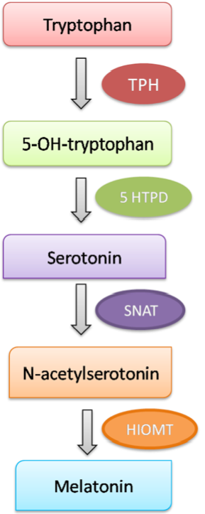
Synthesis of MLT. MLT synthesis takes place in pineal gland. Pineal glands uptake tryptophan and converts it into MLT through five enzymes catalyzed reactions. The diagram represents the sequential reactions and enzymes involved in biosynthesis of MLT. TPH: Tryptophan hydroxylase, 5-HTPD: 5-Hydroxytryptophan decarboxylase, SNAT: Serotonin N-acetyltransferase, HIOMT: hydroxyindole-O-methyl transferase).
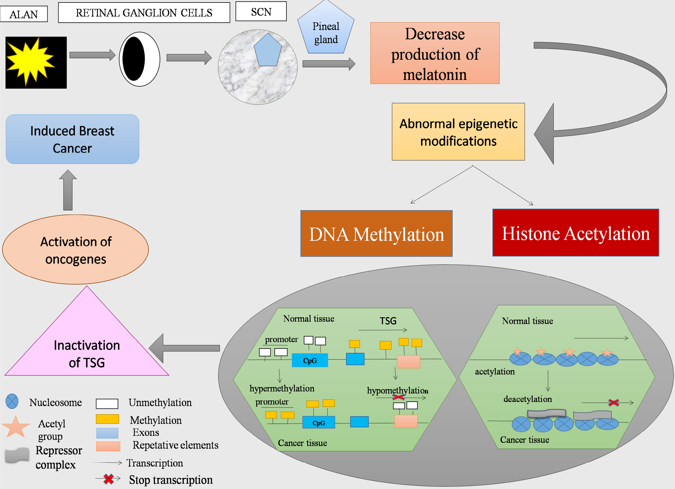
ALAN induced BC: MLT synthesis take place in pineal gland at night, however; ALAN reduced its production which results in abnormal epigenetic changes including DNA methylation and Histone acetylation. The promoter region is unmethylated and acetylated in normal tissue of TSG while it is methylated and deacetylated in cancer tissues. As a result, TSG become inactive and oncogenes become active leading to BC induction.
References
- Amin, N., Shafabakhsh, R., Reiter, R.J. and Asemi, Z., 2019. Melatonin is an appropriate candidate for breast cancer treatment: Based on known molecular mechanisms. J. Cell. Biochem., 120: 12208-12215. https://doi.org/10.1002/jcb.28832[CrossRef]
- Audia, J.E. and Campbell, R.M., 2016. Histone modifications and cancer. Cold Spring Harbor Perspect. Biol., 8: a019521. https://doi.org/10.1101/cshperspect.a019521[CrossRef]
- Basse, C. and Arock, M., 2015. The increasing roles of epigenetics in breast cancer: Implications for pathogenicity, biomarkers, prevention and treatment. Int. J. cancer, 137: 2785-2794. https://doi.org/10.1002/ijc.29347[CrossRef]
- Bauer, S.E., Wagner, S.E., Burch, J., Bayakly, R. and Vena, J.E., 2013. A case-referent study: light at night and breast cancer risk in Georgia. Int. J. Hlth. Geogr., 12: 23. https://doi.org/10.1186/1476-072X-12-23[CrossRef]
- Blakeman, V., Williams, J.L., Meng, Q.J. and Streuli, C.H., 2016. Circadian clocks and breast cancer. Breast Cancer Res., 18: 89. https://doi.org/10.1186/s13058-016-0743-z[CrossRef]
- Blask, D.E., Dauchy, R.T., Sauer, L.A., Krause, J.A. and Brainard, G.C., 2003. Growth and fatty acid metabolism of human breast cancer (MCF-7) xenografts in nude rats: impact of constant light-induced nocturnal melatonin suppression. Breast Cancer Res. Tr., 79: 313-320. https://doi.org/10.1023/A:1024030518065[CrossRef]
- Blask, D.E., Hill, S.M., Dauchy, R.T., Xiang, S., Yuan, L., Duplessis, T. and Sauer, L.A., 2011. Circadian regulation of molecular, dietary, and metabolic signaling mechanisms of human breast cancer growth by the nocturnal melatonin signal and the consequences of its disruption by light at night. J. Pineal Res., 51: 259-269. https://doi.org/10.1111/j.1600-079X.2011.00888.x[CrossRef]
- Bondy, S. and Campbell, A., 2018. Mechanisms Underlying Tumor Suppressive Properties of Melatonin. Int. J. Mol. Sci., 19: 2205. https://doi.org/10.3390/ijms19082205[CrossRef]
- Carugno, M., Maggioni, C., Crespi, E., Bonzini, M., Cuocina, S., Dioni, L., Tarantini, L., Consonni, D., Ferrari, L. and Pesatori, A.C., 2019. Night Shift Work, DNA Methylation and Telomere Length: An Investigation on Hospital Female Nurses. Int. J. Env. Res. Pub. He., 16: 2292. https://doi.org/10.3390/ijerph16132292[CrossRef]
- Cohen, I., Poręba, E., Kamieniarz, K. and Schneider, R., 2011. Histone modifiers in cancer: friends or foes? Genes Cancer, 2: 631-647. https://doi.org/10.1177/1947601911417176[CrossRef]
- Cui, Z., Xie, M., Wu, Z. and Shi, Y., 2018. Relationship Between Histone Deacetylase 3 (HDAC3) and Breast Cancer. Med. Sci. Monit., 24: 2456. https://doi.org/10.12659/MSM.906576[CrossRef]
- Dauchy, R.T., Xiang, S., Mao, L., Brimer, S., Wren, M.A., Yuan, L. and Blask, D.E., 2014. Circadian and melatonin disruption by exposure to light at night drives intrinsic resistance to tamoxifen therapy in breast cancer. Cancer Res., 74: 4099-4110. https://doi.org/10.1158/0008-5472.CAN-13-3156[CrossRef]
- Ehrlich, M., 2002. DNA methylation in cancer: too much, but also too little. Oncogene, 21: 5400. https://doi.org/10.1038/sj.onc.1205651[CrossRef]
- Erdem, J.S., Skare, Ø., Petersen-Øverleir, M., Notø H.Ø., Lie, J.A., Reszka, E., Pepłońska, B. and Zienolddiny, S., 2017. Mechanisms of breast cancer in shift workers: DNA methylation in five core circadian genes in nurses working night shifts. J. Cancer, 8: 2876. https://doi.org/10.7150/jca.21064[CrossRef]
- Fang, J.Y., Lu, J., Chen, Y.X. and Yang, L., 2003. Effects of DNA methylation on expression of tumor suppressor genes and proto-oncogene in human colon cancer cell lines. World J. Gastroenterol., 9: 1976-1980. https://doi.org/10.3748/wjg.v9.i9.1976[CrossRef]
- Ferlay, J., Colombet, M., Soerjomataram, I., Mathers, C., Parkin, D.M., Piñeros, M., Znaor, A. and Bray, F., 2019. Estimating the global cancer incidence and mortality in 2018: GLOBOCAN sources and methods. Int. J. Cancer, 144: 1941-1953. https://doi.org/10.1002/ijc.31937[CrossRef]
- Giudice, A., Crispo, A., Grimaldi, M., Polo, A., Bimonte, S., Capunzo, M. and Botti, G., 2018. The Effect of Light Exposure at Night (LAN) on Carcinogenesis via Decreased Nocturnal Melatonin Synthesis. Molecules, 23:1308. https://doi.org/10.3390/molecules23061308[CrossRef]
- GRØNBÆK, K., Hother, C. and Jones, P.A., 2007. Epigenetic changes in cancer. Apmis, 115:1039-1059. https://doi.org/10.1111/j.1600-0463.2007.apm_636.xml.x[CrossRef]
- Haim, A. and Zubidat, A.E., 2015. Artificial light at night: melatonin as a mediator between the environment and epigenome. Philos. Trans. R. Soc. B. Biol. Sci., 370: 20140121. https://doi.org/10.1098/rstb.2014.0121[CrossRef]
- Hasan, T.N., Shafi, G., Grace, B.L., Tegner, J. and Munshi, A., 2015. DNA methylation: an epigenetic marker of breast cancer influenced by nutrients acting as an environmental factor. In Noninvasive Mol. Markers Gynecol. Cancers, pp. 191-210. https://doi.org/10.1201/b18015-9[CrossRef]
- Hill, S.M., Belancio, V.P., Dauchy, R.T., Xiang, S., Brimer, S., Mao, L. and Frasch, T., 2015. Melatonin: an inhibitor of breast cancer. Endocr. Relat. Cancer, 22: 183-204. https://doi.org/10.1530/ERC-15-0030[CrossRef]
- Hill, S.M., Frasch, T., Xiang, S., Yuan, L., Duplessis, T. and Mao, L., 2009. Molecular mechanisms of melatonin anticancer effects. Integr. Cancer Ther., 8: 337-346. https://doi.org/10.1177/1534735409353332[CrossRef]
- Jovanovic, J., Rønneberg, J.A., Tost, J. and Kristensen, V., 2010. The epigenetics of breast cancer. Mol. Oncol., 4: 242-254. https://doi.org/10.1016/j.molonc.2010.04.002[CrossRef]
- Kaur, R.P., Banipal, R.P.S., Vashistha, R., Dhiman, M. and Munshi, A., 2019. Association of elevated levels of C-reactive protein with breast cancer, breast cancer subtypes, and poor outcome. Curr. Prob. Cancer, 43: 123-129. https://doi.org/10.1016/j.currproblcancer.2018.05.003[CrossRef]
- Keshet-Sitton, A., Or-Chen, K., Yitzhak, S., Tzabary, I. and Haim, A., 2016. Can avoiding light at night reduce the risk of breast cancer? Integr. Cancer Ther., 15 :145-152. https://doi.org/10.1177/1534735415618787[CrossRef]
- Khan, M., Maryam, A., Zhang, H., Mehmood, T. and Ma, T., 2016. Killing cancer with platycodin D through multiple mechanisms. J. Cell. Mol. Med., 20: 389-402. https://doi.org/10.1111/jcmm.12749[CrossRef]
- Kochan, D.Z. and Kovalchuk, O., 2015. Circadian disruption and breast cancer: an epigenetic link? Oncotarget, 6:16866. https://doi.org/10.18632/oncotarget.4343[CrossRef]
- Korkmaz, A. and Reiter, R. J., 2008. Epigenetic regulation: a new research area for melatonin? J. Pineal Res., 44: 41-44.
- Korkmaz, A., Sanchez-Barcelo, E.J., Tan, D.X. and Reiter, R.J., 2009. Role of melatonin in the epigenetic regulation of breast cancer. Breast Cancer Res. Tr., 115:13-27. https://doi.org/10.1007/s10549-008-0103-5[CrossRef]
- Kuo, S.J., Chen, S.T., Yeh, K.T., Hou, M.F., Chang, Y.S., Hsu, N.C. and Chang, J.G., 2009. Disturbance of circadian gene expression in breast cancer. Virchows Arch., 454: 467-474. https://doi.org/10.1007/s00428-009-0761-7[CrossRef]
- Lee, E.Y. and Muller, W.J., 2010. Oncogenes and tumor suppressor genes. Cold Spring Harbor Perspect. Biol., a003236. https://doi.org/10.1101/cshperspect.a003236[CrossRef]
- Lee, S.E., Kim, S.J., Yoon, H.J., Yu, S.Y., Yang, H., Jeong, S.I. and Park, Y.S., 2013. Genome-wide profiling in melatonin-exposed human breast cancer cell lines identifies differentially methylated genes involved in the anticancer effect of melatonin. J. Pineal Res., 54: 80-88. https://doi.org/10.1111/j.1600-079X.2012.01027.x[CrossRef]
- Li, Y., Li, S., Zhou, Y., Meng, X., Zhang, J.J., Xu, D.P. and Li, H.B., 2017. Melatonin for the prevention and treatment of cancer. Oncotarget, 8: 39896-39921. https://doi.org/10.18632/oncotarget.16379[CrossRef]
- Li, Z.H., Zhang, X.B., Han, X.Q., Feng, C.R., Wang, F.S., Wang, P.G. and Shi, Y.K., 2013. Antitumor effects of a novel histone deacetylase inhibitor NK-HDAC-1 on breast cancer. Oncol. Rep., 30:499-505. https://doi.org/10.3892/or.2013.2434[CrossRef]
- Liu, R., Jacobs, D.I., Hansen, J., Fu, A., Stevens, R.G. and Zhu, Y., 2015. Aberrant methylation of miR-34b is associated with long-term shiftwork: a potential mechanism for increased breast cancer susceptibility. Cancer Causes Control, 26: 171-178. https://doi.org/10.1007/s10552-014-0494-z[CrossRef]
- Lujambio, A. and Esteller, M., 2008. CpG Island Hypermethylation, miRNAs, and Human Cancer. Curr. Perspect. microRNAs (miRNA), pp. 367-384. https://doi.org/10.1007/978-1-4020-8533-8_20[CrossRef]
- Mahmood, N. and Rabbani, S.A., 2017. DNA methylation and breast cancer: mechanistic and therapeutic applications. Trends Cancer Res, 12: 1-18.
- Pfeifer, G., 2018. Defining driver DNA methylation changes in human cancer. Int. J. Mol. Sci., 19: 1166. https://doi.org/10.3390/ijms19041166[CrossRef]
- Pouliot, M.C., Labrie, Y., Diorio, C. and Durocher, F., 2015. The role of methylation in breast cancer susceptibility and treatment. Anticancer Res., 35: 4569-4574.
- Proietti, S., Cucina, A., Dobrowolny, G., D’Anselmi, F., Dinicola, S., Masiello, M.G., Pasqualato, A., Palombo, A., Morini, V., Reiter, R.J. and Bizzarri, M., 2014. Melatonin down-regulates MDM 2 gene expression and enhances p53 acetylation in MCF-7 cells. J. Pineal Res., 57: 120-129. https://doi.org/10.1111/jpi.12150[CrossRef]
- Radpour, R., Kohler, C., Haghighi, M.M., Fan, A.X.C., Holzgreve, W. and Zhong, X.Y., 2009. Methylation profiles of 22 candidate genes in breast cancer using high-throughput MALDI-TOF mass array. Oncogene, 28: 2969. https://doi.org/10.1038/onc.2009.149[CrossRef]
- Ropero, S. and Esteller, M., 2007. The role of histone deacetylases (HDACs) in human cancer. Mol. Oncol., 1:19-25. https://doi.org/10.1016/j.molonc.2007.01.001[CrossRef]
- Saha, S. and Sassone-Corsi, P., 2007. Circadian clock and breast cancer: a molecular link. Cell Cycle, 6:1329-1331. https://doi.org/10.4161/cc.6.11.4295[CrossRef]
- Salavaty, A., 2015. Carcinogenic effects of circadian disruption: an epigenetic viewpoint. Chin. J. Cancer, 34: 38. https://doi.org/10.1186/s40880-015-0043-5[CrossRef]
- Shanmugam, V., Wafi, A., Al-Taweel, N. and Büsselberg, D., 2013. Disruption of circadian rhythm increases the risk of cancer, metabolic syndrome and cardiovascular disease. J. Local Glob. Hlth. Sci., 2013: 3. https://doi.org/10.5339/jlghs.2013.3[CrossRef]
- Sharma, S., Kelly, T.K. and Jones, P.A., 2010. Epigenetics in cancer. Carcinogenesis, 31: 27-36. https://doi.org/10.1093/carcin/bgp220[CrossRef]
- Stevens, R.G. and Zhu, Y., 2015. Electric light, particularly at night, disrupts human circadian rhythmicity: is that a problem? Philos. Trans. R. Soc. B. Biol. Sci., 370: 20140120. https://doi.org/10.1098/rstb.2014.0120[CrossRef]
- Stevens, R. G., 2005. Circadian disruption and breast cancer: from melatonin to clock genes. Epidemiology, 16: 254-258. https://doi.org/10.1097/01.ede.0000152525.21924.54[CrossRef]
- Stevens, R.G., 2009. Light-at-night, circadian disruption and breast cancer: assessment of existing evidence. Int. J. Epidemiol., 38: 963-970. https://doi.org/10.1093/ije/dyp178[CrossRef]
- Suzuki, J., Chen, Y.Y., Scott, G.K., DeVries, S., Chin, K., Benz, C.C. and Hwang, E.S., 2009. Protein acetylation and histone deacetylase expression associated with malignant breast cancer progression. Clin. Cancer Res., 15: 3163-3171. https://doi.org/10.1158/1078-0432.CCR-08-2319[CrossRef]
- Torres, D., Lorenzo Bermejo, J., Garcia Mesa, K., Gilbert, M., Briceño, I., Pohl-Zeidler, S., González Silos, R., Boekstegers, F., Plass, C. and Hamann, U., 2019. Interaction between genetic ancestry and common breast cancer susceptibility variants in Colombian women. Int. J. Cancer, 144: 2181-2191. https://doi.org/10.1002/ijc.32023[CrossRef]
- Wajed, S.A., Laird, P.W. and DeMeester, T.R., 2001. DNA methylation: an alternative pathway to cancer. Ann. Surg., 234: 10. https://doi.org/10.1097/00000658-200107000-00003[CrossRef]
- White, A.J., Weinberg, C.R., Park, Y.M., D’aloisio, A.A., Vogtmann, E., Nichols, H.B. and Sandler, D.P., 2017. Sleep characteristics, light at night and breast cancer risk in a prospectivecohort. Int. J. Cancer, 141: 2204-2214. https://doi.org/10.1002/ijc.30920[CrossRef]
- Xiang, S., Dauchy, R.T., Hoffman, A.E., Pointer, D., Frasch, T., Blask, D.E. and Hill, S.M., 2019. Epigenetic inhibition of the tumor suppressor ARHI by light at night-induced circadian melatonin disruption mediates STAT 3-driven paclitaxel resistance in breast cancer. J. Pineal Res., pp. e12586. https://doi.org/10.1111/jpi.12586[CrossRef]
- Yang, X., Yan, L. and Davidson, N.E., 2001. DNA methylation in breast cancer. Endocr. Relat. Cancer, 8: 115-127. https://doi.org/10.1677/erc.0.0080115[CrossRef]
- Zaidi, Z. and Dib, H.A., 2019. The worldwide female breast cancer incidence and survival, 2018. https://doi.org/10.1158/1538-7445.AM2019-4191[CrossRef]
- Zhu, Y., Stevens, R.G., Hoffman, A.E., Tjonneland, A., Vogel, U.B., Zheng, T. and Hansen, J., 2011. Epigenetic impact of long-term shiftwork: pilot evidence from circadian genes and whole-genome methylation analysis. Chronobiol. Int., 28: 852-861. https://doi.org/10.3109/07420528.2011.618896[CrossRef]
- Zubidat, A.E. and Haim, A., 2017. Artificial light-at-night–a novel lifestyle risk factor for metabolic disorder and cancer morbidity. J. Basic Clin. Physiol. Pharmacol., 28: 295-313. https://doi.org/10.1515/jbcpp-2016-0116[CrossRef]
Services
Article usage statistics
Loading…