
Aging is a natural biological process that leads to deterioration in the liver structures and functions due to oxidative stress and the reduction in the oxidative damage’s tolerance. Quercetin, a flavonoid has the ability to face the oxidative injury through its antioxidant activity. Therefore, the present study was designed to investigate the possible role of quercetin in modulating liver’s function and structure in aged male rats. Forty eight male albino rats (3 and 30 months old) were used. Half of the rats at each age received 200 mg quercetin/Kg every other day for 2 weeks. Thirty months old rats showed a decrease in the liver AST, albumin, SOD, CAT and GPx while showed an increase in the liver ALT, plasma cholesterol and MDA comparing with 3 months old rats. The percentages of p53 and bax positive areas/ field were 26.04% and 24.9%, respectively. After quercetin administration, 30 months old rats showed non-significant change in the previous biochemical parameters compared with 3 months old rats and the percentages of p53 and bax positive areas/ field were 0.6% and 0.9%, respectively. In conclusion, quercetin at a dose of 200 mg/kg every other day for 2 weeks could improve liver function, decrease the expression of pro-apoptotic proteins p53 and bax and increase the antioxidant defense of hepatocytes in aged male rats.
Research Article · Volume 54, Issue 6, 19 August 2022 · Open Access
Quercetin Improves Liver Function, Decreases the Expression of Pro-apoptotic Proteins p53 and Bax and Increases the Antioxidant Defense of Hepatocytes in Aged Male Rats
· · ·
Affiliations & Notes ⌄
- 1Department of Biology, Faculty of Science and Arts, King Khaled University, Abha, Kingdom of Saudi Arabia 2Department of Biology, Faculty of Science, Ibb University, Ibb, Yemen 3Biology Department, College of Science for Girls, King Khaled University, Abha, Kingdom of Saudi Arabia 4Department of Zoology, Faculty of Science, Tanta University, Tanta, Egypt * Corresponding author: abeer.eldeen@science.tanta.edu.eg, abeer75875@hotmail.com
Article Info ⌄
- Journal:
- Pakistan Journal of Zoology
- Published:
- 19 August 2022

From the article
- The data indicated that 30 months old rats displayed certain decline in the liver function as it showed a significant decrease in the values of the liver AST and albumin while showed a significant increase in the values of the liver ALT and plasma cholesterol.
- Aged liver cells were more sensitive to oxidative stress and were transformed to apoptotic cells compared to younger rats.
- After quercetin administration, 30 months old rats showed an improvement in the liver architecture and also showed a non-significant change in the liver functions, SOD, CAT, GPx and MDA levels and p53 and bax percentages compared with 3 months old rats.
- Quercetin administration at a dose of 200mg/kg each other day for 15 days improved the liver function and decreased the expression of pro-apoptotic proteins, p53 and bax and increased the antioxidant defense of the aged hepatocytes.
Selected from the article's own text.
Abstract
INTRODUCTION
Aging is a natural biological process that leads to gradual deterioration in the structures and functions of different organs. This is because the ability to maintain homeostasis is lost with aging . Accumulation of the free radical damage in the cells was reported to be the main cause of the aging phenomena. Aging is greatly associated with the increase of the oxidative stress and the reduction to oxidative damage’s tolerance which result in a significant percentage of the oxidized lipids and proteins. It was reported that reactive oxygen species (ROS) cause accumulation of the oxidative damage to cellular elements which result in reducing the cellular survivability . Liver’s volume decreases by 20–40% with aging . Moreover, a decrease in the concentration of the serum albumin and bilirubin and liver cells volume were reported .
P53 has a role in apoptosis and in cellular response to the oxidative stress ; ; . Oxidative stress activates p53 transcriptional response that regulates cellular senescence and aging . P53 regulates several cellular processes such as DNA repair, cell cycle and apoptosis . The deficiency in the function of p53 network leads to aging which in turn suppresses apoptosis. P53 regulates the transcription of pro-apoptotic genes like bax .
Quercetin, a flavonoid detected in some fruits and vegetables has diverse biological properties such as anti-inflammatory, antioxidant and antiviral activities. It was reported that it can also inhibit lipid peroxidation and face the myocardial oxidative injury through its free radical scavenging capacity , decrease the histological signs of acute inflammation via decreasing the levels of lipid peroxidation end-product malondialdehyde (MDA) and increasing the antioxidant activity , modulate hepatic oxidative damage induced by gold nanoparticles via its antioxidant activity and maintain the contractility of bladder tissue against ischemic injury via decreasing the oxidative stress and apoptosis . It was also reported that quercetin had the ability to decrease the hepatotoxicity via its antioxidant, metal-chelating and anti-inflammatory effects. Quercetin could scavenge different reactive oxygen species and it is considered as a good antioxidant agent when cellular damage occurs . Wu et al. (2017) have reported that quercetin ameliorates liver damage and histopathological changes, and attenuate apoptosis and autophagy. Taskan and Gevrek (2020) have reported that quercetin reduced alveolar bone loss by increasing osteoblastic activity and decreasing osteoclastic activity, apoptosis, and inflammation. Consequently, the present study was designed to study the possible role of quercetin on aging related processes at molecular level in aged male rats.
MATERIALS AND METHODS
Animals
Forty eight male albino rats housed in plastic cages and maintained under standard conditions of temperature, humidity, and 12 h light/dark cycle were used in the present study. Half of the rats were 3 months old and the other half was 30 months old. The rats were provided with pellets of concentrated diet containing all the necessary nutritive elements. The rats were acclimatized for two weeks before starting the experiment. All the procedures of the experiment were adhered to the guidelines of the Ethical Committee of King Khaled University, KSA.
Experimental protocol
The 3 and 30 months old rats were subdivided equally into 2 subgroups of 12 rats each; control and quercetin subgroups weighing 98.2 ± 4.9 g and 110.36 ± 7.62 g, 321.64 ± 13.16 g and 346.9 g ± 15.2 g, respectively. The control rats were injected with saline intraperitoneally (i.p.). Quercetin subgroups included 12 rats each were injected i.p. with 200 mg quercetin/Kg body weight every other day for 2 weeks . Quercetin was purchased from Sigma-Aldrich (St. Louis Mo., U.S.A). After two weeks, all rats were sacrificed and samples from the liver were carefully collected under strict hygienic conditions. Some liver samples were quickly storing at -80 ˚C for biochemical analyses and the other samples were fixed in 10% neutral buffered formalin for histopathological and immunohistochemical examinations.
Biochemical parameters
The activities of serum aspartate and alanine aminotransferases (AST and ALT) were determined according to Reitman and Frankel (1957). The levels of the albumin and cholesterol levels were assessed by commercial kits . Superoxide dismutase (SOD) activity was determined by the methods of Paoletti and Mocali (1990) and the data was expressed as U/mg protein. Catalase (CAT) activity was assayed according to the method of Aebi (1984) and the data was expressed as U/mg protein. Glutathione peroxidase (GPx) activity was assayed by the method of Paglia and Valentine (1967) and the data was expressed as U/mg protein. Lipid peroxidation (MDA) content was estimated by the method of Buege and Aust (1978) and expressed as nmole/mg protein.
Histopathological examination
Liver samples that were fixed in the neutral buffered formalin were washed, dehydrated and embedded in paraffin. Serial sections of 5 μm thick were prepared then some were processed for hematoxylin and eosin staining and the others were processed for immunohistochemical staining.
Immunohistochemicl examination
The sections were dewaxed and rinsed in phosphate buffer saline (PBS) then incubated with an aqueous solution of 0.3% hydrogen peroxide for 10 min to block nonspecific background staining due to endogenous peroxidase then washed 2 times in PBS buffer. The section was then incubated with 100 µL of monoclonal mouse anti-p53 (cat. no. M7001, Dako Pty Ltd., Campbellfield, VIC, Australia) or 100 µL of rabbit polyclonal anti- Bax (Oncogene Science, Inc.) at dilution of 1:100 and 1:400 overnight at 4°C, respectively. The slides were washed 4 times in PBS then rinsed in biotinylated secondary antibody for 15 min followed by washing with phosphate buffer and avidin biotin for 15 min. The slides were then rinsed using DAB (3, 3´-diaminobenzidine) for 3 min until appearance of brown color. The slides were counterstained with Mayer’s hematoxylin for 2–5 min, dehydrated and mounted. The positive stained area is brown in color. The sections were photographed. Fifteen random fields from each group were analysed using the objective lens X40 were analyzed for p53 and bax expressions. The percentages of the areas occupied by positive staining in each field in relation to the total area in each field were calculated using image J software. Finally, the percentage area occupied by p53 or bax positive staining in relation to the total area was calculated for each group.
Statistical analysis
All data are the means of 4 replicates. Two-way analysis of variance (ANOVA) was performed followed by Tukey post hoc test. Results were expressed as mean ± standard deviation (mean ± SD). P values ≤ 0.05 was considered to be statistically significant. Statistical analysis were performed using Excel 2013 (Microsoft Corporation, USA), SPSS statistical version 22 software package (SPSS Inc., USA) and Minitab version 18).
RESULTS
Biochemical investigations
To test the liver function before and after quercetin administration, AST, ALT, albumin and cholesterol were assayed. The data showed no significant difference (p ≥ 0.05) in the values of the liver AST, ALT, albumin and plasma cholesterol in 3 months old rats after quercetin administration. However, 30 months old rats exhibited significant (p ≤ 0.05) decline in the liver function as it showed decrease in the values of the liver AST (-30.67%) and albumin (-19.34%) while showed a significant (p ≤ 0.05) increase in the levels of the liver ALT 27.89% and plasma cholesterol 76.03% in 30 months old control rats compared with 3 months rats. After quercetin administration, 30 months old rats showed a non-significant change (p ≥ 0.05) compared with 3 months old rats (Fig. 1).
The results showed that no significant differences (p ≥ 0.05) were noticed in the levels of the liver SOD, CAT, GPx and MDA in the 3 months old rats after quercetin administration. Nevertheless, 30 months old rats showed significant decrease (p ≤ 0.05) in the liver SOD (-31.54%), (CAT -29.07%) and GPx (-51.46%) while showed a significant increase (p ≤ 0.05) in the liver MDA (205.95%) compared with 3 months old rats. After quercetin administration, 30 months old rats showed non-significant differences (p ≥ 0.05) in the liver SOD, CAT, GPx and MDA compared with 3 months old rats (Fig. 2).
Histological investigations
Observation of the liver sections of 3 months old rats before and after quercetin administration showed similar histological structures. The hepatocytes appeared with normal architecture in which their nuclei were vesicular and basophilic and their cytoplasm were eosinophilic (Fig. 3A). However, marked cellular infiltration was recorded in the liver sections of the rats that had 30 months old. Most hepatocytes had small and dark nuclei. Vacuolated hepatocytes were appeared in which the vacuoles appeared with different sizes and shapes. Congestion of the central vein was noticed too (Fig. 3B, C). After quercetin administration, liver sections of the rats that had 30 months old showed an improvement in the hepatocytes morphology. Most cells appeared were vesicular nucleis and their cytoplasm were eosinophilic. Vacuolated hepatocytes were rarely noticed. Moreover, mild cellular infiltration and moderate congestion of the central and portal veins were determined (Fig. 3D).
Immunohistochemical investigations
Observation of the liver sections of the 3 months old rats which were immunostained with either p53 or bax antibodies, before or after quercetin administration, showed almost similar patterns (Fig. 4A, B and Fig 5A, B). The percentage of the p53-positive area/ field were 0.56% and 0.4% and bax-positive area/field were 0.2% and 0.4% in 3 months old rats before and after quercetin administration, respectively. The liver sections of the rats had 30 months old showed an increase in the immunoreactivity with both of p53 and bax antibodies (Fig. 4C and Fig. 5C). The percentages of the p53 and bax positive areas/ field were 26.04% and 24.9%, respectively. Observation of the liver sections 30 months old rats after quercetin administration showed a decrease in the immunoreactivity with either p53 and bax antibodies (Fig. 4). The percentages of the p53 and bax positive areas/ field were 0.6% and 0.9%, respectively.
DISCUSSION
The present study aimed to clarify the possible role of quercetin to minimize aging induced changes in the structure and function of the liver. The data indicated that 30 months old rats displayed certain decline in the liver function as it showed a significant decrease in the values of the liver AST and albumin while showed a significant increase in the values of the liver ALT and plasma cholesterol. They also exhibited a significant decrease in the liver SOD, CAT and GPx activities while exhibited a significant increase in the liver MDA content. The current data together with other previous studies documented the decline in the liver functions with aging. The decrease of the ALT is a good biomarker with the increase of age . Changes in the levels of the albumin, bilirubin, alkaline phosphatase, aminotransferase and cholesterol and neutral fat levels with aging were recorded previously . They added that a decrease in the mass of the functional liver cells resulted in a decrease of the liver capacity to synthesis and detoxify and consequently reduced the bile, urea and albumin.
Aged liver cells were more sensitive to oxidative stress and were transformed to apoptotic cells compared to younger rats. The imbalance in the oxidant-antioxidant system resulted in the incidence of the oxidative stress and accordingly accelerated the occurrence of the aging process . MDA, CAT and Gpx were the most important markers of the oxidative stress . Oxidative stress caused oxidative damage to the lipids of the cellular membranes, proteins and DNA. The increase in the MDA level is considered an important biomarker of lipid peroxidation under the conditions of the oxidative stress . SOD dismutases the toxic superoxide radical into less toxic hydrogen peroxide and GPx metabolizes hydrogen peroxide and lipid hydroperoxides . The increase of the ROS production at aging was associated with changes in the liver functions and structures . Generally, oxidative stress increases with aging and this might be the reason for alterations in the liver functions and structure in the present study.
The present data showed that 30 months old rats showed vacuolated hepatocytes, cellular infiltration and congestion of the central vein. Moreover, an increase in the expressions of p53 and bax proteins was recorded. The present data was comparable with that of Iwaisako et al. (2012) who found that the incidence of the occurrence and the progression of the liver fibrosis was increased with aging. Acute or chronic damage caused the appearance of the connective tissues in the liver and consequently the fibrosis occurs. Poynard et al. (2001) found that fibrosis is an initial step which is followed by a distortion of the liver architecture and function. Zhu et al. (2007) worked on some apoptosis related genes and reported that p53 had a role in apoptosis and the cellular response to the oxidative stress. P53 enhances aging in activated p53 mutant mice . As a compensatory way to reduce the genotoxic stress, Park et al. (2008) reported that p53 imitates apoptosis. The decline in the p53 function may lead to aging and as a result induce apoptosis. Miyashita et al. (1994)recorded the regulation of Bcl2 and Bax by p53 in vitro and in vivo. Roos and Kaina (2013) documented that the transcription of some pro-apoptotic genes such as bax gene is regulated and controllable by p53.
After quercetin administration, 30 months old rats showed an improvement in the liver architecture and also showed a non-significant change in the liver functions, SOD, CAT, GPx and MDA levels and p53 and bax percentages compared with 3 months old rats. Our results are consistent with Abdelhalim et al. (2018) who reported the ability of quercetin at a dose of 200 mg/kg/day for seven consecutive days to protect rats liver from the acute injury via its antioxidant effects. Tieppo et al. (2007) showed that hepatic cells which were pretreated with quercetin at a dose of 20 mg/kg for 21 days displayed less damage when injected with ciprofloxacin. As a result, they concluded that quercetin has the ability to scavenge free-radicals and inhibit injury in hepatic tissues. Taslidere et al. (2016) reported that quercetin at a dose of 50 mg/kg for 14 days could work against the oxidative stress and decrease the fibrosis in liver via its antioxidant activity. Lien et al. (1999) explained that quercetin may diffuse to the membranes and scavenge oxy radicals throughout the lipid bilayer. They also reported that quercetin’s pentahydroxyl flavones structure may chelate metal ions via the orthodihydroxy phenolic structure and as a result scavenge lipid alkoxyl and peroxyl radicals. Polat et al. (2006) reported that quercetin flavonoids administrated at a dose of 50 mg/kg for 3 days may cause indirect stimulation of detoxifying genes. Furthermore, Bao et al. (2017) reported the upregulation of bcl2 and the downregulation p53, bax and caspase 3 against oxidative stress-induced cytotoxicity in rat pheochromocytoma cells after treatment with quercetin. They speculated the ability of quercetin to inhibit H2O2which causes the increase in the production of ROS and to alleviate lipoperoxidation of cell membranes. As a result, the inhibition of the oxidative stress and the cell damage were prevented. Sharma et al. (2016) stated that quercetin could prevent the oxidative stress process that resulted from the induction of the aluminium in the rat hippocampus by reduced production of the ROS and increased SOD. As a result, Bcl-2 was upregulated and Bax, p53 and caspase-3 were upregulaed and DNA fragmentation was reduced. Therefore, the present results together with the previous studies demonstrate the possibility to use quercetin to minimize the deterioration that may occur in the liver functions and structures with aging after continuous exposure to the oxidative stress.
In conclusion, 30 months old rats showed histological liver damage and decline in liver functions, the levels of SOD, CAT, GPx and MDA and the levels of p53 and bax. Quercetin administration at a dose of 200mg/kg each other day for 15 days improved the liver function and decreased the expression of pro-apoptotic proteins, p53 and bax and increased the antioxidant defense of the aged hepatocytes. Therefore, quercetin could be used as a modulator against the deterioration that may occur in the liver as a result of aging.
ACKNOWLEDGEMENT
The authors extend their appreciation to the Deanship of Scientific Research at King Khaled University for funding this work through general project under grant number (G.R.P. 245/1440).
Statement of conflict of interest
The authors have declared no conflict of interest.
Figures
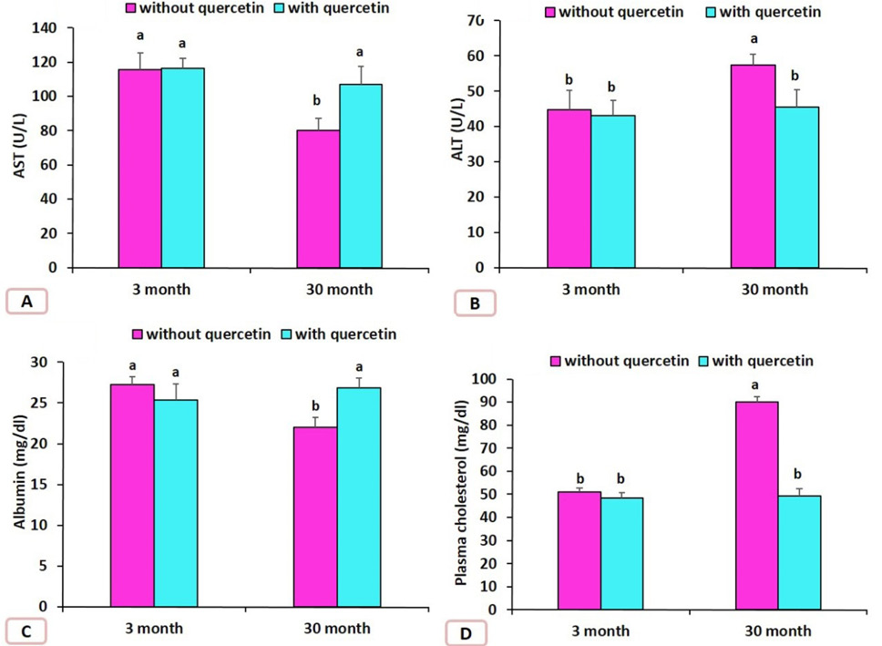
Effect of quercetin on the levels of AST, ALT, Albumin and plasma cholesterol in the rats that had 3 and 30 months old rats. Data are means ± SD with n=4, Different letters indicate significant differences among the columns (p ≤ 0.05).
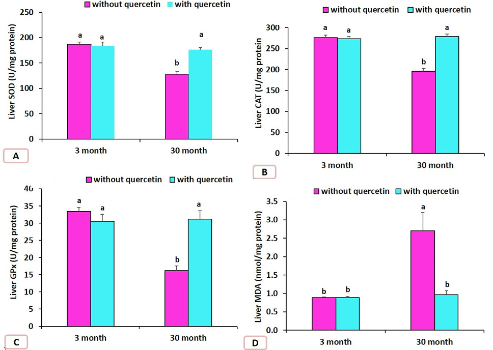
Effect of quercetin on the liver MDA, SOD, CAT, GPx in the rats that had 3 and 30 months old. Data are means ± SD with n=4, Different letters indicate significant differences among the columns (p ≤ 0.05).
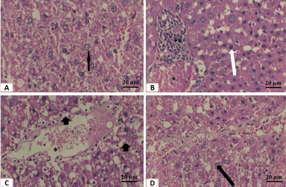
Effect of quercetin on liver of aged male rats. Histological structure of liver of 3 months old control rats (A), 3 months old treated liver (B), 30 months old saline treated rat (C) and 30 months old quercetin-treated rat (D). Note the vesicular nuclei (black arrows), cellular infiltration (**), dark nuclei (white arrow), vacuolated hepatocytes (black arrowhead) and congestion of the central vein (*). Stain: hematoxyline and eosin.
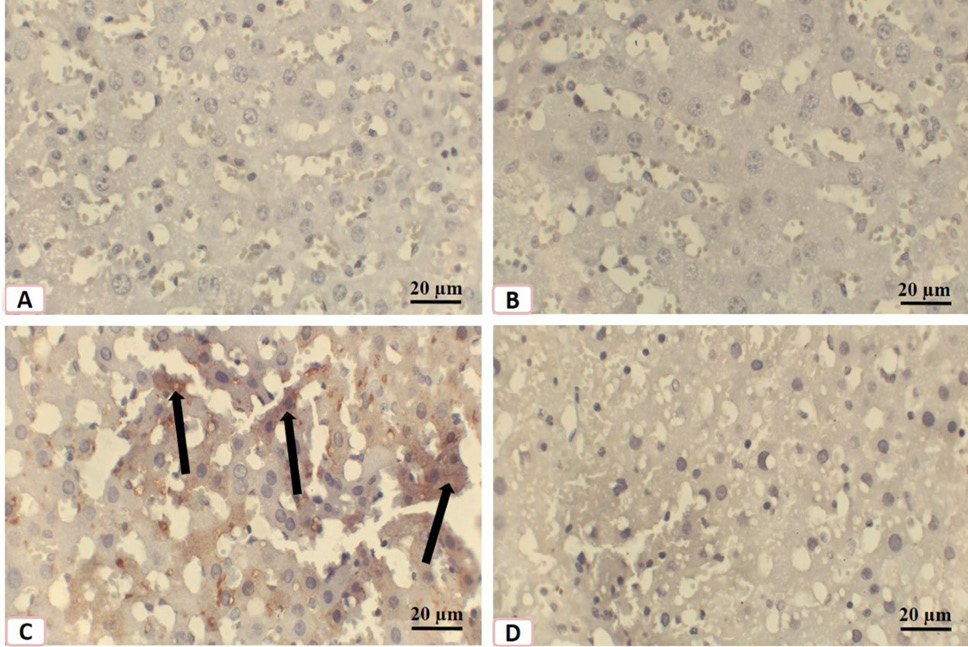
Immunostaining of histological sections of livers of rats with p53 antibodies. A, 3 months old control rats B, 3 months old quercetin-treated rats; C, 30 months old saline-treated rats; D, 30 months old quercetin-treated rats. Note the rarity of p53 expressions in A and B then the increase in its expression in C (arrows) then the rare of its expression in D.
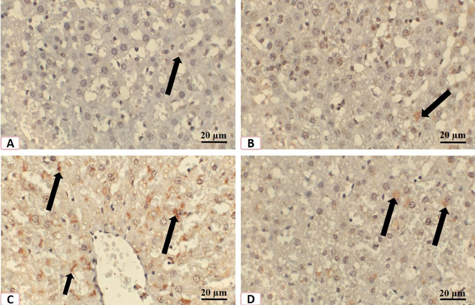
IImmunostaining of histological sections of livers of rats with bax antibodies. A, 3 months old control rat; B, 3 months old quercetin-treated rats; C, 30 months old saline-treated rats; D, 30 months old quercetin-treated rats. Note the rarity of bax expressions (arrows) in A and B then the increase of its expression in C (arrows) then the decrease of its (arrows) expression in D.
References
- Abdelhalim, M.A.K., Moussa, S.A.A. and Qaid, H.A.Y., 2018. The protective role of quercetin and arginine on gold nanoparticles induced hepatotoxicity in rats. Int. J. Nanomed., 13: 2821-2825. https://doi.org/10.2147/IJN.S160995[CrossRef]
- Aebi, H., 1984. Catalase in vitro. Methods Enzymol., 105: 121-126. https://doi.org/10.1016/S0076-6879(84)05016-3[CrossRef]
- Afifi, N.A., Ibrahim, M.A. and Galal, M.K., 2018. Hepatoprotective influence of quercetin and llagicacid on thioacetmide-induced hepatotoxicity in rats. Can. J. Physiol. Pharmacol., 96: 1-6. https://doi.org/10.1139/cjpp-2017-0651[CrossRef]
- Alm-Eldeen, A.A., Basyony, M.A., Elfiky, N.K. and Ghalwash, M.M., 2017. Effect of the Egyptian propolis on the hepatic antioxidant defense and pro-apoptotic p53 and anti-apoptotic bcl2 expressions in aflatoxin B1 treated male mice. Biomed. Pharmacother., 87: 247-255. https://doi.org/10.1016/j.biopha.2016.12.084[CrossRef]
- Alm-Eldeen, A.A., Mona, M.H., Shati, A.A. and El-Mekkawy, H.I., 2015. Synergistic effect of black tea and curcumin in improving the hepatotoxicity induced by aflatoxin B1 in rats. Toxicol. Ind. Hlth., 31: 1269-1280. https://doi.org/10.1177/0748233713491807[CrossRef]
- Alm-Eldeen, A.A., khamis, A.A., Elfiky, N.K. and Ahmad, R.A., 2019. Quercetin modulates age-induced changes in the transcript levels of some apoptosis related genes in the skeletal muscles of male rats. Braz. J. Pharm. Sci., 56: e18861 https://doi.org/10.1590/s2175-979020200003180861[CrossRef]
- Bancroft, J.D., Cook, H.C. and Stirling, R.W., 1994. Manual of histological techniques and their diagnostic application. Churchill Livingstone, Edinburgh, New York.
- Bao, D., Wang, J., Pang, X. and Liu, H., 2017. Protective effect of quercetin against oxidative stress-induced cytotoxicity in rat pheochromocytoma (PC-12) cells. Molecules, 22: 1122-1135. https://doi.org/10.3390/molecules22071122[CrossRef]
- Buege, J.A., Aust, S.D., 1978. Microsomal lipid peroxidation. Methods Enzymol., 52: 302-310. https://doi.org/10.1016/S0076-6879(78)52032-6[CrossRef]
- Dong, Y.S., Wang, J.L., Feng, D.Y., Qin, H.Z., Wen, H., Yin, Z.M., Gao, G. and Li, C., 2014. Protective effect of quercetin against oxidative stress and brain edema in an experimental rat model of subarachnoid hemorrhage. Int. J. med. Sci., 11: 282-290. https://doi.org/10.7150/ijms.7634[CrossRef]
- Dudek, H., Farbiszewski, R., Michno, T. and Kozłowski, A., 2001. Activity of glutathione peroxidase (GSH-Px), glutathione reductase (GSSG-R) and superoxide dismutase in the brain tumors. Przegl. Lek., 58: 504-506.
- Gambino, V., De Michele, G., Venezia, O., Migliaccio, P., Dall’Olio, V., Bernard, L., Minardi, S.P., Della Fazia, M.A., Bartoli, D., Servillo, G., Alcalay, M., Luzi, L., Giorgio, M., Scrable, H., Pelicci, P.G. and Migliaccio, E., 2013. Oxidative stress activates a specific p53 transcriptional response that regulates cellular senescence and aging. Aging Cell, 12: 435–445. https://doi.org/10.1111/acel.12060[CrossRef]
- Hagen, T.M., 2003. Oxidative stress, redox imbalance, and the aging process. Antioxid. Redox. Signal, 5: 503-506. https://doi.org/10.1089/152308603770310149[CrossRef]
- He, L., Hou, X., Fan, F. and Wu, H., 2016. Quercetin stimulates mitochondrial apoptosis dependent on activation of endoplasmic reticulum stress in hepatic stellate cells. Pharm. Biol., 54: 3237-3243. https://doi.org/10.1080/13880209.2016.1223143[CrossRef]
- Hohn, A. and Grune, T., 2013. Lipofuscin: Formation, effects and role of macroautophagy. Redox Biol., 1: 140–144. https://doi.org/10.1016/j.redox.2013.01.006[CrossRef]
- Itzhaki, R.F. and Gil, D.M., 1964. A micro-biuret method for estimating proteins. Anal. Biochem., 9: 401-410. https://doi.org/10.1016/0003-2697(64)90200-3[CrossRef]
- Iwaisako, K., Brenner, D.A. and Kisseleva, T. 2012. What’s new in liver fibrosis? The origin of myofibroblasts in liver fibrosis. J. Gastroenterol. Hepatol., 27: 65–68. https://doi.org/10.1111/j.1440-1746.2011.07002.x[CrossRef]
- Le Couteur, D.G., Blyth, F.M., Creasey, H.M., Handelsman, D.J., Naganathan, V., Sambrook, P.N., Seibel, M.J., Waite, L.M. and Cumming, R.G., 2010. The association of alanine transaminase with aging, frailty and mortality. J. Gerontol. A. Biol. Sci. med. Sci., 65: 712-717. https://doi.org/10.1093/gerona/glq082[CrossRef]
- Lien, E.J., Ren, S., Bui, H.H. and Wang. R., 1999. Quantitative structure-activity relationship analysis of phenolic antioxidants. Free Radic. Biol. Med., 26: 285–294. https://doi.org/10.1016/S0891-5849(98)00190-7[CrossRef]
- Liu, H., Zhang, L. and Lu, SP., 2012. Evaluation of antioxidant and immunity activities of quercetin in isoproterenol-treated rats. Molecules, 17: 4281–4291. https://doi.org/10.3390/molecules17044281[CrossRef]
- López-Otín, C., Blasco, M.A., Partridge, L., Serrano, M. and Kroemer, G., 2013. The hallmarks of aging. Cell, 153: 194-1217. https://doi.org/10.1016/j.cell.2013.05.039[CrossRef]
- Miyashita, T., Krajewski, S., Krajewska, M., Wang, H.G., Lin, H.K., Liebermann, D.A., Hoffman, B. and Reed, J.C., 1994. Tumor suppressor p53 is a regulator of bcl-2 and bax gene expression in vitro and in vivo. Oncogene, 9: 1799-1805.
- Paglia, D.E. and Valentine, W.N., 1967. Studies on the quantitative and qualitative characterization of erythrocyte glutathione peroxidase. J. Lab. clin. Med., 70: 158-169.
- Paoletti, F. and Mocali, A., 1990. Determination of superoxide dismutase activity by purely chemical system based on NAD(P)H oxidation. Methods Enzymol., 186: 209-220. https://doi.org/10.1016/0076-6879(90)86110-H[CrossRef]
- Park, J.Y., Cho, M.O., Leonard, S., Calder, B., Mian, I.S., Kim, W.H., Wijnhoven, S. van Steeg, H., Mitchell, J., van der Horst, G.T., Hoeijmakers, J., Cohen, P., Vijg, J. and Suh, Y., 2008. Homeostatic imbalance between apoptosis and cell renewal in the liver of premature aging Xpd mice. PLoS One, 3: 2346-2355. https://doi.org/10.1371/journal.pone.0002346[CrossRef]
- Park, J.H., Zhuang, J., Li, J. and Hwang, P.M., 2016. p53 as guardian of the mitochondrial genome. FEBS Lett., 590: 924–934. https://doi.org/10.1002/1873-3468.12061[CrossRef]
- Polat, C., Tokyol, C., Kahraman, A., Sabuncuoglu, B. and Yilmaz, S., 2006. The effects of desferrioxamine and quercetin on hepatic ischemia-reperfusion induced renal disturbance. Prostaglandins Leukot. Essent. Fatty Acids, 74: 379–383. https://doi.org/10.1016/j.plefa.2006.03.007[CrossRef]
- Poynard, T., Ratziu, V., Charlotte, F., Goodman, Z., McHutchison, J. and Albrecht, J., 2001. Rates and risk factors of liver fibrosis progression in patients with chronic hepatitis c. J. Hepatol., 34:730–739. https://doi.org/10.1016/S0168-8278(00)00097-0[CrossRef]
- Reitman, S. and Frankel, S., 1957. A colorimetric method for the determination of serum glutamic oxalacetic and glutamic pyruvic transaminases. Am. J. clin. Pathol., 28: 56–63. https://doi.org/10.1093/ajcp/28.1.56[CrossRef]
- Roos, W.P. and Kaina, B., 2013. DNA damage-induced cell death: From specific DNA lesions to the DNA damage response and apoptosis. Cancer Lett., 232: 237-248. https://doi.org/10.1016/j.canlet.2012.01.007[CrossRef]
- Schieber, M. and Chandel, N.S., 2014. ROS function in redox signaling and oxidative stress. Curr. Biol., 24: 453-462. https://doi.org/10.1016/j.cub.2014.03.034[CrossRef]
- Schmucker, D.L., 2005. Age-related changes in liver structure and function: implications for disease? Exp. Gerontol., 40: 650–659. https://doi.org/10.1016/j.exger.2005.06.009[CrossRef]
- Sharma, D.R., Wani, W.Y., Sunkaria, A., Kandimalla, R.J., Sharma, R.K., Verma, D., Verma, D., Bal, A. and Gill, K.D., 2016. Quercetin attenuates neuronal death against aluminum-induced neurodegeneration in the rat hippocampus. Neuroscience, 324:163–176. https://doi.org/10.1016/j.neuroscience.2016.02.055[CrossRef]
- Stahl, E.C., Haschak, M.J., Popovic, B. and Brown, B.N., 2018. Macrophages in the aging liver and age-related liver disease. Front. Immunol., 9: 2795-2808. https://doi.org/10.3389/fimmu.2018.02795[CrossRef]
- Taskan, M.M. and Gevrek, F., 2020. Quercetin decreased alveolar bone loss and apoptosis in experimentally induced periodontitis model in Wistar rats. Antiinflamm. Antiall. Agents Med Chem., [Epub ahead of print]. https://doi.org/10.2174/1871523019666200124114503[CrossRef]
- Taslidere, E., Dogan, Z., Elbe, H., Vardi, N., Cetin, A. and Turkoz, Y., 2016. Quercetin protection against ciprofloxacin induced liver damage in rats. Biotech. Histochem., 91:116–121. https://doi.org/10.3109/10520295.2015.1085093[CrossRef]
- Tieppo, J., Vercelino, R., Dias, A.S., Silva Vaz M.F., Silveira, T.R., Marroni, C.A., Marroni, N.P., Henriques, J.A. and Picada, J.N., 2007. Evaluation of the protective effects of quercetin in the hepatopulmonary syndrome. Fd. Chem. Toxicol., 45: 1140–1146. https://doi.org/10.1016/j.fct.2006.12.020[CrossRef]
- Tietz, N.W., Shuey, D.F. and Wekstein, D.R., 1992. Laboratory values in fit aging individuals–sexagenarians through centenarians. Clin. Chem., 38: 1167–1185. https://doi.org/10.1093/clinchem/38.6.1167[CrossRef]
- Tinay, I., Sener, T.E., Cevik, O., Cadirci, S., Toklu, H., Cetinel, S., Sener, G. and Tarcan, T., 2017. Antioxidant agent quercetin prevents impairment of bladder tissue contractility and apoptosis in a rat model of ischemia/reperfusion injury. Low Urin. Tract Sympt., 9: 117-123. https://doi.org/10.1111/luts.12125[CrossRef]
- Tousson, E., Alm-Eldeen, A. and El-Moghazy M., 2011. p53 and Bcl-2expression in response to boldenone induced liver cells injury. Toxicol. Ind. Hlth., 27: 711-718. https://doi.org/10.1177/0748233710395350[CrossRef]
- Wu, L., Wang, C., Li, J., Li, S., Feng, J., Liu, T., Xu, S., Wang, W., Lu, X., Chen, K., Xia, Y., Fan, X. and Guo, C., 2017. Hepatoprotective effect of quercetin via TRAF6/JNK pathway in acute hepatitis. Biomed. Pharmacother., 96:1137-1146. https://doi.org/10.1016/j.biopha.2017.11.109[CrossRef]
- Zhong, S.Z., Ge, Q.H., Qu, R., Li, Q. and Ma, S.P., 2009. Paeonol attenuates neurotoxicity and ameliorates cognitive impairment induced by d-galactose in ICR mice. J. neurol. Sci., 277: 58-64. https://doi.org/10.1016/j.jns.2008.10.008[CrossRef]
- Zhu, Q., Wani, G., Yao, J., Patnaik, S., Wang, Q.E., El-Mahdy, MA, Praetorius-Ibba, M. and Wani, A.A., 2007. The ubiquitin-proteasome system regulates p53-mediated transcription at p21waf1 promoter. Oncogene, 26: 4199–4208. https://doi.org/10.1038/sj.onc.1210191[CrossRef]
Services
Article usage statistics
Loading…