
The present study investigates the ameliorative effect of quercetin against cadmium-induced toxicity and oxidative stress in a male Sparaque Dawley rat model. There were four experimental groups with 6 rats in each: Group I control orally received normal saline, Group II orally administrated cadmium chloride (5 mg/kg bw/d), Group III orally administrated cadmium chloride (5mg/kg bw/d) and treated with low dose of quercetin (50 mg/kg bw/d), Group IV orally administrated cadmium chloride (5mg/kg bw/d) and treated with high dose of quercetin (70 mg/kg bw/d). Experimental period was four weeks. The protective efficacy of quercetin was evaluated in terms of cadmium (Cd) accumulation in liver and hair, blood profile, catalase (CAT) and superoxide dismutase (SOD) activity in liver, malondialdehyde (MDA) levels in liver and serum, and histopathological evaluation of liver and kidney. Results showed that low dose of quercetin was significantly (p≤0.05) more effective than the high dose. Low dose was more efficacious in reducing Cd accumulation in the tissues, reversing the effects of Cd toxicity on blood profile, and on the CAT and SOD activity in the liver and decreased the MDA levels in both serum and liver. Thus, the key findings suggest a profound antioxidative potential of the low dose of querectin, which could be a prospective approach in the treatment of Cd intoxication.
Research Article · Volume 51, Issue 6, 5 September 2019 · Open Access
Protective Effect of Quercetin Treatment against Cadmium-Induced Oxidative Stress in a Male Rat Model
· ·
Affiliations & Notes ⌄
- 1Department of Zoology, Faculty of Science, King Saud University, Riyadh, Saudi Arabia , * Corresponding author: noufnalharbi@gmail.com
Article Info ⌄
- Journal:
- Pakistan Journal of Zoology
- Published:
- 5 September 2019

From the article
- In the present study, treatment with low dose and high dose of quercetin reversed the toxic effect of Cd on the hematopoietic system, which suggests a strong antioxidative effect of the compound against cadmium-induced toxicity.
- In the current study, treatment with low dose of quercetin showed a significant increase in the hepatic SOD and CAT activity as compared to the group exposed to Cd only, while treatment with the high dose did not show any significant effect on these antioxidant enzymes.
- On the other hand, high dose of quercetin did not show any detectable protective effect on Cd toxicity in the hepatic tissue as compared to the low dose.
- It can be concluded that treatment with quercetin was dose effective in preventing oxidative damage induced by Cd.
Selected from the article's own text.
Abstract
Introduction
Cadmium (Cd) is a pollutant introduced into the environment by both natural and anthropogenic activities, Over the years, Cd concentration has increased in agricultural land due to the use of phosphate fertilizers, pesticides and waste water ; . Cd is a toxic metal classified in group one of the International Agency for Research on Cancer categories of carcinogens . This heavy metal has a long biological half-life in human and a low excretion rate . Cd acts as an endocrine disruptor and causes damage to several body tissues including the hepatic and renal tissue where it accumulates at high concentrations and causes oxidative damage ; . Cd also induces oxidative stress by inhibiting antioxidant enzymes and has toxic effect on the haemopoietic system . Since oxidative stress plays a major role in Cd toxicity, using therapeutic mediators to enhance the tissue antioxidant capacity may be effective against Cd toxicity . Therefore, measurement of lipid peroxidation in terms of MDA and activities of antioxidant enzymes, superoxide dismutase (SOD) and catalase (CAT) in biological samples are widely used biomarkers to determine the state of oxidative stress . Recently, several studies have reported the use of different antioxidants such as vitamins C and E, selenium, and quercetin to determinate their effect on various pathological states ; . Quercetin is a flavonoid found abundantly in many plants such as citrus, kale, onions, blueberries and tea. Quercetin has many properties and diverse functions such as antihypertensive, anticoagulant, antiatherogenic, antibacterial and antiproliferative . Also quercetin is well known for being a strong scavenger for oxygen free radicals. Quercetin treatment during Cd administration has been reported to attenuate the oxidative stress and protect the liver and kidney tissues from the associated oxidative damage . A comparison of the antioxidant activities of different kinds of flavonoids showed that quercetin has more radical scavenger potential than any other structurally related compounds . Hence, the present study aims to assess the ameliorative effect quercetin at two different doses, against cadmium-induced toxicity and oxidative stress in a male rat model.
Materials and Methods
Animals
Twenty-four adult male Spraque Dawley rats weighing 200-250 g were obtained from the Animal House of the College of Science, King Saud University. The animals were acclimated to the laboratory conditions for two weeks before the experiment started. The experimental period was 4 weeks.
Chemicals
Quercetin and cadmium chloride (CdCl2. H2O) were purchased from Sigma Chemical Co., (St. Louis, Mo., USA). Commercial ELISA assay kits were obtained from Cayman (Cayman chemicals, USA) and Bio Vision (Bio Vision, Inc., USA). Other chemicals were locally purchased.
Experimental design
Animals were randomly divided into 4 groups with 6 rats in each cage group: Group 1: Control rats orally received normal saline. Group 2: Rats orally received cadmium chloride (5 mg/kg bw/d) dissolved in distilled water .Group 3: Rats orally received cadmium chloride (5 mg/kg bw/d) and quercetin (50 mg/kg bw/d) . Group 4: Rats orally received cadmium chloride (5 mg/kg bw/d) and quercetin (70 mg/kg bw/d) . At the end of the experimental period, rats were made to fast overnight and sacrificed by decapitation on the next day. Blood was collected in tubes containing EDTA. The blood samples were centrifuged (3500 × g for 15 min at 4°C) for serum separation, which was stored at -20°C until further analysis. On dissection, hair samples were collected, liver and kidneys were excised out. Samples of liver and kidney tissue were fixed in formaldehyde for histological investigation. For analyzing the Cd concentration, the hair and liver samples were stored at -80o C until further analysis. For biochemical analysis in liver, the tissue was homogenized in phosphate buffer and centrifuged (10000×g for 15 min at 4°C). The resulting supernatant was stored at -80°C until further analysis.
Bioaccumulation of cadmium in liver and hair
Cd concentration in liver and hair was analyzed using an Atomic Absorption 280Z AA (Agilent Technologies, USA). Sample digestion was done using a digestion oven (Thermo Scientific, USA).
Blood profile
Blood was collected in tubes containing EDTA for complete blood count (CBC) using Ac·T 5diff CP Hematology analyzer (Beckman Coulter Inc., USA). The following parameters were evaluated; white blood cell count (WBC), red blood cell count (RBC), hemoglobin (HGB) levels and platelet count (PLT).
Oxidative stress biomarkers
SOD and CAT activities were measured in liver tissue using commercial ELISA kits obtained from Cayman (Cayman Chemical Company, USA). MDA levels were measured in lever and serum using a commercial ELISA kit obtained from Bio Vision (Bio Vision, Inc.).
Histopathological studies
Samples of fragments of fresh liver and kidneys were fixed separately in 10 % buffered formalin. Thereafter the tissues were dehydrated through a graded series of ethanol (from 70 to 100 % ethanol in subsequent steps). Xylene was used as a clearing agent. Tissues were embedded in paraffin (58.6 ºC). Sections (5 μm) were stained with haematoxylin and eosin, and were examined and photographed using a photomicroscope.
Statistical analysis
All the data are expressed as mean±SE of (n=6). The statistical significance was evaluated using one way analysis of variance (ANOVA) performed by SPSS statistical software version 22 (SPSS, Cary, NC, USA) followed by Tukey comparative test. Values were considered statistically significant at p≤0.05.
Table I.- Mean (± SE) Cd concentration in the liver and the hair (µg/g) in rats exposed to cadmium and treated with low (Qe50) and high dose (Qe70) of quercetin.
| Experimental groups | Control (n=6) | Cd (n=6) | Cd+Qe50 (n=6) | Cd+Qe70 (n=6) |
| Cd concentration in liver (µg/g) | 8.76 ± 2.87* | 82.45 ± 3.56*a | 89.84 ±1.46a | 117.36 ± 4.97b |
| Cd concentration in hair (µg/g) | 1.99 ± 0.19* | 4.30 ± 0.32*a | 5.11 ± 0.14a | 7.98 ± 0.59b |
*Differ significantly from control group at p≤0.05. Different letters indicate significant difference from Cd group at p≤0.05. n=6.
Results
Bioaccumulation of cadmium in liver and hair
Table I shows that 4 week exposure to Cd caused a significant (p≤0.05) increase in Cd concentration in both liver and hair in the group exposed to Cd only in comparison to the control group. Treatment with low dose of quercetin (Cd+Qe50) showed no significant difference in the Cd concentration as compared to group exposed to Cd only. While, the group treated with high dose of quercetin (Cd+Qe70) showed a significant (p≤0.05) increase in the Cd concentration in comparison to the group exposed to Cd only. Treatment with low dose (Cd+Qe50) was significantly (p≤0.05) more effective as compared to the high dose (Cd+Qe70).
Table II.- Mean (± SE) WBC count (103/µl), RBC count (106/µl), PLT count (103/µl) and HGB levels (g/dL) in rats exposed to cadmium and treated with low (Qe50) and high dose (Qe70) of quercetin.
| Experimental groups | Control (n=6) | Cd (n=6) | Cd+Qe50 (n=6) | Cd+Qe70 (n=6) |
| RBC count (106/μl) | 8.05 ± 0.075* | 7.10 ± 0.11*a | 8.09 ± 0.21b | 8.06 ± 0.11c |
| WBC count (103/μl) | 16.03 ± 0.40* | 26.07 ± 0.40*a | 20.05 ± 1.04b | 18.36 ± 0.87c |
| PLT count (103/μl) | 782.00 ± 20.67* | 494.83 ± 67.47*a | 807.33 ± 21.49b | 704.83 ± 6.23c |
| HGB level (g/dL) | 17.08 ± 0.09* | 16.05 ± 0.21*a | 16.88 ± 0.26a | 17.20 ± 0.28b |
*Differ significantly from control group at p≤0.05. Different letters indicate significant difference from Cd group at p≤0.05. n=6.
Blood profile
Table II shows a significant (p≤0.05) increase in the WBC count and a significant (p≤0.05) decrease in the RBC and PLT counts in the Cd group, in comparison to the control group. The HGB levels also showed a significant (p≤0.05) decrease in Cd group as compared to the control group. Treatment with the low dose of quercetin (Cd+Qe50) showed a significant (p≤0.05) decrease in the WBC count and caused a parallel significant (p≤0.05) increase in the RBC and PLT counts. However, no significant change was observed in the HGB levels as compared to Cd group. On the other hand, treatment with high dose of quercetin (Cd+Qe70) showed a significant (p≤0.05) decrease in the WBC count and a significant (p≤0.05) increase in the RBC and PLT counts. A significant (p≤0.05) increase was also observed in the HGB levels in comparison to the Cd group. The effect of both the low and high dose of quercetin was comparable.
Oxidative stress biomarkers
Table III shows that 4 weeks of exposure to Cd caused a significant (p≤0.05) decrease in the hepatic CAT activity (25.62 ± 1.86 mU/ml) and SOD activity (25.21 ± 1.14 U/ml) in the group exposed to Cd only in comparison to the control group (43.85 ± 1.25 mU/ml; (43.81 ± 2.48 U/ml). Treatment with low dose of quercetin (Cd+Qe50) showed a significant (p≤0.05) induction in both the hepatic CAT activity (42.27 ± 1.56 mU/ml) and SOD activity (31.93 ± 0.48 U/ml) in comparison to the Cd group. In contrast, treatment with high dose of quercetin (Cd+Qe70) showed no significant effect on the hepatic CAT activity and SOD activity as compared to the Cd group. Treatment with low dose (Cd+Qe50) was significantly (p≤0.05) more effective as compared to the high dose (Cd+Qe70).
Lipid peroxidation
For lipid peroxidation of MDA levels were determined in liver and serum. Through the exposure period of 4 weeks hepatic MDA levels were significantly (p≤0.05) increased in the Cd group (33.47 ± 0.72 nmol/mg) when compared to the control group (20.28 ± 2.28 nmol/mg). Administration of the low dose of quercetin (Cd+Qe50) significantly (p≤0.05) decreased the hepatic MDA levels in comparison to Cd group (22.18 ± 1.45 nmol/mg). However, there was no significant difference in hepatic MDA levels in the group treated with the high dose of quercetin (Cd+Qe70) (28.11 ± 1.34 nmol/mg) as compared to the Cd group. Treatment with the low dose was more effective than the high dose (Table III). Serum MDA levels showed a significant (p≤0.05) increase in Cd group (17.42 ± 0.21 nmol/ml) as compared to the control group (12.08 ± 0.28 nmol/ml). Administration of quercetin at both low dose and high dose (Cd+Qe50), (Cd+Qe70) showed a significant (p≤0.05) decrease in the serum MDA levels in comparison to the Cd group (14.49 ± 0.37 nmol/ml, 14.09 ± 0.57 nmol/ml). Treatment with the low dose and the high dose of quercetin were comparable (Table III).
Table III.- Mean (± SE) CAT activity (mU/ml), SOD activity (U/ml), MDA levels (nmol/mg) in liver and serum MDA levels (nmol/ml) in rats exposed to cadmium and treated with low (Qe50) and high dose (Qe70) of quercetin.
| Experimental groups | Control (n=6) | Cd (n=6) | Cd+Qe50 (n=6) | Cd+Qe70 (n=6) |
| CAT activity (mU/ml) | 43.85 ± 1.25* | 25.62 ± 1.86*a | 42.27 ± 1.56b | 30.32 ± 2.20a |
| SOD activity (U/ml) | 43.81 ± 2.48* | 25.21 ± 1.14*a | 31.92 ± 0.48b | 28.81 ± 0.81a |
| MDA level in liver (nmol/mg) | 20.29 ± 2.28* | 33.47 ± 0.72*a | 22.17 ± 1.44b | 28.11 ± 1.34a |
| MDA level in serum (nmol/ml) | 12.07 ± 0.28* | 17.42 ± 0.21*a | 14.48 ± 0.37b | 14.09 ± 0.57b |
*Differ significantly from control group at p≤0.05. Different letters indicate significant difference from Cd group at p≤0.05. n=6.
Histopathological studies
Sections of the liver of rats from the control showed normal structure of the hepatic tissue with hepatic cords radiating from the portal traids with normal appearance of the central vein. At a higher magnification, normal hepatic cord separated by blood sinusoids were observed. Hepatocytes showed normal polygonal shape, spherical nucleus and faintly granular cytoplasm (Fig. 1A, B).
Sections of the liver from group exposed to Cd showed loose arrangement of the tissue with disorganized hepatocytes. At higher magnification, histopathological alterations included degeneration of hepatocytes with necrosis, inflammation and fatty degenerative changes. In addition, dilation was observed in the central vein and blood sinosoids, which were congested with blood cells. An increase in Kupffer cells number was also observed (Fig. 1C, D). Treatment with low dose of quercetin showed a mild restoration in the histological alterations induced by Cd exposure represented by less disintegration and more tissue organization with hepatocytes showing nearly normal shape and arrangement as compared to the Cd group. However, number of Kupffer cells were comparable to the Cd group (Fig. 1E, F).
On the other hand, inflammatory cell infiltration was observed in rats treated with high dose of quercetin with degeneration of the hepatic tissue as comparable to the group exposed to Cd only. At higher magnification, liver sections showed less dilated sinusoids, less necrosis and less central vein disintegration in comparison to Cd group (Fig. 1G, H). The low dose of quercetin was more efficacious in restoring the histopathological alterations in the liver as compared to the high dose.
Sections of the kidney from the control group showed normal renal structure in the cortical region. Higher magnification showed intact glomeruli with epithelium lining, the glomerular capsule and a distinct capsular space. Proximal convoluted tubules and distal convoluted tubule also showed normal structure (Fig. 2A, B). Kidney sections from Cd exposed rats showed structural disarrangement with diffused hemorrhage, infiltration by lymphocytes and collapsed tubular lumina. Higher magnification of the previous section showed collapsed glomeruli, hyper-cellularity, intense hemorrhage and detached brush border of proximal convoluted tubules. In addition, flattened tubular cells, infiltration by lymphocytes and dilated lumina were also observed (Fig. 2C, D).
Treatment with low dose of quercetin showed a mild restoration in the structural organization in terms of less hyper-cellularity of glomerulus and less dilated tubules in comparison to Cd group. High magnification showed an almost normal appearance of glomerular capsule with distinct capsular space in comparable to control group. In addition, infiltration by lymphocytes was also observed (Fig. 2E, F). Treatment with the high dose of quercetin did not show any distinct improvement in the kidney in term of loose arrangement of the tissue with diffused hemorrhage, lymphocytes infiltration and collapsed tubular lumina comparable to the Cd group. Higher magnification showed hyper-cellularity, intense hemorrhage with dilation in the proximal and distal convoluted tubules comparable to the group exposed to Cd only. Furthermore, an increase in the capsular space was also observed (Fig. 2G, H). The low dose of quercetin was more efficacious in restoring the histopathological alterations in the kidney as compared to the high dose.
Discussion
Cd concentration in liver and hair was significantly increased in the group exposed to Cd in comparison to the control group. Previous studies have shown that administration of Cd leads to its accumulation in several body tissues and causing alteration in the hepatic and renal tissue. Liver and kidney are the main target organs that are affected after Cd exposure ; . The present results are in agreement with previous studies, which also reported that Cd administration results in high accumulation of metal in
various tissues especially liver and kidney . Treatment with high dose of quercetin showed a significant increase in Cd concentration. This significant increase was in agreement with the findings of a previous study, which showed significantly high accumulation of Cd in selenium treated rats compared to the group treated with Cd only . These results were explained by the fact that selenium diminished the toxicity of Cd and increased the accumulation of Cd in liver by forming a selenium-cadmium complex ; . Thus, high accumulation of Cd in the present study in the group treated with high dose of quercetin could be explained by a decrease in metallothionein levels. Metallothionein plays a major role in chelating metals specially Cd.
The hematopoietic system is considered as one of the most sensitive systems that can be used as an indicator to evaluate the toxicity of environmental toxins and drugs in humans and animals. The blood profile results in the present study showed a significant decrease in RBC and PLT count with a parallel decrease in HGB levels, on exposure to Cd. This is in agreement with a previous study, which reported that Cd exposure did significantly decrease RBC count and HGB levels in rats . On the contrary, results of a study by Fahim et al. (2012) showed no significant changes in the RBC count and total HGB concentration in the Cd- exposed mice. Anemia has been reported in rats exposed to the Cd as a result of Cd accumulation in kidney, spleen and liver that impairs the activity of these hematopoietic tissues . Moreover, anemia could be also a result of accelerated erythrocyte destruction because of the change in erythrocyte membrane permeability, increased mechanical damage, and/or incompetence of the intestinal uptake of iron because of mucosal lesions. On the other hand, the results on the WBC count showed a significant increase in their number, in the group exposed to Cd only, suggesting a systemic inflammatory response to Cd toxicity. These results are in line with previous studies ; ; .
In the present study, treatment with low dose and high dose of quercetin reversed the toxic effect of Cd on the hematopoietic system, which suggests a strong antioxidative effect of the compound against cadmium-induced toxicity. These findings corroborate with other studies that have reported the protective effect of antioxidants such as vitamin E and selenium against cadmium-induced toxicity on the hematopoietic system . The generation of oxidative stress resulting from increased formation of reactive oxygen species as well as reduced activity of the antioxidant system is recognized as one of the important biochemical mechanisms for Cd toxicity. SOD and CAT are antioxidant enzymes that play an important role against oxidative stress. In the present study, the group exposed to Cd showed a significant inhibition in the hepatic SOD and CAT activity when compared to the control group. Also, rats exposed to Cd showed a marked increase in hepatic and serum MDA levels indicating the escalation of lipid peroxidation, which could be attributed to an increased production of free radicals as a consequence of Cd accumulation in the liver. In line with these results, it has been previously reported that Cd induces oxidative damage by producing ROS and reducing the activity of some antioxidant enzymes, such as SOD and CAT . In line with an earlier study , enhanced lipid peroxidation and altered homeostasis as well as impairment of the antioxidant defense system contribute to the Cd toxicity.
In the current study, treatment with low dose of quercetin showed a significant increase in the hepatic SOD and CAT activity as compared to the group exposed to Cd only, while treatment with the high dose did not show any significant effect on these antioxidant enzymes. Contributing to our findings, previous studies have reported that treatment with antioxidants such as quercetin, naringenin and olive oil, post Cd exposure significantly enhanced SOD and CAT activity when compared in rats exposed to Cd ; . It has been observed in the present study that the hepatic and serum MDA levels were significantly reduced on treatment with quercetin at both low and high dose. Several other studies have shown that treatment with quercetin resulted in a significant decrease in the levels of MDA in cadmium-intoxicated rats ; . This could be attributed to the direct scavenging of free radicals and the quenching of the lipid peroxidative chain by the phytoconstituent. Previous animal studies have shown that flavonoids have a high antioxidant properties including free radical scavenging, metal chelation, antioxidant enzyme activation and oxidase inhibition ; . Further, several studies have demonstrated that treatment with free-radical scavengers and antioxidants have a high protective effect against Cd toxicity ; . It has been shown in both in vivo and in vitro studies that apoptosis and oxidative damage induced by Cd could be prevented by quercetin due to its high antioxidant and cytoprotective properties. By increasing the antioxidant defense system and inhibiting the oxidative status, quercetin has been reported to protect the liver and improve blood abnormalities in endotoxic model of acute liver failure ; .
In the current study, exposure to Cd for 30 days caused severe histopathological alteration in the liver with hepatocellular necrosis, dilation and congestion of blood vessels and sinusoids in the group exposed to Cd as compared to the control rats. However, treatment with low dose of quercetin moderately reversed the alterations in the hepatic tissue. On the other hand, high dose of quercetin did not show any detectable protective effect on Cd toxicity in the hepatic tissue as compared to the low dose. Histological investigation of the kidney revealed that rats exposed to Cd only have shown marked histopathological changes represented as tubular degeneration, collapsed glomerulus and intense hemorrhage Treatment with quercetin at low dose showed reduction in the tubular and the glomerular injury in comparison to Cd group. Following the same pathway in liver and kidney, high dose of quercetin did not show any detectable protective effect against Cd toxicity.
Hepatic and renal damage after Cd exposure has been reported ; . In line with another study, administration of low dose of quercetin reduced the histological alterations produced by Cd. This could be due to the antiradical/antioxidant and metal-chelating efficacy of quercetin, which significantly reduced the oxidative stress leading to the reduction of histopathological damages and restoration to near normal physiological state of the liver and the kidney . Quercetin at low dose lined in the present study, effectively attenuated cadmium-toxicity by eliminating free radicals, inhibiting lipid peroxidation levels and protecting the hepatic tissue from cadmium-induced oxidative damage and by significantly lowering lipid peroxidation concentration.
Conclusion
It can be concluded that treatment with quercetin was dose effective in preventing oxidative damage induced by Cd. Nevertheless, further studies need to be done to determine the efficacy of varied doses of quercetin against Cd toxicity.
Acknowledgement
This research project was supported by a grant from the “Research Center of the Female Scientific and Medical Colleges”, Deanship of Scientific Research, King Saud University.
Statement of conflict of interest
The authors declare that there is no conflict of interests regarding the publication of this article.
Figures
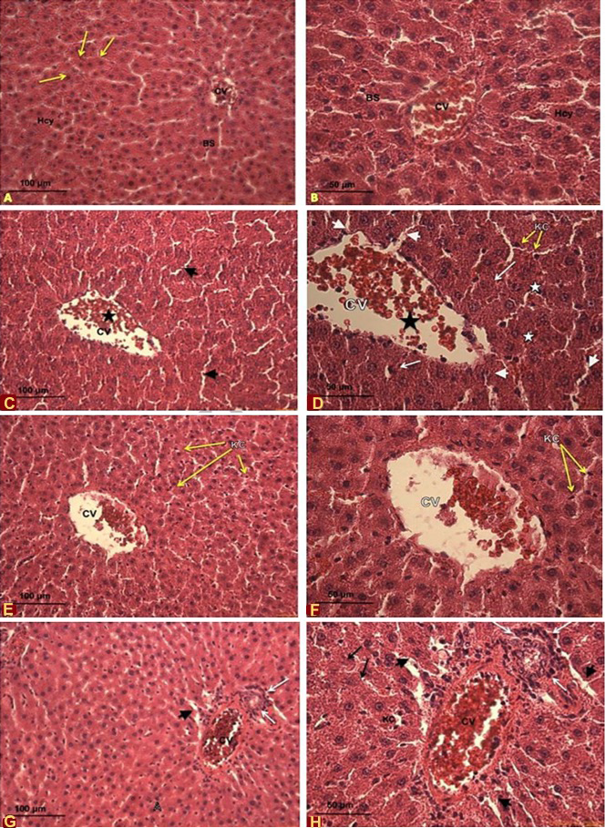
Effects of cadmium and quercetin on the histological sections of male Sprague-Dawley rate. A, B, control rates livers at 10x and 20x magnification, respectfully. A shows the central vein (CV), normal arrangement of hepatocytes (Hcy) along the hepatic cord (arrows) with normal blood sinusoids (BS) and B shows the central vein (CV), normal arrangement of hepatocytes (Hcy) along the hepatic cord (arrows) with normal blood sinusoids (BS); C, D, liver of rates oraly administrated with CdCl2 at 5mg/kg bw/d at 20x (C) and 40x (D) magnification. C shows disorganized hepatic architecture, dilated blood sinusoids (arrow heads), and dilated central vein (CV) (star); D shows dilated central vein (CV) (black star), dilated blood sinusoids (Arrow heads) congested with blood cells. Notice red blood cells (black arrow), Kupffer cells (KC) (yellow arrows), the disintegration in the tissue (white star), and degenerated hepatocytes and necrosis (arrows). E, F, liver of rates oraly administrated with quercetin at 5mg/kg bw/d at 20x (E) and 40x (F) magnification. E shows mild restoration of hepatic tissue. Notice Kupffer cell (KC) (yellow arrows), less disintegration in the central vein (CV) and more organized hepatocytes (star); F shows mild restoration of hepatic tissue. Notice red blood cells (black arrow), Kupffer cells (KC) (yellow arrows), less disintegration in the central vein (CV) and more organized hepatosytes. G, H, liver of rates oraly administrated with quercetin at 70mg/kg bw/d at 20x (G) and 40x (H) magnification. G shows disorganized hepatic architecture with disintegrated and highly congested central vein (CV) (star), inflammatory cell infiltration (white arrows), H shows dilated blood sinusoids (arrow heads) and increase in number of Kupffer cells (yellow arrows); B shows dilated blood sinusoids (arrow heads), necrosis (black arrows) and disintegration and congestion in the central vein (CV) (star) and inflammatory cell infiltration (white arrows). Notice the increase in number of Kupffer cells (yellow arrows).
Magnification: A, 10x; B, C, E, G, 20x; D,F, H, 40x.
Stain: Haematoxylin and Eosin
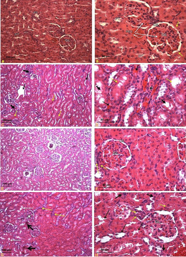
Effects of oral administration of cadmium and quercetin on the histological section of male Sprague-Dawley rate. A, B, control rates kidneys at 20x (A) and 40x (B) magnification. A shows the normal structure in the cortical region. Notice the renal glomerulus (GL), the proximal convoluted tubules (PT) and distal convoluted tubules (DT); B shows the normal intact cortical region. Notice the red blood cells (RBC), renal glomerulus (GL) with epithelium cells (arrows), proximal convoluted tubules (PT) and the distal convoluted tubules (DT) showed normal structure. C, D, kidneys of rats oraly administrated with CdCl2 at 5mg/kg bw/d at 10x (C) and 40x (D) magnification, respectively. C shows kidney of rats exposed to cadmium (5 mg/kg bw/d) (10x) showing collapsed glomerulus (star), tubular dilation (black arrows), diffuse hemorrhage (yellow arrows) and infiltration by lymphocytes (thick arrow), collapsed glomerulus (GL), hyper-cellularity and collapsed lumina of tubules; D shows collapsed glomerulus (GL) (star), intense hemorrhage (arrow), detached brush border of proximal convoluted tubules (yellow arrows), hyper-cellularity and flattened tubular cells and lumina are relatively dilated (thick arrows). E, F, kidneys of rates oraly administrated with quercetin at 5mg/kg bw/d at 10x (E) and 40x (F) magnification. E shows mild restoration with near normal appearance in renal glomeruli (G), glomerular capsule (BC) with distinct capsular space (CS); F shows reduced the damage induced by cadmium. Notice lymphocytes infiltration (yellow arrow), renal glomeruli (GL) and glomerular capsule with distinct capsular space (CS). G, H, kidneys of rats oraly administrated with quercetin at 70mg/kg bw/d at 10x (G) and 40x (H) magnification. G shows tubular dilation (white arrows), collapsed glomerulus (GL) (star), lymphocytes infiltration (yellow arrow) and intense hemorrhage (black arrow); H shows collapsed glomeruli (GL), glomerular space (CS), tubular dilation (arrows), hyper-cellularity (thick arrows) and lymphocytes infiltration (yellow arrow).
Magnification: A, 20x; B, D, F, G, 40x; C, E, G, 10x.
Stain: Haematoxylin and Eosin
References
- Abdalla, F., Schmatz, R., Cardoso, A., Carvalho, F., Baldissarelli, J., de Oliveira, J., Rosa, M., Gonçalves Nunes, M., Rubin, M., da Cruz, I., Barbisan, F., Dressler, V., Pereira, L., Schetinger, M., Morsch, V., Gonçalves, J. and Mazzanti, C., 2014. Quercetin protects the impairment of memory and anxiogenic-like behavior in rats exposed to cadmium: Possible involvement of the acetylcholinesterase and Na+, K+-ATPase activities. Physiol. Behav., 135: 152-167.
- Abo-Salem, O., Abd-Ellah, M. and Ghonaim, M., 2011. Hepatoprotective activity of quercetin against acrylonitrile-induced hepatotoxicity in rats. J. Biochem. mol. Toxicol., 25: 386-392.
- Aktoz, T., Kanter, M., Uz, Y., Aktaş, C., Erboğa, M. and Atakan, İ., 2011. Protective effect of quercetin against renal toxicity induced by cadmium in rats. Balkan med. J., 29: 56-61.
- Alía, M., Mateos, R., Ramos, S., Lecumberri, E., Bravo, L. and Goya, L., 2005. Influence of quercetin and rutin on growth and antioxidant defense system of a human hepatoma cell line (HepG2). Eur. J. Nutr., 45: 19-28.
- Amamou, F., Nemmiche, S., Meziane, R., Didi, A., Yazit, S. and Chabane-Sari, D., 2015. Protective effect of olive oil and colocynth oil against cadmium-induced oxidative stress in the liver of Wistar rats. Fd. Chem. Toxicol., 78: 177-184. https://doi.org/10.1016/j.fct.2015.01.001[CrossRef]
- Ashour, T., 2014. Preventative effects of caffeic Acid phenyl ester on cadmium intoxication induced hematological and blood coagulation disturbances and hepatorenal damage in rats. ISRN Hematol., 2014: 764-754. https://doi.org/10.1155/2014/764754[CrossRef]
- Bhatt, K. and Flora, S., 2009. Oral co-administration of α-lipoic acid, quercetin and captopril prevents gallium arsenide toxicity in rats. Environ. Toxicol. Pharmacol., 28: 140-146. https://doi.org/10.1016/j.etap.2009.03.012[CrossRef]
- Błasiak, J., 2001. DNA-damaging effect of cadmium and protective action of quercetin. Polish J. environ. Stud., 10: 437-442.
- Boots, A., Haenen G. and Bast, A., 2008. Health effects of quercetin: From antioxidant to nutraceutical. Eur. J. Pharmacol., 585: 325-337. https://doi.org/10.1016/j.ejphar.2008.03.008[CrossRef]
- Chen, L., Liu, L. and Huang, S., 2008. Cadmium activates the mitogen-activated protein kinase (MAPK) pathway via induction of reactive oxygen species and inhibition of protein phosphatases 2A and 5. Free Radical Biol. Med., 45: 1035-1044. https://doi.org/10.1016/j.freeradbiomed.2008.07.011[CrossRef]
- Chlebda, E., Magdalan, J., Merwid-Lad, A., Trocha, M., Kopacz, M., Kuźniar, A., Nowak, D. and Szelag, A., 2010. Influence of water-soluble flavonoids, quercetin-5′-sulfonic acid sodium salt and morin-5′-sulfonic acid sodium salt, on antioxidant parameters in the subacute cadmium intoxication mouse model. Exp. Toxicol. Pathol., 62: 105-108. https://doi.org/10.1016/j.etp.2009.02.118[CrossRef]
- El-Boshy, M., Risha, E., Abdelhamid, F., Mubarak, M. and Hadda, T., 2014. Protective effects of selenium against cadmium induced hematological disturbances, immunosuppressive, oxidative stress and hepatorenal damage in rats. J. Trace Elem. Med. Biol., 29: 104-110. https://doi.org/10.1016/j.jtemb.2014.05.009[CrossRef]
- Eybl, V., Kotyzova, D., Cerna, P. and Koutenský, J., 2008. Effect of melatonin, curcumin, quercetin, and resveratrol on acute ferric nitrilotriacetate (Fe-NTA)-induced renal oxidative damage in rats. Hum. exp. Toxicol., 27: 347-353. https://doi.org/10.1177/0960327108094508[CrossRef]
- Fahim, M., Nemmar, A., Dhanasekaran, S., Singh, S., Shafiullah, M., Yasin, J., Zia, S. and Hasan, M., 2012. Acute cadmium exposure causes systemic and thromboembolic events in mice. Physiol. Res., 61: 73-80.
- Fang, Y., Yang, S. and Wu, G., 2002. Free radicals, antioxidants and nutrition. Nutr. J., 18: 872-879. https://doi.org/10.1016/S0899-9007(02)00916-4[CrossRef]
- Farombi, E., Adedara, I., Akinrinde, S., Ojo, O. and Eboh, A., 2012. Protective effects of kolaviron and quercetin on cadmium-induced testicular damage and endocrine pathology in rats. Andrologia, 44: 273-284. https://doi.org/10.1111/j.1439-0272.2012.01279.x[CrossRef]
- Ferrandino, I., Favorito, R., Annunziata, M. and Grimaldi, M., 2009. Cadmium induces apoptosis in the pituitary gland of Podarcis sicula. Annls N.Y. Acad. Sci., 1163: 386-388. https://doi.org/10.1111/j.1749-6632.2008.03667.x[CrossRef]
- Ikediobi, C., Badisa, V., Ayuk-Takem, L., Latinwo, L. and West, J., 2004. Response of antioxidant enzymes and redox metabolites to cadmium-induced oxidative stress in CRL- 1439 normal rat liver cells. Int. J. mol. Med., 14: 87-92. https://doi.org/10.3892/ijmm.14.1.87[CrossRef]
- Jihen, E., Imed, M., Fatima, H. and Abdelhamid, K., 2009. Protective effects of selenium (Se) and zinc (Zn) on cadmium (Cd) toxicity in the liver of the rat: Effects on the oxidative stress. Ecotoxicol. environ. Safe., 72: 1559-1564. https://doi.org/10.1016/j.ecoenv.2008.12.006[CrossRef]
- Khandelwal, S., Pathak, N. and Singh, M., 2008. Protective role of piperine against cadmium induced hepatic and renal toxicity in mice. Toxicol. Int., 15: 85-89.
- Korish, A., 2010. Effect of caffeic acid phenethyl ester on the hemostatic alterations associated with toxic-induced acute liver failure. Blood Coagul. Fibrinolysis, 21: 158-163. https://doi.org/10.1097/MBC.0b013e32833678be[CrossRef]
- Krishnakumar, N., Prabu, S. and Sulfikkarali, N., 2012. Quercetin protects against cadmium-induced biochemical and structural changes in rat liver revealed by FT-IR spectroscopy. Biomed. Prevent. Nutr., 2: 179-185. https://doi.org/10.1016/j.bionut.2012.03.010[CrossRef]
- Kuester, R., Waalkes, M., Goering, P., Fisher, B., McCuskey, R. and Sipes, I., 2002. Differential hepatotoxicity induced by cadmium in Fischer 344 and Sprague-Dawley rats. Toxicol. Sci., 65: 151-159. https://doi.org/10.1093/toxsci/65.1.151[CrossRef]
- Limei, Z., Xiaoyong, L., Tongbin, C., Xiulan, Y., Hua, X., Bin, W. and Lixia, W., 2008. Regional assessment of cadmium pollution in agricultural lands and the potential health risk related to intensive mining activities. J. environ. Sci. (China), 20: 696-703. https://doi.org/10.1016/S1001-0742(08)62115-4[CrossRef]
- Ognjanović, B., Marković, S., Pavlović, S., Zikić, R., Stajn, A. and Saicić, Z., 2008. Effect of chronic cadmium exposure on antioxidant defense system in some tissues of rats: Protective effect of selenium. Physiol. Res., 57: 403-411.
- Renugadevi, J. and Prabu, M., 2009. Ameliorative effect of quercetin against cadmium induced toxicity in liver of Wistar rats. Cell Tissue Res., 9: 1665-1672.
- Singh, S., Kumar, M., Singh, R., Singh, S. and Chandra, K., 2011. Ameliorative potential of quercetin against paracetamol-induced oxidative stress in mice blood. Toxicol. Int., 18: 140-145. https://doi.org/10.4103/0971-6580.84267[CrossRef]
- Wahba, Z., Coogan, T., Rhodes, S. and Waalkes, M., 1993. Protective effects of selenium on cadmium toxicity in rats: Role of altered toxicokinetics and metallothionein. J. Toxicol. environ. Hlth., 38: 171-182.
- Wang, L., Lin, S., He, Y., Liu, G. and Wang, Z., 2013. Protective effects of quercetin on cadmium- induced cytotoxicity in primary cultures of rat proximal tubular cells. Biomed. environ. Sci., 26: 258-267.
- Yuan, G., Dai, S., Yin, Z., Lu, H., Jia, R., Xu, J., Song, X., Li, L., Shu, Y. and Zhao, X., 2014. Toxicological assessment of combined lead and cadmium: Acute and sub-chronic toxicity study in rats. Fd. Chem. Toxicol., 65: 260-268. https://doi.org/10.1016/j.fct.2013.12.041[CrossRef]
Cited by 1 article
In journals published by ResearchersLinks.
Services
Article usage statistics
Loading…