
In this experiment, the effects of naringenin (Nar) on cadmium (Cd)-induced changes in blood physiological and biochemical indices and kidney damage among rats were studied. Twenty-four male Sprague–Dawley rats were randomly divided into four groups: control group, Cd-treated group (1 mg/kg b.w. CdCl2), Nar-treated group (100 mg/kg b.w.), and Cd + Nar-treated group (1 mg/kg b.w. CdCl2 + 100 mg/kg b.w. Nar). They were treated for 4 weeks, and then the blood physiological and biochemical indicators were assessed. The levels of glutathione (GSH), malondialdehyde (MDA), catalase (CAT), and total superoxide dismutase (T-SOD) were measured. Histopathological sections of the kidney were made to observe the structural damage. Results showed that the red blood cell count (RBC), hemoglobinconcentration (HGB), and hematocrit(HCT) of serum were significantly reduced after Cd exposure for 4 weeks. Cd increased the expression of GSH and MDA and decreased the activities of CAT and T-SOD. Furthermore, in the Cd-treated rats, the glomerulus contracted, the renal tubular structure was damaged, the epithelial cells were disordered and degenerated, and a large area of lesions occurred. However, treatment with Nar slowed down the occurrence of oxidative stress induced by Cd. Therefore, as an effective antioxidant, Nar has a certain protective effect on oxidative stress and kidney injury induced by Cd.
Research Article · Volume 55, Issue 2, 19 January 2023 · Open Access
Effects of Naringenin on Cadmium-Induced Changes in Blood Physiological and Biochemical Indices and Kidney Damage Among Rats
· · · · · ·
Affiliations & Notes ⌄
- 1College of Animal Science and Technology, Henan University of Science and Technology, No.263, Kaiyuan Avenue, 471023, Luoyang, PR China. * Corresponding author: wangjicang@126.com
Article Info ⌄
- Journal:
- Pakistan Journal of Zoology
- Published:
- 19 January 2023

From the article
- Therefore, the present study determined the effect of Cd and Nar on rat blood by detecting changes in blood physiological indicators, such as RBC, HGB, and HCT.
- The decrease in hemoglobin content may be related to the increase in the rate of red blood cell damage or the decrease in the production rate of red blood cells.
- The renal tissue structure of Cd + Nar co treatment group was relatively complete, indicating that Nar can effectively reduce the renal damage caused by Cd by alleviating oxidative stress.
- This study confirmed that Cd could lead to changes in the blood physiological and biochemical indexes of rats, the kidney toxicity induced by Cd in rats is related to oxidative stress, and Nar could inhibit the toxic effect of Cd by reducing the occurrence of oxidative damage.
Selected from the article's own text.
Abstract
Introduction
Cadmium (Cd) is a highly toxic heavy metal that occurs widely in nature from agriculture, industry, and other production activities, and it is widely found in the air, food, and water ; . Cd is a non-essential element with a long half-life . Kidney is the organ most vulnerable to Cd accumulation . Excessive Cd accumulation in the body could cause tissue damage in this organ and harm to human body ; ; . With the development of economy, industry, and agriculture, Cd pollution has become increasingly serious in recent years, and the harm caused by Cd pollution has attracted the attention of more people than before. Naringenin (Nar), which is mainly distributed in oranges, grapefruits, citrus, and other fruits, is a monomer of flavonoids extracted from citrus plants in the Rutaceae family . Existing in tomato and lemon peel, Nar is a representative flavonoid with antioxidant, antiallergic, antihepatotoxic, antithrombotic, and anti-inflammatory effects .
Blood plays an important role in metabolic regulation and maintaining the balance between the internal and external environments of the body. Studies have found that Cd-poisoned rats appeared to have different degrees of anemia, which may be related to the change in red blood cell permeability. Therefore, the changes of blood physiological and biochemical indexes in rats exposed to Cd were detected in our experiment to judge the relationship between Cd poisoning and blood injury. Studies have also shown that Cd-induced liver and kidney damage in rats may be related to oxidative stress and autophagy ; ; . Studying the toxic effect of Cd and its mechanism and establishing methods to alleviate Cd pollution are highly important. However, the mechanism of Nar in Cd-induced renal injury in rats remains unclear. Therefore, we determined the antioxidant indexes to explore whether Cd induced kidney injury is related to oxidative stress . Given the anti-inflammatory effect of Nar, it is often used to treat arthritis. Its anti-damage and antioxidant effects have received widespread attention. For this, we chose Nar as a protective agent to explore the protective effect of Nar on Cd induced kidney injury in rats, so as to provide a theoretical basis for the clinical use of Nar .
In this study, red blood cell count (RBC), hemoglobin concentration (HGB), hematocrit (HCT), mean erythrocyte hemoglobin concentration (MCHC), and mean platelet volume (MPV) were examined to explore the effect of Cd on blood physiological and biochemical indexes of rats. The kidney damage caused by Cd was assessed by measuring the changes in antioxidant indicators glutathione (GSH), malondialdehyde (MDA), catalase (CAT), and total superoxide dismutase (T-SOD). Histological evaluation of rat kidney revealed the damage brought by Cd to the kidney’s tissue structure. In addition, the protective effect of Nar on Cd-induced kidney damage in rats was studied.
MATERIALS AND METHODS
Chemicals
CdCl2(99.95%) (No. 7790-78-5) was purchased from Aladdin Biochemical Technology Co., Ltd (Shanghai, China), Nar (97%) (No. 67604-48-2) was purchased from Beijing Bailingwei Technology Co., Ltd (Beijing, China).
Animals
Six-week-old Sprague–Dawley rats weighing approximately 150 g were used and kept on 12 h light:12 h dark cycles with controlled temperature (24°C ± 2°C). They were fed with a commercial standard diet, with free access to drinking water under standard laboratory conditions.
Experimental design
After 1 week of adaptive feeding, the rats were randomly divided into four groups, with six rats per group. They were treated everyday as follows: The control group received distilled water and 0.9% NaCl by oral gavage, the Cd-treated group received 1 mg/kg b.w. of CdCl2 intraperitoneally, the Nar-treated group received 100 mg/kg b.w. of Nar daily by oral gavage, and the Cd + Nar-treated group received 1 mg/kg b.w. of CdCl2 intraperitoneally and 100 mg/kg b.w. of Nar by oral gavage. The doses of CdCl2 and Nar were used were selected from previous studies ; ; ; Wang et al., 2020a).
The experiment lasted for 4 weeks. Afterwards, respiratory anesthesia was performed on the rats in each group with diethyl ether. Blood was collected and placed in test tubes containing anticoagulant substances. Then, the rats were sacrificed by neck dissection, and the kidneys were collected. A part of the kidney tissue was placed into the grinder, PBS buffer solution was added, and the mixture was grinded in a low-temperature environment to prepare a tissue homogenate with a concentration of 10%. The supernatant was collected after centrifugation and placed in the refrigerator at −20°C for later use. Then, a non-destructive kidney was placed in 10% formaldehyde solution for fixation, and a kidney section was prepared for later use.
Blood physiological, biochemical and antioxidant indices analysis
The blood physiological indicators were measured using a blood cell analyser, animal blood cell analyser was purchased from Nanjing pulang medical equipment co., LTD (Nanjing, China). The MDA, GSH, CAT, and T-SOD levels in the kidney were analyzed according to the manufacturer’s protocols. GSH (No. A006-2-1), MDA (No. A003-1-1), CAT (No. A007-1-1), and T-SOD (No. A001-1-1) kits were obtained from Nanjing Jiancheng Bioengineering Institute (Nanjing, China).
Histopathological analysis
Fresh kidneys were immersed in formaldehyde solution for 24 h and fixed. Then, they were cut into 1 × 1 × 0.5 cm-rectangular parallelepiped, placed in the embedding box, and rinsed with running water for 12 h to remove the formaldehyde solution attached to the surface. Afterwards, they were dehydrated with different concentrations of ethanol, removed with xylene, and embedded in paraffin. A slice with a thickness of approximately 5 μm was cut and placed in 42°C water. It was stretched out and placed in the center of the glass slide. Subsequently, it was dried in a 45°C oven for approximately 40 minutes. The prepared slices were placed under 40 × lenses for their photos to be taken and observed, and then the photos were saved.Next, according to the histopathological scoring method described above ; , the experimental team members scored the renal histopathological sections. Renal pathological lesions were exhibited as follows: Destruction of renal tissue structure, glomerular atrophy, deformation and necrosis of renal tubules. According to the distribution range of the above lesions, the scores are recorded as follows: 0, no lesions; 1, the distribution range of lesions is less than 10%; 2, the distribution of lesions is 10%-50%; 3, the distribution of lesions is 50%-90%; 4, the distribution of lesions is more than 90%, respectively.
Statistical analysis
All data were analyzed on SPSS 20.0 software. One-way analysis of variance (ANOVA) and the least significant difference (LSD) post hoc test were used to analyze the data.
Table I. Effect of Nar on Cd-induced changes in blood physiological indices.
| Treated | RBC(×1012/L) | HGB(g/L) | HCT(%) | MCV(fl) | MCHC(g/L) | MPV(fL) |
| Control | 6. 81±0. 26 | 114. 17±4. 19 | 34. 52±0. 98 | 50. 80±0. 71 | 330. 33±2. 79 | 7. 85±0. 07 |
| Cadmium | 5. 49±0. 17* | 98. 5±2. 53* | 30. 04±6. 67* | 51. 73±0. 72 | 327. 75±0. 76 | 8. 77±0. 16 |
| Naringenin | 7. 53±0. 11 | 126. 6±3. 44 | 38. 66±1. 02 | 51. 40±1. 66 | 327. 70±0. 87 | 8. 42±0. 21 |
| Cadmium + Naringenin | 6. 74±0. 16# | 120. 5±2. 25# | 36. 63±0. 67 | 51. 83±1. 26 | 328. 75±1. 93 | 8. 13±0. 34 |
*P< 0.05 compared with control group (non-Cd-exposed group); #P< 0.05 compared with Cd-exposed group, the same as follow.
All measurement results were expressed as mean ± standard deviation. The value was not considered significant when P> 0.05 and extremely significant when P < 0.05.
Results
Blood physiological indices of rats
As shown in Table I, in the Cd-treated group, the serum RBC, HGB, and HCT were significantly reduced (P< 0.05) compared with those in the control group after Cd exposure for 4 weeks. However, the Nar-treated group (100 mg/kg b.w.) did not show any significant difference in blood physiological indices (P> 0.05) compared with the control group. The Cd + Nar-treated group (100 mg/kg b.w.) exhibited significantly reduced serum RBC and HGB (P< 0.05) compared with the Cd-treated group after Cd exposure for 4 weeks, but the HCT, MCHC, and MPV indices did not significantly differ (P> 0.05).
GSH and MDA contents of rat kidney
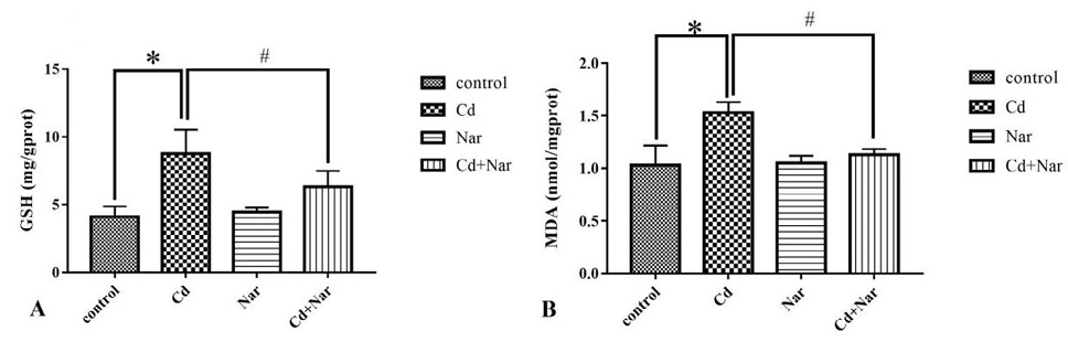
As shown in Figure 1, the content of GSH and MDA in the Cd-treated group significantly (P< 0.05) increased compared with that in the control group, whereas this content the Cd + Nar-treated group was significantly (P< 0.05) reduced compared with that in the Cd-treated group.
Rats were treated with CdCl2 and/or Nar for 4 weeks. Then, the kidney tissues were used to determine the levels of GSH (A) and MDA (B). All measurement results were expressed as mean ± standard deviation. *, indicates significant difference compared with the corresponding control (*P < 0.05). #, indicates statistically significant difference between the Cd and Cd + Nar groups (#P < 0.05).
CAT and T-SOD activity in rat kidney tissues
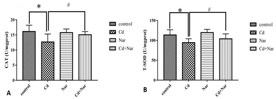
As shown in Figure 2, the Cd-treated group showed significantly (P< 0.05) reduced activities of CAT and T-SOD compared with the control group. In the Cd + Nar-treated group, the CAT activity significantly (P< 0.05) increased and the T-SOD activity increased, but the difference was not significant (P<0.05) compared that in the Cd-treated group.
Rats were treated with CdCl2 and/ or Nar for 4 weeks. Then, the kidney tissues were used to determine the levels of CAT (A) and T-SOD (B). All measurement results were expressed as mean±standard deviation. *,indicates significant difference compared with the corresponding control (*P < 0.05). #, indicates statistically significant difference between the Cd and Cd + Nar groups (#P < 0.05).
Histological evaluation of rat kidney
After 4 weeks of treatment, compared with the control group (Fig. 3A and C), the renal tissue of Nar treatment group had no abnormal changes, the tissue structure was relatively complete, and the boundary between the spheroid and renal tubular epithelial cells was clear, no obvious lesions were found. Compared with the control group (Fig. 3A and B), the Cd exposed group, the renal tissue structure was obviously destroyed, the glomerulus was atrophied, the renal tubules were deformed and vacuolated. The cell structure was disordered and degenerated, and the boundary of cell membrane was blurred. In the Cd + Nar co treatment group, the renal tissue structure was basically intact compared with the Cd treatment group, a small part of the cell structure damage (Fig. 3B and D), normal globular, no obvious disease. Furthermore, the histopathological changes of renal tissue were scored and quantified by the members of the experimental team (Fig. 4). In comparison to the control group, the renal pathological score in the Cd-treated group increased remarkably (p< 0.05); in comparison to the Cd-treated group, the renal pathological score in the Cd + Nar co-treated group decreased remarkably (p< 0.05). These results suggest that Nar has a protective effect on Cd induced renal injury.
Discussion
Cd enters the human body in several manners, and the effect of Cd pollution on human health is increasingly serious . As one of the important storage locations, the kidney is seriously damaged by Cd . In addition, Cd could cause oxidative damage to the body, and Nar is an antioxidant . Previous studies have shown that Cd could cause kidney damage in rats, and it was related to the occurrence of oxidative stress . However, no evidence could explain if Nar could reduce the oxidative stress caused by Cd.
Cd could cause various symptoms of anemia, leading to changes in the morphology of red blood cells, which could be detected by blood physiological indicators . Therefore, the present study determined the effect of Cd and Nar on rat blood by detecting changes in blood physiological indicators, such as RBC, HGB, and HCT. Compared with the control group, the Cd-treated group showed a significant decrease in RBC, HGB, and HCT in the blood, indicating that Cd could inhibit the production of red blood cells . The significant decrease of RBC indicated that Cd exposure had an adverse effect on erythropoiesis . The decrease in hemoglobin content may be related to the increase in the rate of red blood cell damage or the decrease in the production rate of red blood cells. Therefore, the decrease in HCT indicates that Cd is one of the causes of anemia . Compared with the Cd-treated group, the Cd + Nar-treated group exhibited significantly increased blood RBC and HGB levels, indicating that Nar can resist the damage of blood physiological function caused by Cd to a certain extent by increasing the number of blood cells and the content of haemoglobin .
GSH is an important antioxidant found in cells. It could decompose antioxidant substances in the body, remove excess free radicals, and maintain normal immunity in the body . Some studies have shown that Cd can interact with -SH in GSH , which is the main intracellular defense substance, leading to ROS generation, lipid peroxidation and oxidative damage. SH consumption is one of the important mechanisms of Cd induced oxidative stress, especially in oxidative phosphorylation and detoxification. These processes are the final cause of ROS accumulation (Wang et al., 2020b). Therefore, changes in GSH content could reflect the degree of oxidative damage to cells . MDA is an important product of lipid peroxidation, which is related to oxidative damage to the body . The increase of MDA content is a marker of membrane lipid peroxidation and cell damage . Therefore, the content of MDA could reflect the degree of oxidative damage suffered by the body, and antioxidant drugs could reduce the content of MDA in damaged tissues ; . In this experiment, compared with the control group, the GSH and MDA content in the kidney of the Cd treated group was significantly increased, which confirmed that the toxicity of Cd caused excessive accumulation of free radicals, resulting in oxidative stress, and the increase of lipid peroxides in the tissue, leading to renal damage and dysfunction . The 4-hydroxyl group on Nar β ring is electron-donating, which can contact with the target free radical and quench the free radical. Therefore, it can protect the cell membrane from the attack of free radicals, and reduce lipid peroxidation . In addition, the lipophilic properties of Nar effectively help it adhere to the lipid bilayer, reduce the formation of free radicals and protect cell membranes . The significant decrease in the content of GSH and MDA in the Cd + Nar-treated group compared with that in the Cd-treated group proved that Nar could enhance the antioxidant capacity of tissues by reducing the formation of free radicals and reduce the oxidative damage caused by Cd .
CAT is the most important antioxidant protein that removes free radicals in the body . It could catalyze the decomposition of H2O2 to produce H2O and O2, hence often used to judge the oxidation status in the body . T-SOD is an antioxidant that could remove free radicals in the body and protect the body from the influence of oxygen free radicals . Under normal circumstances, the enzyme system in the body plays an important role in scavenging free radicals and maintaining the balance of tissue cells ; however, when the body is subjected to harmful stimuli from the external environment, the oxidation and antioxidant balance of the tissues in the body is broken , and oxidative stress occurs ; . In the present study, compared with the control group, the Cd-treated group demonstrated significantly reduced contents of CAT and T-SOD in the kidney tissue, proving that Cd exposure caused oxidative damage to the body, consumed a large amount of CAT and T-SOD produced by the body, and destroyed the balance of oxidation and antioxidant in the body. Compared with the Cd-treated group, the Cd + Nar treatment group, CAT and SOD contents were significantly increased in kidney tissue, indicating that Nar can inhibit oxidative stress induced by H2O2, relieve the consumption of T-SOD, increase CAT activity, and finally reduce the damage caused by Cd .
In the pathological section of rat kidney tissue, various structures in the kidney tissue of the Cd-treated group showed varying degrees of damage ; . Kidney sections from control rats showed normal glomerular and tubular histomorphology. Rats in the Cd exposed group, the renal structure of was damaged, glomerular shrinkage, tubular structure was deformed and vacuolated (marked by arrows) . The structural damage of glomeruli and tubules was alleviated in Nar and Cd co treated group . This finding may be caused by the increased lipid peroxidation reaction caused by free ions in the kidney tissue due to Cd exposure, and the accumulation of free radicals eventually leads to extensive kidney disease ; . The renal tissue structure of Cd + Nar co treatment group was relatively complete, indicating that Nar can effectively reduce the renal damage caused by Cd by alleviating oxidative stress.
Conclusions
This study confirmed that Cd could lead to changes in the blood physiological and biochemical indexes of rats, the kidney toxicity induced by Cd in rats is related to oxidative stress, and Nar could inhibit the toxic effect of Cd by reducing the occurrence of oxidative damage. This study provides a theoretical basis for the treatment of Nar on Cd poisoning.
Acknowledgements
This study was supported by grants from the National Nature Science Foundation of China (No.31972753), National Innovation and Entrepreneurship Training Program for College Students (No.202010464068), Henan Provincial Department of Science and Technology Research Project (No. 222102110340 ).
Statement of conflicts of interest
The authors have declared no conflict of interest.
Figures
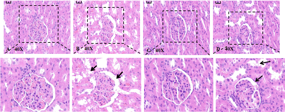
Effect of naringenin on cadmium induced kidney injury in rats (H and E, 400×, scale bar = 50 μm). Kidney sections from control rats showed normal glomerular and tubular histomorphology. In the cadmium exposure group, the glomerulus was shrunk and the tubular structure was damaged. The structural damage of glomeruli and renal tubules was alleviated in Nar and Cd co treated group. (A) control group; (B) Cd-treated group; (C) Nar-treated group; (D) Cd + Nar-treated group.
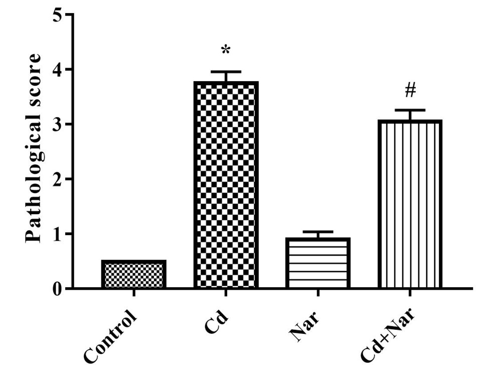
The pathological scores of kidney tissues in different treatment groups were evaluated by members of the experimental group, and three independent experiments were repeated. *, indicates significant difference compared with the corresponding control (*P < 0.05). #, indicates statistically significant difference between the Cd and Cd + Nar groups (#P < 0.05).
References
- Abaza, M.S., Orabi, K.Y., Al-Quattan, E. and Al-Attiyah, R.J., 2015. Growth inhibitory and chemo-sensitization effects of naringenin, a natural flavanone purified from Thymus vulgaris, on human breast and colorectal cancer. Cancer Cell Int., 15: 46. https://doi.org/10.1186/s12935-015-0194-0[CrossRef]
- Abdeen, A., Abou-Zaid, O.A., Abdel-Maksoud, H.A., Aboubakr, M., Abdelkader, A., Abdelnaby, A., Abo-Ahmed, A.I., El-Mleeh, A., Mostafa, O., Abdel-Daim, M. and Aleya, L., 2019. Cadmium overload modulates piroxicam-regulated oxidative damage and apoptotic pathways. Environ. Sci. Pollut. Res. Int., 26: 25167-25177. https://doi.org/10.1007/s11356-019-05783-x[CrossRef]
- Ahmed, Z.A., Abtar, A.N., Othman, H.H. and Aziz, T.A., 2019. Effects of quercetin, sitagliptin alone or in combination in testicular toxicity induced by doxorubicin in rats. Drug Des. Dev. Ther., 13: 3321-3329. https://doi.org/10.2147/DDDT.S222127[CrossRef]
- Almeer, R.S., Soliman, D., Kassab, R.B., AlBasher, G.I., Alarifi, S., Alkahtani, S., Ali, D., Metwally, D. and Abdel Moneim, A.E., 2018. Royal jelly abrogates cadmium-induced oxidative challenge in mouse testes: Involvement of the Nrf2 pathway. Int. J. mol. Sci., 19: 3979. https://doi.org/10.3390/ijms19123979[CrossRef]
- Arab-Nozari, M., Mohammadi, E., Shokrzadeh, M., Ahangar, N., Amiri, F.T. and Shaki, F., 2020. Co-exposure to non-toxic levels of cadmium and fluoride induces hepatotoxicity in rats via triggering mitochondrial oxidative damage, apoptosis, and NF-kB pathways. Environ. Sci. Pollut. Res. Int., 27: 24048-24058. https://doi.org/10.1007/s11356-020-08791-4[CrossRef]
- Badr, G.M., Elsawy, H., Sedky, A., Eid, R., Ali, A., Abdallah, B.M., Alzahrani, A.M. and Abdel-Moneim, A.M., 2019. Protective effects of quercetin supplementation against short-term toxicity of cadmium-induced hematological impairment, hypothyroidism, and testicular disturbances in albino rats. Environ. Sci. Pollut. Res. Int., 26: 8202-8211. https://doi.org/10.1007/s11356-019-04276-1[CrossRef]
- Brzóska, M., Kamiński, M., Dziki, M. and Moniuszko-Jakoniuk, J., 2004. Changes in the structure and function of the kidney of rats chronically exposed to cadmium. II. Histoenzymatic studies. Arch. Toxicol., 78: 226-231. https://doi.org/10.1007/s00204-003-0500-9[CrossRef]
- Canales, B.K., Reyes, L., Reinhard, M.K., Khan, S.R., Goncalves, C.G. and Meguid, M.M., 2012. Renal glomerular and tubular injury after gastric bypass in obese rats. Nutrition, 28: 76-80. https://doi.org/10.1016/j.nut.2011.03.003[CrossRef]
- Chen, H., Chen, J., Shi, X., Li, L. and Xu, S., 2021a. Naringenin protects swine testis cells from bisphenol A-induced apoptosis via Keap1/Nrf2 signaling pathway. BioFactors (Oxford, England). https://doi.org/10.1002/biof.1814[CrossRef]
- Chen, H., Li, P., Shen, Z., Wang, J. and Diao, L., 2021b. Protective effects of selenium yeast against cadmium-induced necroptosis through miR-26a-5p/PTEN/PI3K/AKT signaling pathway in chicken kidney. Ecotoxicol. environ. Saf., 220: 112387. https://doi.org/10.1016/j.ecoenv.2021.112387[CrossRef]
- Cheng, C.H., Ma, H.L., Deng, Y.Q., Feng, J., Jie, Y.K. and Guo, Z.X., 2021. Oxidative stress, cell cycle arrest, DNA damage and apoptosis in the mud crab (Scylla paramamosain) induced by cadmium exposure. Chemosphere, 263: 128277. https://doi.org/10.1016/j.chemosphere.2020.128277[CrossRef]
- Edwards, H., 2003. Environmental health criteria (Book). Int. J. environ. Stud., 1: 67.
- Egger, A.E., Grabmann, G., Gollmann-Tepekoylu, C., Pechriggl, E.J., Artner, C., Turkcan, A., Hartinger, C.G., Fritsch, H., Keppler, B.K., Brenner, E., Grimm, M., Messner, B. and Bernhard, D., 2019. https://doi.org/10.1039/C9MT00178F. Chemical imaging and assessment of cadmium distribution in the human body. Metallomics, 11: 2010-2019.[CrossRef]
- El-Demerdash, F.M., Yousef, M.I., Kedwany, F.S., and Baghdadi, H.H., 2004. Cadmium-induced changes in lipid peroxidation, blood hematology, biochemical parameters and semen quality of male rats: protective role of vitamin E and beta-carotene. Fd. Chem. Toxicol., 42: 1563-1571. https://doi.org/10.1016/j.fct.2004.05.001[CrossRef]
- El-Desoky, A.H., Abdel-Rahman, R.F., Ahmed, O.K., El-Beltagi, H.S. and Hattori, M., 2018. Anti-inflammatory and antioxidant activities of naringin isolated from Carissa carandas L: In vitro and in vivo evidence. Phytomedicine, 42: 126-134. https://doi.org/10.1016/j.phymed.2018.03.051[CrossRef]
- El-Nekeety, A.A., El-Kady, A.A., Soliman, M.S., Hassan, N.S. and Abdel-Wahhab, M.A., 2009. Protective effect of Aquilegia vulgaris (L.) against lead acetate-induced oxidative stress in rats. Fd. Chem. Toxicol., 47: 2209-2215. https://doi.org/10.1016/j.fct.2009.06.019[CrossRef]
- Fujiwara, Y., Lee, J-Y., Banno, H., Imai, S., Tokumoto, M., Hasegawa, T., Seko, Y., Nagase, H. and Satoh, M., 2020. Cadmium induces iron deficiency anemia through the suppression of iron transport in the duodenum. Toxicol. Lett., 332: 130-139. https://doi.org/10.1016/j.toxlet.2020.07.005[CrossRef]
- Fukumoto, M., Kujiraoka, T., Hara, M., Shibasaki, T., Hosoya, T. and Yoshida, M., 2001. Effect of cadmium on gap junctional intercellular communication in primary cultures of rat renal proximal tubular cells. Life Sci., 3: 247-254. https://doi.org/10.1016/S0024-3205(01)01063-3[CrossRef]
- Genchi, G., Sinicropi, M.S., Lauria, G., Carocci, A. and Catalano, A., 2020. The effects of cadmium toxicity. Int. J. environ. Res. Publ. hlth., 17: 100372. https://doi.org/10.3390/ijerph17113782[CrossRef]
- Geng, H.X., and Wang, L., 2019. Cadmium: Toxic effects on placental and embryonic development. Environ. Toxicol. Pharmacol., 67: 102-107. https://doi.org/10.1016/j.etap.2019.02.006[CrossRef]
- Gong, Z., Zhao, Y., Wang, Z., Fan, R., Liu, Z. and Wang, L., 2021. Epigenetic regulator BRD4 is involved in cadmium-induced acute kidney injury via contributing to lysosomal dysfunction, autophagy blockade and oxidative stress. J. Hazard. Mater., 423: 127110. https://doi.org/10.1016/j.jhazmat.2021.127110[CrossRef]
- He, L., He, T., Farrar, S., Ji, L., Liu, T. and Ma, X., 2017. Antioxidants maintain cellular redox homeostasis by elimination of reactive oxygen species. Cell. Physiol. Biochem. Int. J. exp. Cell. Physiol., Biochem. Pharmacol., 44: 532-553. https://doi.org/10.1159/000485089[CrossRef]
- Honohan, T., Hale, R., Brown, J. and Wingard, R., 1976. Synthesis and metabolic fate of hesperetin-3-14C. J. Agric. Fd. Chem., 24: 906-911. https://doi.org/10.1021/jf60207a031[CrossRef]
- Jihenel, H., Imed, M., Fatima, H. and Abdelhamid, K., 2008. Protective effects of selenium (Se) and zinc (Zn) on cadmium (Cd) toxicity in the liver and kidney of the rat: Histology and Cd accumulation. Fd. Chem. Toxicol., 46: 3522-3527. https://doi.org/10.1016/j.fct.2008.08.037[CrossRef]
- Kamiyama, T., Miyakawa, H., Li, J.P., Akiba, T., and Sato, C., 1995. Effects of one-year cadmium exposure on livers and kidneys and their relation to glutathione levels. Res. Commun. mol. Pathol. Pharmacol., 88: 177.
- Kanaze, F.I., Bounartzi, M.I., Georgarakis, M. and Niopas, I., 2007. Pharmacokinetics of the citrus flavanone aglycones hesperetin and naringenin after single oral administration in human subjects. Eur. J. clin. Nutr., 61: 472-477. https://doi.org/10.1038/sj.ejcn.1602543[CrossRef]
- Khan, M.S., Qais, F.A., Rehman, M.T., Ismail, M.H., Alokail, M.S., Altwaijry, N., Alafaleq, N.O., AlAjmi, M.F., Salem, N. and Alqhatani, R., 2020. Mechanistic inhibition of non-enzymatic glycation and aldose reductase activity by naringenin: Binding, enzyme kinetics and molecular docking analysis. Int. J. Biol. Macromol., 159: 87-97. https://doi.org/10.1016/j.ijbiomac.2020.04.226[CrossRef]
- Koyu, A., Gokcimen, A., Ozguner, F., Bayram, D. and Kocak, A., 2006. Evaluation of the effects of cadmium on rat liver. Mol. Cell. Biochem., 284: 81-85. https://doi.org/10.1007/s11010-005-9017-2[CrossRef]
- Lemarie, A., Lagadic-Gossmann, D., Morzadec, C., Allain, N., Fardel, O. and Vernhet, L., 2004. Cadmium induces caspase-independent apoptosis in liver Hep3B cells: Role for calcium in signaling oxidative stress-related impairment of mitochondria and relocation of endonuclease G and apoptosis-inducing factor. Free Radic. Biol. Med., 36: 1517-1531. https://doi.org/10.1016/j.freeradbiomed.2004.03.020[CrossRef]
- Liu, F., Wang, X.Y., Zhou, X.P., Liu, Z.P., Song, X.B., Wang, Z.Y. and Wang, L., 2017. Cadmium disrupts autophagic flux by inhibiting cytosolic Ca(2+)-dependent autophagosome-lysosome fusion in primary rat proximal tubular cells. Toxicology, 383: 13-23. https://doi.org/10.1016/j.tox.2017.03.016[CrossRef]
- Mir, I.A. and Tiku, A.B., 2015. Chemopreventive and therapeutic potential of Naringenin, a flavanone present in citrus fruits. Nutr. Cancer (Nutr. Cancer), 1: 27-42.
- Mouro, V.G.S., Ladeira, L.C.M., Lozi, A.A., de Medeiros, T.S., Silva, M.R., de Oliveira, E.L., de Melo, F. and da Matta, S.L.P., 2021. Different routes of administration lead to different oxidative damage and tissue disorganization levels on the subacute cadmium toxicity in the liver. Biol. Trace Elem. Res. Epub., Ahead of Print 2021/01/06. https://doi.org/10.1007/s12011-020-02570-5[CrossRef]
- Nemmiche, S., Chabane-Sari, D. and Guiraud, P., 2007. Role of α-tocopherol in cadmium-induced oxidative stress in Wistar rat’s blood, liver and brain. Chem. Biol. Interact., 170: 221-230. https://doi.org/10.1016/j.cbi.2007.08.004[CrossRef]
- Odewumi, C.O., Badisa, V.L., Le, U.T., Latinwo, L.M., Ikediobi, C.O., Badisa, R.B. and Darling-Reed, S.F., 2011. Protective effects of N-acetylcysteine against cadmium-induced damage in cultured rat normal liver cells. Int. J. mol. Med., 27: 243-248. https://doi.org/10.3892/ijmm.2010.564[CrossRef]
- Possik, E. and Pause, A., 2015. Measuring oxidative stress resistance of Caenorhabditis elegans in 96-well microtiter plates. J. Visual. Exp., 99: e52746. https://doi.org/10.3791/52746[CrossRef]
- Sies, H., 2015. Oxidative stress: A concept in redox biology and medicine. Redox Biol., 4: 180-183. https://doi.org/10.1016/j.redox.2015.01.002[CrossRef]
- Sinha, M., Manna, P. and Sil, P.C., 2009. Induction of necrosis in cadmium-induced hepatic oxidative stress and its prevention by the prophylactic properties of taurine. J. Trace Elem. Med. Biol., 23: 300-313. https://doi.org/10.1016/j.jtemb.2009.03.010[CrossRef]
- Sinha, N. and Dabla, P., 2015. Oxidative stress and antioxidants in hypertension. A current review. Curr. Hypert. Rev., 11: 132-142. https://doi.org/10.2174/1573402111666150529130922[CrossRef]
- Skipper, A., Sims, J., Yedjou, C. and Tchounwou, P., 2016. Cadmium chloride induces DNA damage and apoptosis of human liver carcinoma cells via oxidative stress. Int. J. environ. Res. Publ. Hlth., 13: 88. https://doi.org/10.3390/ijerph13010088[CrossRef]
- Testai, L., Da Pozzo, E., Piano, I., Pistelli, L., Gargini, C., Breschi, M.C., Braca, A., Martini, C., Martelli, A. and Calderone, V., 2017. The citrus flavanone naringenin produces cardioprotective effects in hearts from 1 year old rat, through activation of mitoBK channels. Front. Pharmacol., 8: 71. https://doi.org/10.3389/fphar.2017.00071[CrossRef]
- Tsikas, D., 2017. Assessment of lipid peroxidation by measuring malondialdehyde (MDA) and relatives in biological samples: Analytical and biological challenges. Anal. Biochem., 524: 13-30. https://doi.org/10.1016/j.ab.2016.10.021[CrossRef]
- Vicente-Sanchez, C., Egido, J., Sanchez-Gonzalez, P.D., Perez-Barriocanal, F., Lopez-Novoa, J.M. and Morales, A.I., 2008. Effect of the flavonoid quercetin on cadmium-induced hepatotoxicity. Fd. Chem. Toxicol., 46: 2279-2287. https://doi.org/10.1016/j.fct.2008.03.009[CrossRef]
- Wang, J., Zhu, H., Wang, K., Yang, Z. and Liu, Z., 2020a. Protective effect of quercetin on rat testes against cadmium toxicity by alleviating oxidative stress and autophagy. Environ. Sci. Pollut. Res. Int., 27: 25278-25286. https://doi.org/10.1007/s11356-020-08947-2[CrossRef]
- Wang, L., Fan, R., Yang, D., Zhang, D. and Wang, L., 2019. Puerarin reverses cadmium-induced lysosomal dysfunction in primary rat proximal tubular cells via inhibiting Nrf2 pathway. Biochem. Pharmacol., 162: 132-141. https://doi.org/10.1016/j.bcp.2018.10.016[CrossRef]
- Wang, X., Wang, T., Pan, T., Huang, M., Ren, W., Xu, G., Amin, H.K., Kassab, R.B. and Abdel Moneim, A.E., 2020b. Senna alexandrina extract supplementation reverses hepatic oxidative, inflammatory, and apoptotic effects of cadmium chloride administration in rats. Environ. Sci. Pollut. Res. Int., 27: 5981-5992. https://doi.org/10.1007/s11356-019-07117-3[CrossRef]
- Wang, X.Y., Yang, H., Wang, M.G., Yang, D.B., Wang, Z.Y. and Wang, L., 2017. Trehalose protects against cadmium-induced cytotoxicity in primary rat proximal tubular cells via inhibiting apoptosis and restoring autophagic flux. Cell Death Dis., 8: e3099. https://doi.org/10.1038/cddis.2017.475[CrossRef]
- Wang, Y., Wu, Y., Luo, K., Liu, Y., Zhou, M., Yan, S., Shi, H. and Cai, Y., 2013. The protective effects of selenium on cadmium-induced oxidative stress and apoptosis via mitochondria pathway in mice kidney. Fd. Chem. Toxicol., 58: 61-67. https://doi.org/10.1016/j.fct.2013.04.013[CrossRef]
- Wu, C., Zhang, Y., Chai, L. and Wang, H., 2017. Histological changes, lipid metabolism and oxidative stress in the liver of Bufo gargarizans exposed to cadmium concentrations. Chemosphere, 179: 337-346. https://doi.org/10.1016/j.chemosphere.2017.03.131[CrossRef]
- Xu, D., Han, H., He, Y., Lee, H., Wu, D., Liu, F., Liu, X., Liu, Y., Lu, Y. and Ji, C., 2018. A hepatocyte-mimicking antidote for alcohol intoxication. Adv. Mater. (Deerfield Beach, Fla.), 30: e1707443. https://doi.org/10.1002/adma.201707443[CrossRef]
- Xu, S., Xiaojing, L., Xinyue, S., Wei, C., Honggui, L. and Shiwen, X., 2021. Pig lung fibrosis is active in the subacute CdCl exposure model and exerts cumulative toxicity through the M1/M2 imbalance. Ecotoxicol. environ. Saf., 225: 112757. https://doi.org/10.1016/j.ecoenv.2021.112757[CrossRef]
- Zhang, D., Li, Y., Zhang, T., Liu, J., Jahejo, A.R., Yang, L., Chen, P., Ning, G., Huo, N., Ma, H., Yan, F. and Tian, W., 2018. Protective effects of zinc and N-acetyl-L-cysteine supplementation against cadmium induced erythrocyte cytotoxicity in Arbor Acres broiler chickens (Gallus gallus domesticus). Ecotoxicol. environ. Saf., 163: 331-339. https://doi.org/10.1016/j.ecoenv.2018.07.069[CrossRef]
- Zhang, Q., Yang, W., Liu, J., Liu, H., Lv, Z., Zhang, C., Chen, D. and Jiao, Z., 2020. Identification of six flavonoids as novel cellular antioxidants and their structure-activity relationship. Oxid. Med. Cell. Longev., 2020: 4150897. https://doi.org/10.1155/2020/4150897[CrossRef]
Services
Article usage statistics
Loading…