
Approximately 350 million patients of hepatitis B and 170 million patients of Hepatitis C are present worldwide according to WHO. Many risk factors are involved in the transmission of theses deadly viral infections but blood transfusion in Beta thalassemic patients is working with two faces, one as remedy and the other is key risk factor in the spread of silent killers. Thalassemia patients registered in Combine Military Hospital (CMH) Rawalakot and Sheikh Khalifa Bin Zayed Al-Nahyan Hospital, Muzaffarabad Azad Jammu and Kashmir Pakistan were studied for the viral hepatitis B and C prevalence. A total of 303 (including 164 males and 139 females) individuals, aged between 1 and 12 years were studied. All the understudy participants were interviewed through questionnaire method. After taking written consent from each participant or guardian, 5 ml of blood was collected from each participant and brought to the working laboratory for HBV and HCV screening through ICT kit method. All ICT positive samples were further confirmed through ELISA. Individuals 25(8.2%) were found positive for both hepatitis B surface Antigen (HBsAg) and Anti hepatitis C antibody (Anti-HCV antibody) after initial screening with no coinfection of both diseases. Out of 25 total infected individuals, 05(1.6%) were found HBsAg positive and 20(6.6%) were found anti-HCV positive. All the ICT positive individuals were further confirmed by quantitative Enzyme Linked Immunosorbent Assay (ELISA) and 23(7.6%) individuals were confirmed for both hepatitis B and C including 05(1.6%) HBsAg positive as well as 18(5.9%) anti-HCV antibody positive individuals. We can conclude that 8.2% prevalence of hepatitis B and C among thalassemic patients is an alarming health concern which directly indicates to pay attention for ensuring 100% safe blood transfusion.
Short Communication · Volume 52, Issue 2, 14 February 2020 · Open Access
Prevalence of Hepatitis B and C and Assessment of Responsible Risk Factors among the Vulnerable β-Thalassemic Patients of Azad Kashmir, Pakistan
· · · · · · · · · · · · ·
Affiliations & Notes ⌄
- 1Department of Zoology, University of Azad Jammu and Kashmir, Muzaffarabad 2Department of Zoology, Government College University, Lahore 3Health Care Diagnostics and Research Centre, Allama Iqbal Town, Lahore 4Department of Biotechnology, University of Azad Jammu and Kashmir, Muzaffarabad 5Department of Chemistry, University of Azad Jammu and Kashmir, Muzaffarabad , * Corresponding author: shuakatali134@yahoo.com
Article Info ⌄
- Journal:
- Pakistan Journal of Zoology
- Published:
- 14 February 2020

From the article
- Out of 25 total infected individuals, 05(1.6%) were found HBsAg positive (3 male and 2 female) and 20(6.6%) were found anti-HCV positive (3 male and 17 female).
- In case of Anti-HCV prevalence, the females were found more prevalent with 5.6% prevalence as compare to 1% of HBsAg in male population (Fig. 1A).
- Moreover, the age group 7-12 years was found with higher prevalence of both hepatitis B (4.4%) and hepatitis C (14.4%) as compare to age groups of 1-6 years.
- The high (8.2%) prevalence of hepatitis B and C in a vulnerable population such as thalassemic patients is an alarming health concern that directly indicates that attention should be paid to ensure pre-screened blood transfusion.
Selected from the article's own text.
Abstract
Materials and Methods
The study was approved by the Directorate of Advance Studies and Research (DASR), which have authority to approve research and deal ethical issues in University of Azad Jammu and Kashmir, Muzaffarabad.
Thalassemia patients registered in Combine Military Hospital (CMH) Rawalakot and Sheikh Khalifa Bin Zayed Al-Nahyan Hospital, Muzaffarabad, Azad Jammu and Kashmir Pakistan were studied for the viral hepatitis B and C prevalence. A total of 303 (including 164 males and 139 females) individuals, aged between 1 and 12 years were studied. All the understudy participants were interviewed through questionnaire method. A filled and signed consent was collected from each participant or guardian. The consent form based upon collection of general information (age, sex and locality) and for risk factor assessment.
Blood samples (5ml) were collected from each participant and injected in the gel tube (Biotube Gel Clot Activator). Serum was separated by centrifugation. Each serum sample was labeled and stored at -20°C. The hepatitis B surface antigen (HBsAg) and hepatitis C virus antibody (anti-HCV) were detected by using ICT kit method through ICT devices (SD BIOLINE HBsAg and Anti-HCV kits). Moreover, all the initially HBsAg and anti-HCV positive individuals were further quantified for viral antigens and antibodies respectively through quantitative ELISA with the cut-off value (Non-Reactive < 0.10, Equivocal 0.10 - 0.50 and Reactive > 0.50) provided with diagnostic kit for HBsAg and anti-HCV by the Linear Chemicals S.L., Spain.
Data was analyzed through GraphPad Prism (Version 5.0) to calculate the mean age (with 95% Confidence Interval), standard deviation and Chi-square test was used to detect the significance level of different risk factors involved in the spread of hepatitis B and C in thalassemia patients.
Results
For the present study total, 303 β-thalassemia patients were screened through immune-chromatography devices (ICT) including 164 (52.1%) male and 139 (45.9%) female individuals. 25 (8.2%) individuals were found positive for both hepatitis B surface antigen (HBsAg) and Anti hepatitis C antibody (anti-HCV antibody) after initial screening with no coinfection of both diseases. Out of 25 total infected individuals, 05(1.6%) were found HBsAg positive (3 male and 2 female) and 20(6.6%) were found anti-HCV positive (3 male and 17 female).
All the ICT positive individuals were further confirmed by quantitative enzyme linked immunosorbent assay (ELISA) and we found 23(7.6%) individuals confirmed for both hepatitis B and C including 05 (1.6%) HBsAg positive (3 male and 3 female) as well as 18 (5.9%) anti-HCV antibody positive (3 male and 15 female) individuals.
The male population was found with 1% HBsAg prevalence as compare to 0.6% prevalence of HBsAg in female population. In case of Anti-HCV prevalence, the females were found more prevalent with 5.6% prevalence as compare to 1% of HBsAg in male population (Fig. 1A).
For determining prevalence of hepatitis B and C among β-thalassemia patients, the age-wise data was analyzed by using GraphPad Prism (Version 5.0) software and the population was divided in to two age groups, 1-6 years and 7-12 years, respectively. Standard deviation for the total population was found 3.13 and mean age with 95% Confidence Interval was found 4.80±0.35 for the total population. Moreover, the age group 7-12 years was found with higher prevalence of both hepatitis B (4.4%) and hepatitis C (14.4%) as compare to age groups of 1-6 years. This is due to the increase rate of multi transfusion with the increase of age in the case of thalassemia because the blood transfusion is the key risk factor involve in the spread of hepatitis B and C world-wide (Table I; Fig. 1B).
For the present study risk factors were assessed through a questions based proforma from each participant or from the guardian on the behalf of participants. Data was analyzed through two sided chi-square test (at p 0.05) by using GraphPad Prism (version 5.0). We found significant relationship of hepatitis B and C spread with family hepatitis history and dental treatment. Treatment from a local Practician was found not significant with p value of 0.0625 regarding to spread of hepatitis B and C in thalassemia patients. Table II shows all the details related to risk factors assessment in the thalassemia patients other than blood transfusion.
Table I.- Age-wise prevalence of hepatitis B and C among β-thalassemia patients.
| Age | No. of Ind. | SD | Mean age with 95% CI | HBsAg +ve | Anti-HCV +ve |
| 01-06 | 213 | 1.89 | 3.11±0.25 | 01 (0.5%) | 07 (3.3%) |
| 07-12 | 90 | 1.37 | 8.80±0.29 | 04 (4.4%) | 13 (14.4%) |
| Total Ind. | 303 | 3.13 | 4.80±0.35 | 05 (1.6%) | 20 (6.6%) |
Ind., individuals, SD, standard deviation.
Discussion
In the present study, the sero-prevalence of HBV markers (HBsAg and anti-HBC) was 22.8% in beta-thalassemia patients living in Azad Kashmir which indicated a high exposure rate. While it was observed that the prevalence of HBsAg was 1.6%. No anti-HBc detection procedure was used to study the selected population.
In a similar study conducted by Jang et al. (2017) 140 patients were tested, out of which only one patient was found positive for HBsAg that accounts for a prevalence of 6.4% and anti-HCV was found to be 45.7%. While in the present study 303 β-thalassemia patients were screened in total through immune-chromatography devices (ICT), in which 164 (52.1%) were male and 139 (45.9%) were female individuals. Among 303 beta thalassemia patients 25(8.2%) individuals were found to be positive for both hepatitis B surface antigen (HBsAg) and anti-hepatitis C antibody (anti-HCV antibody) that were believed to be uninfected after initial screening. Out of 25 infected individuals, 05 (1.6%) were confirmed as HBsAg positive (3 males and 2 females) and 20 (6.6%) were found anti-HCV positive (3 males and 17 females). While the average age of patient was 1 to 12 years. A higher prevalence of both hepatitis B (4.4%) and hepatitis C (14.4%) infections was observed in the age group of 7-12 years. Another study was conducted in Pakistan in which total 95 patients observed were found β-thalassemia major (96%) and β-thalassemia intermedia (4%). Among these, 47 (49%) were found positive for anti-HCV antibodies and three for hepatitis B surface antigen .
Table II.- Risk factors assessment regarding to spread of hepatitis B and C.
| Risk factors | Response options | Overall responses | Responses of diseased | Responses of non-diseased | Chi-square value P-value (two sided) | Statistical significance (at P-0.05) |
| Hepatitis history in the family? | Yes | 2 | 1 | 1 | 0.0313 | Yes |
| No | 301 | 24 | 277 | |||
| Dental treatment? | Yes | 1 | 1 | 0 | 0.0008 | Yes |
| No | 302 | 24 | 278 | |||
| Treatment from a local practician? | Yes | 106 | 13 | 93 | 0.0625 | No |
| No | 197 | 12 | 185 |
It is revealed from other studies that the chances of infection significantly rise with the increasing age as the demand of blood transfusions increases with the passage of time . Similar findings we observed in our study where a higher prevalence of HVC (14.4%) and HBV (4.4%) infections were observed in 7-12-year age group due to the increase rate of multiple transfusions to meet the demand in case of thalassemia.
According to a study, 206 individuals were tested for anti-HCV through ELISA. 28.15% of the 206 patients were found anti-HCV positive . While in the present study the total population was 303, the standard deviation for the total population was found 3.13 and mean age with 95% Confidence Interval was found 4.80±0.35. Moreover, after ELISA, 5.9% individuals were confirmed for hepatitis C infection which is too low as compared to 28.1% prevalence of hepatitis C found in the study of Boroujerdnia et al. (2009).
Conclusions and recommendations
The high (8.2%) prevalence of hepatitis B and C in a vulnerable population such as thalassemic patients is an alarming health concern that directly indicates that attention should be paid to ensure pre-screened blood transfusion.
Acknowledgements
We are grateful to the team of Hepatitis Society, University of Azad Jammu and Kashmir, Muzaffarabad and Health Care Diagnostics and Research Centre (HCD&RC), Allama Iqbal Town, Lahore, Pakistan for their support for the present study.
Statement of conflict of interest
The authors declare no conflict of interest.
Figures
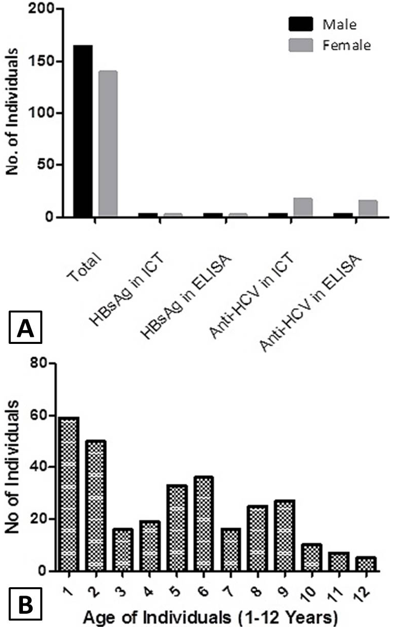
Comprehensive prevalence (A) and age wise frequency distribution (B) of hepatitis B and C in thalassemia patients of Azad Kashmir, Pakistan.
References
- Ahmadi-Ghezeldasht, S., Badiei, Z., Sima, H.R., Hedayati-Moghaddam, M.R., Habibi, M., Khamooshi, M. and Azimi, A., 2018. Middle East J. Dig. Dis., 10: 35-39. https://doi.org/10.15171/mejdd.2017.88[CrossRef]
- Ahmed Kiani, R., Anwar, M., Waheed, U., Asad, M.J., Abbasi, S. and Abbas Zaheer, H., 2016. J. Blood Transf., 2016: 8135649. https://doi.org/10.1155/2016/8135649[CrossRef]
- Ali, M.A., Arif, M.M., Arif, A. and Fatima, T., 2016. Annls Punjab med. Coll., 10: 20-25.
- Ataei, B., Hashemipour, M., Kassaian, N., Hassannejad, R., Nokhodian, Z. and Adibi, P., 2012. Int. J. Prevent. Med., 3: S118-123.
- Boroujerdnia, M.G., Zadegan, M.A.A., Zandian, K.M. and Rodan, M.H., 2009. Pak. J. med. Sci., 25: 113-117.
- Din, G., Malik, S., Ali, I., Ahmed, S. and Dasti, J.I., 2014. Asian Pac. J. Trop. Med., 7: 127-133.
- Edgren, G., Hjalgrim, H., Rostgaard, K., Dahl, V., Titlestad, K., Erikstrup, C., Wikman, A., Norda, R. and Majeed, A., 2018. J. Intern. Med., 284: 92-103. https://doi.org/10.1111/joim.12762[CrossRef]
- Farooq, A., Waheed, U., Zaheer, H. A., Rauf, A., Arshad, A. and Arshad, M., 2018. Pakistan J. Zool., 50: 1191-1194. https://doi.org/10.17582/journal.pjz/2018.50.3.sc3[Full text][CrossRef]
- Hussain, H., Iqbal, R., Khan, M.H., Iftikhar, B., Aziz, S. and Burki, F.K., 2008. Gomal J. med. Sci., 6: 87-90.
- Jafroodi, M., Davoudi-Kiakalayeh, A., Mohtasham-Amiri, Z., Pourfathollah, A.A. and Haghbin, A., 2015. Int. J. Prevent. Med., 6: 89. https://doi.org/10.4103/2008-7802.164832[CrossRef]
- Jang, T.Y., Lin, P.C., Huang, C.I., Liao, Y.M., Yeh, M.L., Zeng, Y.S., Liang, P.C., Hsu, W.Y., Tsai, S.P., Lin, Z.Y. and Chen, S.C., 2017. PLoS One, 12: e0178883. https://doi.org/10.1371/journal.pone.0178883[CrossRef]
- Irshad, S., Muhammad, A., Muazzam, A., Anmol, F.S. and Shahzad, R., 2019. Pakistan J. Zool., 51: 295-300. http://dx.doi.org/10.17582/journal.pjz/2019.51.1.295.300[Full text][CrossRef]
- Kadhim, K.A., Baldawi, K.H. and Lami, F.H., 2017. Hemoglobin, 41: 164-168.
- Manisha, S., Sanjeev, K., Seema, N., Dilip, C. and Rashmi, D., 2015. Indian J. Hematol. Blood Transf., 31: 367-373. https://doi.org/10.1007/s12288-014-0462-5[CrossRef]
- Mehta, R., Kabrawala, M., Nandwani, S., Desai, P., Bhayani, V., Patel, S. and Parekh, V., 2017. J. clin. exp. Hepatol., 8: 3-6. https://doi.org/10.1016/j.jceh.2017.06.002[CrossRef]
- Moatter, T., Adil, S., Haroon, S., Azeemuddin, S., Hassan, F. and Khurshid, M., 1999. Indian J. Pathol. Microbiol., 42: 475-482.
- Sajjad, S.F., Ahmad, W., Jaffery, S.S.H., Asif, M. and Alam, S.E., 2017. J. Pak. med. Assoc., 67: 926-928.
- Tafesse, T.B., Gebru, A.A., Gobalee, S., Belay, G.D., Belew, M.T., Ataro, D., Ebrahim, B.A., Shebeshi, G.M. and Yimam, Y., 2017. Human Antibodies, 25: 39-55. https://doi.org/10.3233/HAB-160304[CrossRef]
- Twagirumugabe, T., Swaibu, G., Bergström, T., Walker, T.D., Gahutu, J.B. and Norder, H., 2017. Transfusion, 57: 2420-2432. https://doi.org/10.1111/trf.14204[CrossRef]
- WHO, 2000. Hepatitis C: Fact sheet. World Health Organization, Geneva. Available at: https://www.who.int/news-room/fact-sheets/detail/hepatitis-c (Accessed on March 11, 2019).
Cited by 3 articles
In journals published by ResearchersLinks.
- Knowledge, Attitudes, and Perceptions Towards Beta-Thalassemia among Residents of Azad Kashmir, PakistanPunjab University Journal of Zoology · 2023
- Methanolic Extract of Neem Plant Inhibits NS3 and NS5A Nonstructural Proteins of HCV 3A GenotypePakistan Journal of Zoology · 2023
- Bacterial Expression and Purification of Antigenic Recombinant Protein Encoded by Hepatitis Delta Virus Antigen of Pakistani IsolatePakistan Journal of Zoology · 2022
Services
Article usage statistics
Loading…