
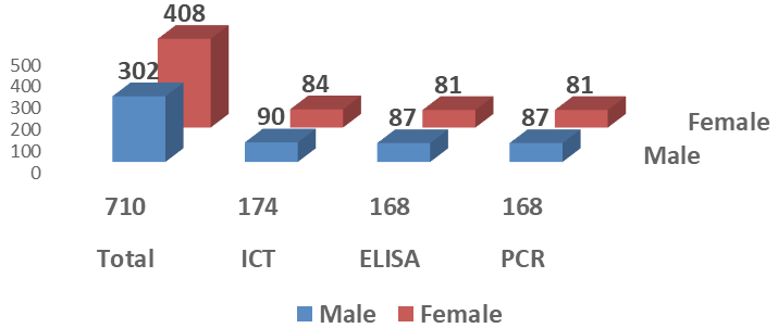
This is a cross sectional study to determine the prevalence of hepatitis B and C infection among the general population of union council Sena Daman district Jhelum Valley, Azad Kashmir and to assess the risk factors for their transmission. From approximately 16000 total individuals of population of Union Council Sena Daman 710 (4.4%) individuals were randomly screened for hepatitis B and C prevalence using Immuno-Chromatography Kits, ELISA and further confirmed by PCR. Among the tested individuals 174 (24.5%) including 90(51.7%) male and 84(48.3%) were found positive for both HBsAg and anti-HCV including 4.6% HBsAg positive, 19.7% anti-HCV positive. After ELISA 168 individuals were found positive which were also found positive after PCR. Out of these 29 were HBV positive and 138 were HCV positive. One individual was confirmed after PCR for co-infection of HBV and HCV. Out of 168 total individuals, 65(38.7%) individuals were found with an abnormal range of LFTs. Visit to local clinic, major or minor surgery and tattooing or piercing of nose, ears etc., were assessed significant risk factors in the spread of hepatitis B and C. Awareness of the general population was carried out through organizing awareness lectures and distribution of awareness material.
Short Communication · Volume 55, Issue 1, 13 November 2022 · Open Access
Prevalence of Hepatitis B and C Along with Associated Risk Factors in General Population of Sena Damin District Jhelum Valley Azad Jammu and Kashmir
· · · · · · · ·
Affiliations & Notes ⌄
- 1Department of Public Health, University of Azad Jammu and Kashmir, Muzaffarabad 2Department of Zoology, University of Azad Jammu and Kashmir, Muzaffarabad 3Healthcare Diagnostics and Research Centre (HD and RC) Muzaffarabad, Azad Kashmir, Pakistan 4Department of Biotechnology, University of Azad Jammu and Kashmir, Muzaffarabad, Pakistan * Corresponding author: ayaz.biotech.uajk@gmail.com
Article Info ⌄
- Journal:
- Pakistan Journal of Zoology
- Published:
- 13 November 2022

From the article
- From among hepatitis positive individuals 33(4.6%) were positive for HBsAg, 140(19.7%) were found positive for anti-HCV and 01(0.14%) had co-infection of HBsAg and anti-HCV.
- All of 168(23.6%) ELISA positive individuals were found positive after PCR for viral DNA (HBV) or RNA (HCV).
- Out of 29 HBV positive individuals 17(58.6%) were male and 12(41.2%) were female.
- One individual was confirmed after PCR for co-infection of both HBV and HCV and was a female (Fig. 1).
Selected from the article's own text.
Abstract
Materials and methods
A total of 710 from approximately 16000 total individuals of population of Union Council Sena Daman including 302(42.5%) males and 408(57.5%) females actively participated in the present study. After consent, 5ml blood was collected, serum was separated which was used for estimition of hepatitis B surface antigen (HBsAg) and hepatitis C virus antibody (anti-HCV) by ICT kit method using ICT devices (SD BIOLINE HBsAg and Anti-HCV kits). All HBsAg and Anti-HCV ICT positive samples were further quantified for viral antibodies and antigens using quantitative ELISA method.
From serum samples of HBsAg positive persons, viral DNA was isolated using the modified DNAzol method . The amplification of HBV DNA was performed by using Favor-PrepTM Viral DNA purification kit (Favorgen®, Biotech Corp, USA) and Thermo-Start Taq DNA Polymerase Kit (Thermo Scientific®, USA) with quantification range between 100IU/ml to 150,000,000IU/ml. PCR products were exmained by gel electrophoresis.
Viral RNA was also isolated from the serum of anti-HCV antibody positive individuals through TRIzol RNA Isolation Protocol followed by Chomczynski and Mackey (1995). The real-time amplifications of 5` UTR of HCV genomes were performed using Favor-PrepTM Viral purification kit (Favorgen®, Biotech Corp, USA) and Verso 1-Step RT-PCR Hot Start Kit (Thermo Scientific®, USA) using a set of specific primers already reported by Casanova et al. (2014). The quantification range of the assay was 100IU/ml to 350,000,000IU/ml. HCV RNA of genotypes 1a, 1b, 2a, 2b, 2c, 3a, 3b, 4, 5a and 6 were amplified with similar efficiency. PCR amplification was examined by gel electrophoresis.
Data was statistically analyzed to find out the mean age with 95% confidence interval. The significant relation between risk factors and spread of hepatitis B and C was analyzed using GraphPad Prism (Version 7.04) software.
Results and discussion
Out of total of 710 individuals, 174 (24.5%) including 90(51.7%) male and 84(48.3%) female were found positive for both HBsAg and anti-HCV individuals. From among hepatitis positive individuals 33(4.6%) were positive for HBsAg, 140(19.7%) were found positive for anti-HCV and 01(0.14%) had co-infection of HBsAg and anti-HCV. After ELISA out of 174 total positive individuals 168(23.65) individuals were found positive 29(4.4%) were HBsAg positive, 138(19.4%) anti-HCV, positive and one (0.14%) was co-infected both with HBV and HCV. All of 168(23.6%) ELISA positive individuals were found positive after PCR for viral DNA (HBV) or RNA (HCV). Out of 29 HBV positive individuals 17(58.6%) were male and 12(41.2%) were female. Similarly, out of 138 HCV positive individuals 70(50.7%) were male and 68(49.3%) were female. One individual was confirmed after PCR for co-infection of both HBV and HCV and was a female (Fig. 1).
Sandesh et al. (2006) conducted a study in Kerala and concluded that the prevalence of hepatitis B and C in the normal population of Calicut in the northern part of Kerala is 0.52% and 0.24%. Compared to other areas of India, the seroprevalence of hepatitis B and C are low in the normal population of Calicut. Among the high-risks drug users had high prevalence of Anti-HCV.
Table I shows age wise prevalence of hepatitis B and C. The age group 35-54 years was found more prevalent for hepatitis as compared to the other two age groups but the prevalence difference between all these age groups is not highly significant (Table I).
Table I. Age wise prevalence of hepatitis B and C.
| Age groups | No. of individuals±SD | Mean age with 95% CI | No. of HBsAg and anti-HCV positive individuals |
| 15-34 | 190±15.4 | 27.9 ± 2.2 | 46(24.2%) |
| 35-54 | 379±5.9 | 44.7 ± 0.6 | 94(24.8%) |
| 55-74 | 141±15.7 | 58.4 ± 2.6 | 34(24.1%) |
| Total | 710±11.5 | 42.9 ± 0.8 | 174(24.5%) |
Behal et al. (2008) reported that the age specific prevalence of hepatitis B and C rose from 1.78% (108/6058) in donors aged 19-25 years to a maximum of 3.03% (96/3161) in donors aged 35-45 years and decreased in older age groups. Yang et al. (2017) reported that the mean age of the 16601 participants (7881 males and 8720 females) who completed the survey for viral hepatitis B screening was 40.28±19.47 years. The positive rate of hepatitis B surface antigen (HBsAg) was 4.04% (95% CI 3.74% to 4.35%), and 3.85% when standardized by age and gender. However, in the present study, the population size was 710, having mean age of 42.9 ± 0.8 with 95% Confidence Interval. Out of 174 total hepatitis positive individuals 33(4.6%) were positive for HBsAg.
Qureshi et al. (2010) reported 2.5% and 4.8%, prevalence of hepatitis B surface antigen (HBsAg) and anti-hepatitis C virus (HCV), respectively, in Pakistan. It also reflected a combined infection rate of 7.6% in the general population. There was significant association of these viral infections with a range of risk factors led by reuse of syringes.
Rauf et al. (2013) conducted a study in Hill Surang Area, Azad Jammu and Kashmir, Pakistan and reported that 7.5% of participants were positive for HCV antibody and only 0.96% were positive for HBV surface antigen. Among the individuals positive for HCV-PCR, the genotype 3a was the most prevalent, whereas genotypes 2a, 1b, 3b and an unidentified strain were also found.
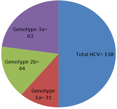
In the present study we found 138 individuals positive for hepatitis C virus. From the positive of these 31(22.5%) individuals had 1a genotype, 44(31.9%) had 2b genotype and 63(45.6%) individuals had 3a genotype. Genotype 3a was more prevalent genotype of HCV compared to other genotypes (Fig. 2).
Out of 168 total PCR positive individuals 103(61.3%) individuals were found with normal LFTs and 65(38.7%) individuals were found with abnormal range of LFTs. The abnormal LFTs level indicates liver damage in 38.7 percent PCR positive individuals (Table I).
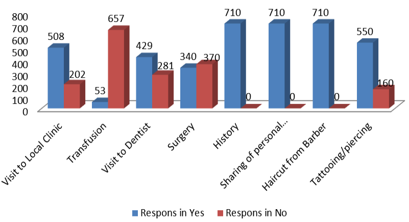
Risk factors were assessed through the use of questionnaires and personal interviews. Visit to local clinic, major or minor surgery and tattooing or piercing of nose, ears etc., were assessed significant risk factors in spread of hepatitis B and C in union council Sena Daman (Fig. 3).
In the present study, we found 4.6% individuals positive for HBsAg. On visiting to local clinic, major or minor surgery and tattooing or piercing of nose, ears etc., were assessed significant risk factors in spread of hepatitis B and C in union council Sena Daman. Olayinka et al. (2016) reported 12.2% prevalence of hepatitis B infection was (confidence interval [CI] = 10.3-14.5). Of the participants, 54.6% had evidence of previous exposure to HBV, 31.7% showed no serologic evidence of infection or vaccination. Only 76 (7.9%) participants showed serologic evidence of immunity to HBV through vaccination. Factors associated with testing positive for HBV infection were dental procedure outside the health facility (odds ratios [OR] = 3.4, 95% CI = 1.52-7.70), local circumcision (OR = 1.73, 95% CI = 1.17-2.57), and uvulectomy (OR = 1.65, 95% CI = 1.06-2.57).
The literacy rate and preventive awareness about diseases of Sena Daman is lower than the Hill Surang area. Both these studies also differ in the sense of hepatitis B and C prevalence because we found 4.6% of HBsAg prevalence and 19.7% prevalence of Anti-HCV antibody, which is far higher than the study conducted in Hill Surang area. Regarding HCV genotype prevalence both studies are on the same conclusion, we both found genotype 3a of HCV with highest prevalence among all other genotypes of HCV.
Conclusion
Dangerously high Infection rate of hepatitis B and C in the general population indicates an alarming health situation. So the authorities should pay attention on ever increasing hepatitis. People should be given a proper awareness, timely diagnosis, treatment and prevention against these deadly viral diseases. All the quacks should be banned by the health authorities because they the major contributors in the spread of viral hepatitis B and C in the village areas.
Ethical approval and consent to participate
The study was approved by the Directorate of Advanced Studies and Research (DASandR), with letter number F-BASR/ (47TH M)/26-29/5689-5713/2018, which have authority to approve research topics and it also deals with ethical issues in University of Azad Jammu and Kashmir, Muzaffarabad. A filled and signed consent was collected from each participant and all the information of the participants kept confidentially and laboratory testing and results provision were free of cost.
Acknowledgments
We acknowledge teachers and students of Department of Zoology, University of Azad Jammu and Kashmir Muzaffarabad, Hepatitis Society UAJ&K and Health Care Diagnostic and Research Centre Muzaffarabad for their active involvement in this study. This study was solely supported by Department of Public Health, University of Azad Jammu and Kashmir Muzaffarabad.
Statement of conflict of interest
The authors have declared no conflict of interest.
Figures
Overall prevalence of hepatitis B and C in a union counicl in AJK valley.
Prevalence of various genotypes of HCV.
Risk factor assessment for hepatitis B and C in the studied population.
References
- Attaullah, S., Khan, S., Naseemullah, A.S., Khan S.N., Ali, I., Hoti, N. and Siraj, S., 2011. Virol. J., 8: 275. https://doi.org/10.1186/1743-422X-8-275[CrossRef]
- Behal, R., Jain, R., Behal, K.K., Bhagoliwal, A., Aggarwal, N. and Dhole, T.N., 2008. Arq Gastroenterol., 45: 137-40. https://doi.org/10.1590/S0004-28032008000200009[CrossRef]
- Bosch, F.X., Ribes, J., Díaz, M. and Clérie,s R., 2004. Gastroenterology, 127: S5-S16. https://doi.org/10.1053/j.gastro.2004.09.011[CrossRef]
- Casanova, Y.S., Boeira, T.R., Sisti, E., Celmer, A., Fonseca, A.S.K., Ikuta, N., Simon, D. and Lunge, V.R., 2014. Rev. Soc. Bras. Med. Trop., 47: 287-294. https://doi.org/10.1590/0037-8682-0040-2014[CrossRef]
- Chomczynski, P. and Mackey, K., 1995. Biotechniques, 19: 942-925.
- Ferlay, J., Shin, H.R., Bray, F., Forman, D., Mathers, C. and Parkin, D.M., 2010. Int. J. Cancer, 127: 2893-917. https://doi.org/10.1002/ijc.25516[CrossRef]
- Kumar, V., Fausto, N. and Abbas, A., 2003. Robbins & Cotran pathologic basis of disease (7th ed.), Saunders. pp. 914–917.
- Lok, A.S. and McMahon, B.J., 2007. Hepatology, 45: 507-539. https://doi.org/10.1002/hep.21513[CrossRef]
- Naz, S., Ahmad, M. and Asghar, H., 2002. Int. J. Agric. Biol., 4: 227-230.
- Olayinka, A.T., Oyemakinde, A., Balogun, M.S., Ajudua, A., Nguku, P., Aderinola, M., Egwuenu-Oladejo, A., Ajisegiri, S.W., Sha’aibu, S., Musa, B.O., Gidado, S. and Nasidi, A., 2016. Am. J. trop. Med. Hyg., 95: 902-907. https://doi.org/10.4269/ajtmh.15-0874[CrossRef]
- Palitzsch, K.D., Hottenträger, B., Schlottmann, K., Frick, E., Holstege, A., Schölmerich, J. and Jilg, W., 1999. Eur. J. Gastroenterol. Hepatol., 11: 1215–1220. https://doi.org/10.1097/00042737-199911000-00005[CrossRef]
- Qureshi, H., Bile, K.M., Jooma, R., Alam, S.E. and Afridi, H.U., 2010. E. Mediterr. Hlth. J., 16: 15-23. https://doi.org/10.26719/2010.16.Supp.15[CrossRef]
- Rauf, A., Nadeem, M.S., Arshad, M., Riaz, H., Latif, M.M., Iqbal, M., Latif, M.Z., Ahmed, N. and Shakoori, A.R., 2013. Pakistan J. Zool., 45: 543-548.
- Sandesh, K., Varghese, T., Harikumar, R., Beena, P., Sasidharan, V.P., Bindu, C.S., Tony, J., Harish, K., Sunilkumar, K. and Ramachandran, T.M., 2006. Trop. Gastroenterol., 27: 80-83.
- Sarwar, J., Ahmad, W., Saleem, M., Jamshed, F., Gul, N. and Idrees, M., 2010. J. Ayub Med. Coll. Abbottabad, 22: 139-142.
- Wanderlei-Silva, D., Nobre, M., Gonzaga, R.S., Viana, L.S. and Neto, E.R., 2005. Braz. Arch. Biol. Technol., 48: 37-40. https://doi.org/10.1590/S1516-89132005000100006[CrossRef]
- Yang, S., Ding, C., Cui, Y., Wu, J., Yu, C., Chen, P., Xu, K., Deng, M., Li, Y., Liu, J., Yin, P., Ren, W., Qiu, Y., Cao, Q., Zhou, Y., Yao, J., Ruan, B., Ren, J. and Li, L., 2017. BMJ Open, 7: e014947. https://doi.org/10.1136/bmjopen-2016-014947[CrossRef]
Services
Article usage statistics
Loading…