
The study was designed to evaluate the efficacy of the Peste des Petits Ruminants (PPR) vaccine administered through different routes so that other effective and alternate routes may be opted for vaccination to control and eradicate PPR from both, domestic and wild small ruminants. A total of 75 bucks were divided into five equal groups, four of which were vaccinated through subcutaneous, oral, intra-nasal and intra-ocular routes with live attenuated Nigerian strain 75/1 vaccine (Pestivac, Jordan) and one was kept as negative control (non-vaccinated). Based on cELISA, the oral route presented the highest titer, followed by intraocular and subcutaneous routes. Only in the animals vaccinated via intranasal and oral routes, the presence of viral antigen was found up to 2 weeks post-vaccination through Hemagglutination Assay (HA) and RT-PCR.
Short Communication · Volume 58, Issue 4, 25 June 2026 · Open Access
Post-Inoculation Antibodies Titer and Vaccine Virus Shedding in Goats Administered Live Attenuated Peste des Petits Ruminants Vaccine through Prospective Routes
· · · · · ·
Affiliations & Notes ⌄
- 1University of Veterinary Medicine, Hannover, Germany 2Pakistan Agricultural Research Council, Park Road, Islamabad 3Livestock and Dairy Development Department, Punjab, Pakistan 4National Veterinary Laboratory, Park Road, Islamabad, Pakistan 5Progressive Control of Peste des Petits Ruminants (PPR) in Pakistan, FAO, Islamabad 6Department of Zoology, Quaid-i-Azam University, Islamabad, Pakistan * Corresponding author: saeedkhan@qau.edu.pk
Article Info ⌄
- Journal:
- Pakistan Journal of Zoology
- Published:
- 25 June 2026

From the article
- The GM of PI values for Group A significantly rose and reached the protective level (50); there was no shift in the protective level of antibodies in any of the animals, as they were not vaccinated.
- In the following two weeks, 50% of animals in Group A, 25% in Group B and 25% of animals in Group D attained the protective level of antibodies.
- Oral and intra-ocular routes can be used for PPR vaccination, as these routes effectively trigger the humoral response by producing antibodies against PPRV.
- HA and RT-PCR confirmed the vaccine virus (Nigerian strain 75/1) shedding through oral and intra-nasal routes.
Selected from the article's own text.
Abstract
Materials and methods
The trial was conducted on a non-descriptive goat breed sero-negative for PPR. A total of 75 animals (12-18 months of age) were selected for trial and divided into five equal groups: Group A, Group B, Group C, Group D and Group E. The four different routes of vaccination chosen for the first four groups were subcutaneous (control), oral, intranasal and intraocular, respectively, while Group E was left unvaccinated. Prior to vaccination, all animals were dewormed with Levamisole HCl and Oxyclozanide. Vaccination was carried out with freeze-dried live attenuated vaccine of Pestivac Jovac, Jordan containing Nigerian 75/1 strain belonging to the lineage I, dispensed at the dose rate of 1 ml per animal. In group A, the vaccine was injected subcutaneously, in group B, it was given per oral by mixing in feed, in group C, it was sprinkled in the nostrils, in group D, it was applied to the eyes with the help of a dropper and the negative control group (group E) was not vaccinated at all. Rectal temperature and clinical signs were recorded every day. Post-vaccinal dynamics were recorded for 21 days.
First blood, nasal and fecal swabs were collected before vaccination and then every 7th day post-vaccination. ID Screen® PPR Competition ELISA kit for cELISA of harvested serum from blood samples was used. For Haemagglutination (HA), all fecal and nasal swabs were pooled in Phosphate buffer saline (PBS) and then centrifuged to obtain the supernatant as described by Osman et al. (2008). RT-PCR with fecal and nasal swabs was performed by first purification of RNA with QIAamp Viral RNA Mini Spin kit (Cat-52904, Qiagen) based on the procedure as suggested by Forsyth and Barret (1995). It was followed by a one-step RT-PCR with the help of a QIAGEN one-step RT-PCR kit (Cat-210210) performed as described by Couacy-Hymann et al. (2002), targeting the amplification of N protein-coding genes. The product obtained after PCR amplification was electrophoresed and photographed in a gel documentation system (BioDoc It TM Bio Imaging System, U.S.A).
Mean, standard deviation (SD) and geometric means (GM) were applied to all PI values obtained from cELISA by using the Excel sheet Microsoft version 2010.
Results and discussion
All groups achieved the protective titer of antibodies against PPRV except Group C and Group E (Fig. 1). In the case of the subcutaneous route (Group A), the PI value of cELISA rapidly increased after the 1st week of vaccination, this being maintained in most of the animals in the 2nd and 3rd-week post-vaccination, similar to the results of Intizar et al. (2009). The GM of PI values for Group A significantly rose and reached the protective level (<50) of immunity just after the 1st week of vaccination but there was a more significant rise in GM of PI values for Group B and Group C which had not been previously studied or seen. In the case of the oral route (Group B), the PI value of cELISA showed a significant rise and attained a higher and better protective level of antibodies in the 2nd and 3rd-week post-vaccination (Table I). Most of the animals in this group attained a protective level of antibodies just one-week post-vaccination, this persisting for the remaining weeks of the trial. Similar results were achieved for the intraocular route (Group D); the level of the antibody titer was also maintained in all of the animals during the trial period. In Group E, the GM of the PI values for all animals remained low (>50); there was no shift in the protective level of antibodies in any of the animals, as they were not vaccinated. These results were in agreement with Abubakar et al. (2012). The zero seroconversion of negative control also indicated the poor transmission of the PPR vaccine . 1First week post-vaccination, 50% of animals in Groups A and B, 25% in Group C and 75% in Group D developed a protective level of antibodies. In the following two weeks, 50% of animals in Group A, 25% in Group B and 25% of animals in Group D attained the protective level of antibodies. All these findings were similar to those of Intizar et al. (2009) who also found a gradual increase in antibody titer 14 days post-vaccination.
Table I. Mean and Standard deviation values of cELISA for different vaccination routes in goats.
| Sampling time | Subcutaneous Mean±SD (Group A) | Oral Mean± SD (Group B) | Intranasal Mean± SD (Group C) | Intraocular Mean± SD (Group D) | Non-vaccinated Mean± SD (Group E) |
| 1st-week Post-vaccination | 44.25±31.91 | 56.75±36.30 | 72.63±32.47 | 37.88±32.63 | 100±4.5 |
| 2nd-week Post-vaccination | 28.50±16.79 | 47.88±43.14 | 71±34.02 | 33.63±29.46 | 98.5±4.83 |
| 3rd-week Post-vaccination | 24±11.29 | 27.25±33.89 | 68.5±31.98 | 19.75±9.13 | 96±4.95 |
Within the study period, 100%, 75% and 100% of animals in Group A, B and D, respectively acquired immunity. Group C and Group E remained at 25% and zero percent, respectively by the end of this study. Overall highest titer (15.60) was achieved three weeks post-vaccination through the oral route (Group B). Moreover, Group B also achieved maximum protection for the scattered livestock populations, as suggested by El-Yuguda et al. (2014).
The shedding of the vaccinal virus after vaccination was also detected with HA and RT-PCR. Nasal and fecal swabs were tested by HA and shedding of the vaccinal virus was not seen in Group A, Group D and Group E animals at any stage after vaccination, while in Group B and Group C animals, post-vaccinal virus shedding was observed. Similar results with poor transmission were reported by Abubakar et al. (2012) and El-Yuguda et al. (2014) in small ruminants and wild animals. Shedding of PPRV in fecal samples from PPR-recovered animals was confirmed by Ullah et al. (2016) and Ezeibe et al. (2008) through rRT-PCR and the HA test, respectively which, in turn, are transmitted to naïve in contact animals provoking the immune response .
RT-PCR for detecting the PPRV N-gene was also applied to confirm the HA results for the shedding of PPRV but not because of any other artifact. Through RT-PCR, the presence of viral antigen was confirmed in Group B and Group C. The presence of viral antigen in feces and nasal discharge in orally and intranasal vaccinated animals was an indication of the spreading of live attenuated virus from vaccine through the gastrointestinal tract (GIT) and respiratory tract by triggering the immune response and protecting the animals against PPRV.
Conclusion
Oral and intra-ocular routes can be used for PPR vaccination, as these routes effectively trigger the humoral response by producing antibodies against PPRV. HA and RT-PCR confirmed the vaccine virus (Nigerian strain 75/1) shedding through oral and intra-nasal routes.
Declarations
Acknowledgement
The authors are indebted to the administration of the National Veterinary Laboratory, Islamabad for supporting this work.
Funding
This research work was funded by the National Veterinary Laboratory, Islamabad.
IRB approval
This study was approved by the IRB of Quaid-i-Azam University, Islamabad.
Ethical statement
This study was carried our as per the guidelines of the Bioethics Committee of Quaid-i-Azam University, Islamabad.
Generative AI and AI-assisted technology statement
The authors declare that no generative AI and AI assisted technology was used in the creation of this manuscript.
Statement of conflict of interest
The authors have declared no conflict of interest.
Figures
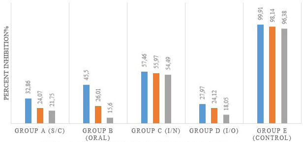
Antibody titers for different routes of vaccine administration.
References
- Abubakar, M., Arshed, M.J., Zahur, A.B., Ali, Q. and Banyard, A.C., 2012. Virus Res. 167: 43-47. https://doi.org/10.1016/j.virusres.2012.03.018[CrossRef]
- Couacy-Hymann, E., Roger, F., Hurard, C., Guillou, J.P., Libeau, G. and Diallo, A., 2002. J. Virol. Methods, 100: 17-25. https://doi.org/10.1016/S0166-0934(01)00386-X[CrossRef]
- El-Yuguda, A.D., Baba, S.S., Ambali, A.G. and Egwu, G.O., 2014. World J. Vaccines, 4: 1-6. https://doi.org/10.4236/wjv.2014.41001[CrossRef]
- Ezeibe, M.C.O., Okoroafor, O.N., Ngene, A.A., Eze, J.I., Eze, I.C. and Ugonabo, J.A.C., 2008. Trop. Anim. Hlth. Pro, 40: 517-519. https://doi.org/10.1007/s11250-008-9128-3[CrossRef]
- Forsyth, M.A. and Barrett, T., 1995. Virus Res., 39: 151-163. https://doi.org/10.1016/0168-1702(95)00076-3[CrossRef]
- Intizar, M., Ahmad, M.D., Anjum, A.A. and Hanif, A., 2009. Pak. Vet. J., 29: 202-205.
- Osman, N.A., A/Rahman, M.E., Ali, A.S. and Fadol, M.A., 2008. Trop. Anim. Hlth. Prod, 40: 363-368. https://doi.org/10.1007/s11250-007-9106-1[CrossRef]
- Ullah, R.W., Zahur, A.B., Latif, A., Dasti, J.I., Irshad, H., Afzal, M., Rasheed, T., Malik, A.R. and Qureshi, Z.U.A., 2016. Biomed. Res. Int., pp. 1-5. https://doi.org/10.1155/2016/1486824[CrossRef]
- Wang, Z., Bao, J., Wu, X., Liu, Y., Li, L., Liu, C., Suo, L., Xie, Z., Zhao, W., Zhang, W., Yang, N., Li, J., Wang, S. and Wang, J., 2009. Emerg. Infect. Dis., 15: 299-301. https://doi.org/10.3201/eid1502.080817[CrossRef]
- Zahur, A.B., Irshad, H., Hussain, M., Ullah, A., Jahangir, M., Khan, M.Q. and Farooq, M.S., 2008. Rev. Sci. Tech. Off. Int. Epiz., 27: 877. https://doi.org/10.20506/rst.27.3.1848[CrossRef]
Services
Article usage statistics
Loading…