
Peste des petits ruminant (PPR) is a highly infectious viral disease affecting mainly small ruminants i.e. sheep and goats. It exerts socioeconomic impacts on livestock in the developing countries. The current study was designed to investigate the genetic relationships of the PPRV strains circulating in some districts of Punjab, Pakistan after detecting by PCR. Samples showing definite clinical signs, were collected from reported disease outbreaks and subjected to RT-PCR based on detecting the nucleoprotein (N) gene of the virus which showed 63% positivity rate in the collected samples. It has been shown, based on phylogenetic analysis, that the circulating strains were related to the viruses present within lineage IV with close relationship to the Asian and Middle Eastern isolates. Serologically, the competitive ELISA (cELISA) was used to test antibodies in the same animals which showed 50 % animals with PPRV specific antibodies. The high presence of the virus indicated the exposure of PPRV in animals where vaccination had not been practiced previously. The study also provides understanding of the frequent virus circulation and aids to adopt the suitable preventive procedures in the region.
Short Communication · Volume 56, Issue 6, 9 November 2024 · Open Access
Molecular Detection and Genetic Characterization of Peste des Petits Ruminant Virus in Punjab, Pakistan
· ·
Affiliations & Notes ⌄
- 1College of Biological Sciences, Nuclear Institute for Agriculture and Biology College, Pakistan Institute of Engineering and Applied Sciences, Nilore, 44000, Islamabad. 2Vaccine Development Group, Animal Sciences Division, Nuclear Institute for Agriculture and Biology, Jhang Road, P.O box 128, Faisalabad, 38000. Pakistan. * Corresponding author: javeriaqamarguj@gmail.com
Article Info ⌄
- Journal:
- Pakistan Journal of Zoology
- Published:
- 9 November 2024

From the article
- Out of 30, 19 (63.3%) samples were positive with RT-PCR indicating the presence of active PPRV infection in the sampled regions.
- Phylogenetic analysis based on N gene sequencing, showed that all the four PPRV circulating strains from the region belong to lineage IV in two different groups.
- One group of viruses including three field strains i.e., Pak-Bwp (MN759256.1), Pak-Ksb (MN514980), Pak-Rwp (MN759254.1) made a cluster with isolates reported from Iraq while other group i.e., Pak-Fsd (MN759255.1) made a cluster with field isolates reported from Iranian and chinese region (Fig. 1).
- In this study, the detection of PPRV was carried out by using cELISA and RT-PCR based on N-gene detection from outbreaks reported from different regions.
Selected from the article's own text.
Abstract
Materials and methods
The susceptible samples were collected from disease outbreaks available at different regions i.e. Khushab, Faisalabad, Bahawalpur and Rawalpindi of Punjab province, Pakistan. These regions exhibit the traditional production systems in which mixed agro-livestock manner is practised. The outbreaks infected with PPRV, affect the entire population of the herd regardless of the species and breed . These outbreaks were reported from October to December and infected animals were characterized by high fever, ocular and nasal discharges, diarrhea and abortions in pregnant animals. The animals were aged between 6-12 months and presented no vaccination history. Clinical samples (nasal swabs, blood, ocular swabs, and tissues from intestine, lungs and lymph nodes) were collected from infected herds in these four regions.
Nasal, ocular and tissues samples were collected in sterile collection tubes and transported on ice. The samples were mixed by brief vortexing and centrifugation to decant the supernatant which was stored at -80 ⁰C until RNA extraction. Blood was collected to separate the serum to be used for detection of antibodies through cELISA.
Competitive enzyme-linked immunosorbent assay (cELISA) was used for the detection of antibodies against PPRV. The ELISA was carried out by using commercially available kit, (ID.vet Innovative diagnostics ID Screen® PPR Competition, Louis Pasteur-Garbels- FRANCE). This kit utilizes recombinant N gene as antigen to detect antibodies specific to PPRV and results were analyzed according to the manufacturer’s guidelines. Competition percentage was calculated from OD values and the sample having PI value less than 50% was declared as positive.
RNA extraction from virus suspension (200µl) was performed using Nucleic Acid Extraction kit (GF-1, Vivantis Technology, Malaysia) according to the manufacturer’s protocol. Finally, total RNA was eluted using 30 µl nuclease free water. Extracted RNA was subjected to reverse transcription (cDNA synthesis) which was carried out using EasyScript™ cDNA Synthesis Kit with manufacturer’s guidelines. Briefly, the reaction was incubated at 25°C for 5 min followed by incubation at 42°C for 50 min. Finally, the reaction was terminated by incubating the reaction at 85°C for 5 min and stored at -20°C until used as template in PCR.
PCR was performed to detect PPRV infection using previously reported gene specific primers: NP3 (Forward-5′-GTCTCGGAAATCGCCTCACAGACT-3′ and NP4 (Reverse-5′-CCTCCTCCTGGTCCTCCAGAATCT-3′) targeting partial Nucleoprtoein (N) gene . PCR was carried out by using Thermo fisher scientific kit in a total 50 µL reaction volume which included 1X Taq DNA polymerase buffer, 5mM MgCl2, 10mM dNTPs, 10uM each primer, 1 unit of Taq DNA polymerase and 2 uL cDNA as template. The reaction was performed using VWR thermal cycler (VWR®, USA) to amplify N gene under following conditions: one incubation cycle at 94 °C for 4 min followed by thirty five cycles of incubations with each incubation at 94°C for 45 sec, 56°C for 45 sec, and 72°C for 45 sec. Final extension step was performed for 10 min at 72⁰C.
The amplified PCR products were analyzed on 1.5 % Tris-borate-EDTA (TBE) agarose gel followed by staining with ethidium bromide solution (50mg/ml). PCR product from each tube was mixed with appropriate ratio with 6X DNA loading buffer and loaded with 10 kb DNA ladder (GeneRuler, MBI Fermentas). The DNA was allowed to separate in 1X TBE buffer at 80 V for 45 min. Amplified products were visualized under a UV-transilluminator. The positive amplified products were purified by using Gene All® gel purification kit (Gene All® Expin ComboTM, GeneAll Biotechnology, Korea) according to the manufacturer’s recommendations. The purified DNA was sequenced at least twice in both directions using ABI PRISM BigDye Terminator version 3.1 (Applied Biosystems) with manufacturer’s directions. The analysis of sequences was performed using an automated nucleic acid analyzer (ABI PRISM 3100; Applied Biosystems).
Results
Total 30 samples were collected from the infected animals showing the clinical signs which were present in different herds. All 30 serum samples were analyzed using cELISA from which 15 (50%) samples were found positive showing the presence of PPRV specific antibodies in their blood. Out of 30, 19 (63.3%) samples were positive with RT-PCR indicating the presence of active PPRV infection in the sampled regions. The presence PPRV genome was confirmed by analyzing the expected 351 bp band corresponding to the N gene following agarose gel electrophoresis. It was also revealed that maximum samples were found positive in Faisalabad district followed by Khushab, Rawalpindi and Bahawalpur (Table I).
Table I. Details of the samples used in the study. All these animals had clinical signs of fever, severe diarrhea, weakness, nasal discharges, sneezing, coughing.
| District | Number of samples/ total animals in herd | RT-PCR | |
| Positive | Negative | ||
| Faisalabad | 8/26 | 6 | 2 |
| Khushab | 8/24 | 5 | 3 |
| Bahawapur | 8/39 | 4 | 4 |
| Rawalpindi | 6/25 | 4 | 2 |
The positive samples from each region were selected as representatives and used for sequencing to analyze their genetic characterization for downstream studies. The analyzed sequences submitted to the GenBank under accession numbers: MN759254.1, MN759256.1, MN514980 and MN759255.1 for Rawalpindi, Bahawalpur, Khushab and Faisalabad respectively, based on partial N gene sequence.
Phylogenetic analysis based on N gene sequencing, showed that all the four PPRV circulating strains from the region belong to lineage IV in two different groups. Interestingly, samples collected from these districts are in close proximity. One group of viruses including three field strains i.e., Pak-Bwp (MN759256.1), Pak-Ksb (MN514980), Pak-Rwp (MN759254.1) made a cluster with isolates reported from Iraq while other group i.e., Pak-Fsd (MN759255.1) made a cluster with field isolates reported from Iranian and chinese region (Fig. 1). Therefore, it was observed that two different groups of PPRV were circulating in the country.
Discussion
The control of PPRV outbreaks mainly depends on the vaccination. These outbreaks are generally detected by clinical manifestation of the disease and laboratory confirmation by either using antigen or antibody detection ELISA. Use of ELISA tests does not necessarily indicate the failure of vaccination . Therefore, the use of molecular characterization based on genetics of the virus is essential for rapid diagnosis to study the epidemiology of the disease and to devise the appropriate control measures. For the molecular characterization of PPR virus, PCR based detection of fusion (F) and nucleoprotein (N) genes were used followed by sequencing to trace the circulating virus strains. Among these, N based detection has been considered sensitive and reliable for the detection and genetic characterization of PPRV than based on F gene. N protein is an internal structural protein and its mRNAs are attractive targets for developing diagnostic assay with higher sensitivity . In this study, the detection of PPRV was carried out by using cELISA and RT-PCR based on N-gene detection from outbreaks reported from different regions.
One sample from each district was sequenced for phylogenetic analysis. Phylogenetic analysis on N gene based detection described the circulating PPR viruses were related to two different groups in lineage IV. One group of viruses containing three field strains closely relate to Iraq and second group with one field strain was clustered with Iran and china. Our study are in close agreement with previous studies revealed that the lineage IV is highly circulating in Pakistan by clustering into different groups such as Saudi Arabia, Tajikistan, Iran, China, Iran and Iraq ; ; ; . The emergence of Iraq isolates in the circulating viruses can be associated with uncontrolled and unrestricted movement of animals between the countries . Iran and Kurdistan isolates have also been reported previously in Pakistan. Moreover, the movement of small ruminants on the occasion of religious festival, Eid-ul-Azha is common in Punjab province which also aggravates the susceptibility of the disease in the region .
The study also describes the high sero-positivity rate in the sampled region as detected by cELISA. The absence of PPRV specific antibodies in remaining samples may be associated with initial phase of PPRV infection or virus shedding at the active infection stage. Thus, the collection of oculo-nasal swabs at the start of infection is suitable for the detection of PPRV infection . The positivity rate with oculo-nasal swabs was 63.3% which found suitable for the detection of PPRV. Vaccination in Pakistan is generally based on Nigeria 75/1 strain which belongs to lineage I while the reported sequences are related to the viruses of Lineage IV. This fact favors the development of new vaccines using the domestic field strains in order to control the disease outbreaks more effectively . Therefore, this study also helps in understanding the outcomes of vaccines failure in the endemic regions with the combined use of ELISA and RT-PCR.
Conventional and real-time PCR systems have been considered reliable for genetic characterization of PPR viruses. Now, these tests have been transferred to FTA cards for suitable transportation of the biological samples from field to specific diagnostic laboratory, most importantly, where the maintenance of cold chain is not possible . The diagnostic specificity and sensitivity can be tested by using large number of samples from different geographical locations which would be favorable in determining the accurate epidemiology and control of the disease.
Acknowledgments
The authors are grateful to Mr. Hafiz Muhammad Muzammil, Mr. Irfanullah Khan, and Mr. Asad Javed for providing technical assistance during the study.
Funding
The study was supported by IAEA project (IAEA RAS 5085).
Statement of conflict of interest
The authors have declared no conflict of interest.
Figures
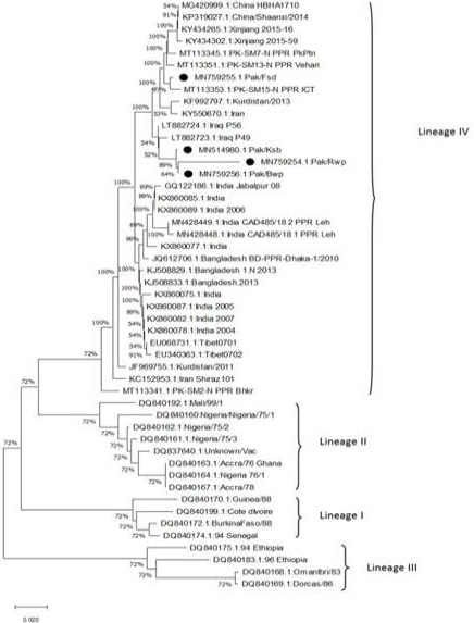
Phylogenetic tree for N gene 351bp of PPRV. The evolutionary history was inferred using the Neighbor-joining method. The bootstrap consensus tree inferred from 1000 replicates. The evolutionary distances were computed using the maximum composite likelihood method. This analysis involved 49 nucleotide sequences. Evolutionary analyses were conducted in MEGA ×(10.2.6).
References
- Abubakar, M. and Munir, M., 2014. Transbound. Emerg. Dis., 61: 7-10. https://doi.org/10.1111/tbed.12192[CrossRef]
- Abubakar, M., Jamal, S.M., Hussain, M. and Ali, Q., 2008. Small Rumin. Res., 75: 256-259. https://doi.org/10.1016/j.smallrumres.2007.12.001[CrossRef]
- Ahmad, K., Jamal, S., Ali, Q. and Hussain, M., 2005. Pak. Vet. J., 209: 146-148.
- Amjad, H., Forsyth, M., Barrett, T. and Rossiter, P., 1996. Vet. Rec. Open, 139: 118-119. https://doi.org/10.1136/vr.139.5.118[CrossRef]
- Anees, M., Shabbir, M.Z., Muhammad, K., Nazir, J., Shabbir, M.A.B., Wensman, J.J. and Munir, M., 2013. BMC Vet. Res., 9: 1-5. https://doi.org/10.1186/1746-6148-9-60[CrossRef]
- Assefa, A., Tibebu, A., Bihon, A. and Yimana, M., 2021. Transbound. Emerg. Dis., 68: 3601-3610. https://doi.org/10.1111/tbed.13967[CrossRef]
- Banyard, A.C., Parida, S., Batten, C., Oura, C., Kwiatek, O. and Libeau, G., 2010. J. Gen. Virol., 91: 2885-2897.
- Clarke, B., Mahapatra, M., Friedgut, O., Bumbarov, V. and Parida, S., 2017. PLoS One, 12: 1-17. https://doi.org/10.1371/journal.pone.0177028[CrossRef]
- Couacy-Hymann, E., Roger, F., Hurard, C., Guillou, J., Libeau, G. and Diallo, A., 2002. J. Virol. Methods, 100: 17-25. https://doi.org/10.1016/S0166-0934(01)00386-X[CrossRef]
- De Vries, R.D., Duprex, W.P. and De Swart, R.L., 2015. Viruses, 7: 699-706.
- Idoga, E.S., Armson, B., Alafiatayo, R., Ogwuche, A., Mijten, E., Ekiri, A.B., Varga, G. and Cook, A.J., 2020. Front. Vet. Sci., 7: 1-10. https://doi.org/10.3389/fvets.2020.592662[CrossRef]
- Kerur, N., Jhala, M. and Joshi, C., 2008. Res. Vet. Sci., 85: 176-183. https://doi.org/10.1016/j.rvsc.2007.07.007[CrossRef]
- Manzoor, S., Abubakar, M., Zahur, A.B., Yunus, A.W., Ullah, A. and Afzal, M., 2020. Transbound. Emerg. Dis., 67: 2126-2132.
- Munir, M., Zohari, S., Saeed, A., Khan, Q., Abubakar, M., LeBlanc, N. and Berg, M., 2012. Transbound. Emerg. Dis., 59: 85-93. https://doi.org/10.1111/j.1865-1682.2011.01245.x[CrossRef]
- Munir, M., Zohari, S., Suluku, R., LeBlanc, N., Kanu, S., Sankoh, F.A.R., Berg, M., Barrie, M.L. and Ståhl, K., 2012. Emerg. Infect. Dis., 18: 193.
- Parida, S., Muniraju, M., Mahapatra, M., Muthuchelvan, D., Buczkowski, H. and Banyard, A., 2015. Vet. Microbiol., 181: 90-106. https://doi.org/10.1016/j.vetmic.2015.08.009[CrossRef]
- Pestil, Z., Sait, A., Ozbaser, F.T. and Bulut, H., 2020. Pak. Vet. J., 40: 494-498. https://doi.org/10.29261/pakvetj/2020.042[CrossRef]
- Sharawi, S., Yousef, M., Al-Hofufy, A. and Al-Blowi, M., 2010. Vet. World, 3: 489-494.
Services
Article usage statistics
Loading…