
HR-HPVs, or high-risk human papillomaviruses, are the cause of cervical cancer, ranks as the second leading cause of female mortality worldwide, following breast cancer. HPV16 is globally recognized as the most common high-risk type. This research sought to examine sequence differences between the local HPV16 genotypes’ E6 and E7 oncogenes and those with foreign genotypes of HPV16. Additionally, it aimed to establish phylogenetic relationships according to the nucleotide sequence comparisons between the variants found in the present study and isolates from different parts of the world that have already been reported.Cervical cancer patients provided samples for the study. Following DNA extraction, HPV16 E6 and E7 amplification and sequencing were performed. Using the obtained E6 and E7 sequences, HPV16 phylogenetic trees were generated. The analysis revealed that Pakistani isolates belong to the A1 sub lineage within the European branch. The study incorporated locations of the first, second and third codons. The final dataset encompassed 477 positions for E6 and 297 positions for E7. Phylogenetic analysis confirmed that Pakistani isolates are situated within the sub lineage A1 of HPV16’s lineage A.
Short Communication · Volume 58, Issue 1, 2 January 2026 · Open Access
Phylogenetic Analysis of E6 and E7 Gene of Human Papillomavirus 16 Variants Isolated from Pakistani Population
· · · ·
Affiliations & Notes ⌄
- 1Centre for Applied Molecular Biology, University of Punjab, Thoker Niaz Beg, Lahore, Pakistan 2Centre of Excellence in Molecular Biology, University of Punjab, Thoker Niaz Beg, Lahore, Pakistan * Corresponding author: shazia.rafique@cemb.edu.pk
Article Info ⌄
- Journal:
- Pakistan Journal of Zoology
- Published:
- 2 January 2026

From the article
- Among 132 women with cervical cancer, HPV16 was identified as the most common type, present in 61 cases.
- While HPV is a significant factor in cervical cancer development, it is not the sole cause.
- The Pakistani HPV16 isolate showed 99% sequence similarity with Brazilian isolates (the sequence of reference KU298880 for the A1 sublineage).
- A phylogenetic study of the genes E6 and E7 sequences about Pakistani HPV16 isolates of CC associated with HPV were this study’s findings.
Selected from the article's own text.
Abstract
Materials and methods
Cervical swabs were obtained from 132 cervical cancer patients aged 35-72 years at various hospitals in Lahore, Pakistan. These samples were processed at the Centre of Excellence in Molecular Biology’s Virology Laboratory at Lahore.
HPV DNA extraction was conducted using ethanol precipitation. A real time PCR method, employing th14 HPV types of detection kit (Healgen) was used to identify high-risk HPV types. This kit can detect 14 HPV types (16, 18, 31, 33, 35, 39, 45, S1, 52, 56, 58, 59, 66 and 68). Samples testing positive for HPV-16 were chosen for further sequence study.
The specific coding area primers were used to amplify the HPV E6 and E7 genes independently (Table I) at an annealing temperature of 58°C. Samples negative for both E6 and E7 were excluded from subsequent analysis.
Table I. Primer sequences for the amplification of HPV16 E6 and E7 genes.
| Primer | Primer sequence |
| HPV-16 E6 | F 5ʹ -ATGCACCAAAAGAGAACTGC- 3ʹ |
| R 5ʹ-TTACAGCTGGGTTTCTCTACG-3ʹ | |
| HPV-16 E7 | F 5ʹ - ATGCATGGAGATACACCTAC- 3ʹ |
| R 5ʹ -TTATGGTTTCTGAGAACAGATGGG-3ʹ |
PCR products underwent purification using the PureLink PCR purification kit. Sequencing was performed bidirectionally using the BigDye Terminator Sequencing Kit on an ABI PRISM 3100 genetic analyzer (applied biosystems) to confirm the PCR-amplified product. The resulting sequences were submitted to the NCBI gene bank database.
Sequence analysis was conducted using BLAST from PubMed. Phylogenetic trees for HPV16 were constructed using E6 and E7 sequences acquired throughout this investigation.The evolutionary history was inferred using the Maximum Likelihood method based on the general time reversible model . Reference sequences for 10 sublineages were taken from the Gene Bank database (www.ncbi.nlm.nih.gov). These included: A1: K02718 and KU298880, A2: AF536179, FJ610152 and KU053892, A3: HQ644236, A4: AF534061, HQ644234 and HQ644261, B1: AF472508, AF536180 and HQ644296, B2: HQ644298, C: AB818690 and AF472509, D1: HQ644257, D2: Ay686579, D3: AF402678, HQ644285 and HQ644289.
Results and discussion
Among 132 women with cervical cancer, HPV16 was identified as the most common type, present in 61 cases. Pakistani HPV16 isolates E6 and E7 gene sequences were analyzed for similarity and submitted to the NCBI Genebank database. The local E7 gene was assigned the accession number MH884521, while MT955329 was given to the local E6 gene.
The maximum likelihood method and general time reversible model were employed to infer evolutionary history. Figure 1 illustrates potential nucleotides at each ancestral node based on their inferred likelihood. The heuristic search’s initial trees were obtained using the Maximum Parsimony approach. Site rates were treated as a Gamma distribution with invariant sites, utilizing 5 Gamma categories. The analysis involved 21 nucleotide sequences, including all codon positions. The final dataset contained 477 positions for E6 and 297 for E7. MEGA11 was used for evolutionary analyses.
The Papillomaviridae family includes HPV, comprising 29 genera with 189 PV types isolated from various species . The double stranded DNA that makes up HPV genome of approximately 8kb, containing 8 genes: E1, E2, E4, E5, E6, E7, L1, and L2 ; ; ; . Oncoproteins, E6 and E7 remain crucial for initiating plus maintaining HPV-associated malignancies and, are expressed themselves in cells that have undergone transformation .
Identifying variants of HPV are crucial for creating testing methods, vaccinations, and therapies . Previous research has linked HPV variant
distribution to geographic or racial factors ; . The purpose of this study was to show how various HPV16 lineages are distributed throughout the Pakistani population, following Yamada et al. (1997) classification.
Cervical cancer ranks as the third most prevalent cancer among women globally, with an estimated 569,847 new cases and 311,365 deaths in 2018. Squamous cell carcinoma is the most common type, followed by adenocarcinomas . In Pakistan, 2012 estimates revealed that the prevalence of HPV related cancers was 7.9%, whereas the combined prevalence of ano-genital and head and neck malignances was 0.5% . The ICO/IARC HPV information centre reports that approximately 5,601 new cervical cancer cases are diagnosed annually in Pakistan. Cervical cancer is the third leading cause of cancer and cancer-related deaths among Pakistani women overall, and the second most common in the 15-44 age group .
While HPV is a significant factor in cervical cancer development, it is not the sole cause. Several other contributing factors have been identified, including cigarette smoking, extended use of hormonal contraceptives, multiple childbirths, and HIV co-infection. Additional risk factors encompass co-infections with Chlamydia trachomatis and herpes simplex virus type-2, compromised immune systems, and specific nutritional deficiencies. The development of cancer can also be influenced by genetic and immunological host factors, as well as viral characteristics such as the type of virus, viral load, and viral integration .
The samples we collected were classified within the sublineage A1 of HPV16’s alpha9 species. The Pakistani HPV16 isolate showed 99% sequence similarity with Brazilian isolates (the sequence of reference KU298880 for the A1 sublineage). This grouping was supported by a strong 91% bootstrap value. In Pakistan, HPV16 was also categorized into the alpha9 species group in a previous comprehensive study on HPV classification .
Given that sexual contact remains the primary mode of HPV transmission, socio-cultural obstacles hinder the accurate assessment of HPV prevalence in Pakistan. Limited awareness results in few women opting for HPV screenings, allowing HPV infections to go undetected until they progress to more severe conditions. Phylogenetic analyses play a crucial role in mapping the groups to which HPV isolates belong, as they can help identify regions that may be potential sources of HPV spread within a country.
Conclusion
The main cause of CC in females, HPV, is estimated to be responsible for 3,000 fatalities annually. A phylogenetic study of the genes E6 and E7 sequences about Pakistani HPV16 isolates of CC associated with HPV were this study’s findings. The HPV16 strain from Pakistan shared 99% of its sequence with isolates from Brazil. In order to create tools for molecular diagnosis in the Pakistani population, phylogenetic analyses are essential.
Declarations
Acknowledgement
We are thankful to the administration and healthcare professionals at INMOL Hospital, Jinnah Hospital, and Mayo Hospital in Lahore for their support in collecting clinical data and samples from cervical cancer patients.
Funding
The study received no external funding.
Statement of conflict of interest
The authors have declared no conflict of interest.
Figures
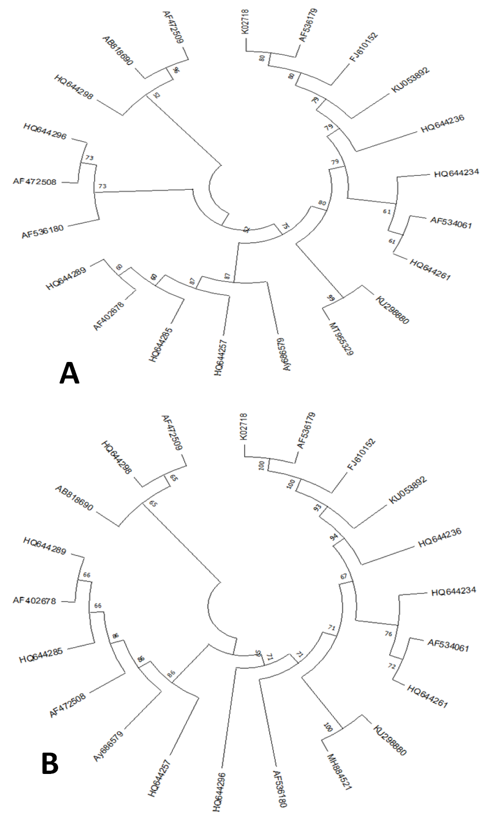
Phylogenetic tree of E6 gene (A) and E7 gene (B) of different HPV16 isolates.
References
- Abdullah, A., Qasim, M., Shafiq, M., Ijaz, M., Parveen, S., Murtaza, S., Javed, Q., Malik, S.A., Tarar, S.H., Mehmood, S., Sami, A., Naqvi, S.M.S. and Hyder, M.Z., 2016. Infect. Agent Cancer, 11: 1. https://doi.org/10.1186/s13027-016-0047-z[CrossRef]
- Amador-Molina, A., Hernández Valencia, J.F., Lamoyi, E., Contreras-Paredes, A. and Lizano, M., 2013. Viruses, 5: 2624-2642. https://doi.org/10.3390/v5112624[CrossRef]
- Asiaf, A., Ahmad, S.T., Mohammad, S.O. and Zargar M.A., 2014. Eur. J. Cancer Prevent., 23: 206. https://doi.org/10.1097/CEJ.0b013e328364f273[CrossRef]
- Bruni, L., Albero, G., Serrano, B., Mena, M., Collado, J., Gómez, D., Muñoz, J., Bosch, F. and Sanjosé, S., 2023. Human papillomavirus and related diseases in the world. Summary report. ICO/IARC information centre on HPV and cancer.
- de Araujo Souza, P.S., Sichero, L. and Maciag, P.C., 2009. Future Oncol., 5: 359. https://doi.org/10.2217/fon.09.8[CrossRef]
- De Martel, C., Plummer, M., Vignat, J. and Franceschi S., 2017. Int. J. Cancer, 141: 664. https://doi.org/10.1002/ijc.30716[CrossRef]
- Dunne, E.F. and Park, I.U., 2013. Infect. Dis. Clin., 27: 765. https://doi.org/10.1016/j.idc.2013.09.001[CrossRef]
- Ferlay, J., Ervik, M., Lam, F., Colombet, M., Mery, L., Piñeros, M., Znaor, A., Soerjomataram, I., and Bray, F., 2018. Cancer Today, 23: 323.
- Fernandes, J.V. and de Medeiros Fernandes, T.A.A., 2012. Human papillomavirus: Biology and pathogenesis. In: Human papillomavirus and related diseases-from bench to bedside-a clinical perspective. IntechOpen.
- Forman, D., de Martel, C., Lacey, C.J., Soerjomataram, I., Lortet-Tieulent, J., Bruni, L., Vignat, J., Ferlay, J., Bray, F., Plummer, M. and Franceschi, S., 2012. Vaccine., 30: F12. https://doi.org/10.1016/j.vaccine.2012.07.055[CrossRef]
- Graham, S.V., 2010. Future Microbiol., 5: 1493-1506. https://doi.org/10.2217/fmb.10.107[CrossRef]
- Gul, S., Murad, S. and Javed, A., 2015. Int. J. Infect. Dis., 34: 14-19. https://doi.org/10.1016/j.ijid.2015.02.018[CrossRef]
- Hafkamp, H.C., Manni J.J. and Speel, E.J., 2004. Acta Otolaryngol., 124: 520-526. https://doi.org/10.1080/00016480310016893[CrossRef]
- Khan, S., 2010. Molecular epidemiology of human immunodeficiency virus (HIV) and Human Papillomavirus (HPV) in Pakistan. University of Karachi, Pakistan.
- Morshed, K., Polz-Gruszka, D., Szymański, M. and Polz-Dacewicz, M., 2014. Otolaryngol. Pol., 68: 213-219. https://doi.org/10.1016/j.otpol.2014.06.001[CrossRef]
- Muñoz, N., Castellsagué, X., de González A.B. and Gissmann L., 2006. Vaccine, 24: S1. https://doi.org/10.1016/j.vaccine.2006.05.115[CrossRef]
- Nei, M., 2000. Molecular evolution and phylogenetics. Oxford University Press, New York. https://doi.org/10.1093/oso/9780195135848.001.0001[CrossRef]
- Pande, S., Jain, N.,Prusty, B.K., Bhambhani, S., Gupta, S., Sharma, R., Batra S. and Das, B.C., 2008. J. clin. microbiol., 46:1060-1066. https://doi.org/10.1128/JCM.02202-07[CrossRef]
- Ramas, V., Mirazo, S., Bonilla, S., Ruchansky, D. and Arbiza, J., 2018. Gene, 654: 103-109. https://doi.org/10.1016/j.gene.2018.02.023[CrossRef]
- Tamura, K., Stecher, G. and Kumar S., 2021. Mol. Biol. Evol., 38: 3022. https://doi.org/10.1093/molbev/msab120[CrossRef]
- Van Doorslaer, K., Tan, Q., Xirasagar, S., Bandaru, S., Gopalan, V., Mohamoud, Y., Huyen Y. and McBride, A.A., 2012. Nucl. Acids Res., 41: D571–D578. https://doi.org/10.1093/nar/gks984[CrossRef]
- Xi, L.F., Kiviat, N.B., Hildesheim, A., Galloway, D.A., Wheeler, C.M., Ho J. and Koutsky L.A., 2006. J. Natl. Cancer Inst., 98: 1045-1052. https://doi.org/10.1093/jnci/djj297[CrossRef]
- Yamada, T., Manos, M.M., Peto, J., Greer, C.E., Munoz, N., Bosch, F.X. and Wheeler, C.M., 1997. J. Virol., 71: 2463-2472. https://doi.org/10.1128/jvi.71.3.2463-2472.1997[CrossRef]
- Yang, A., Farmer, E., Wu, T. and Hung, C.F., 2016. J. Biomed. Sci., 23: 75. https://doi.org/10.1186/s12929-016-0293-9[CrossRef]
- Yousuf, S., Syed, S., Moazzam, A. and Lucky, M.H., 2010. J. Pak. med. Assoc., 60: 193-196.
- Zheng, Z.M. and Baker C.C., 2006. Front. Biosci J. Virtual Libr., 11: 2286-2302. https://doi.org/10.2741/1971[CrossRef]
Services
Article usage statistics
Loading…