
The lower recovery of competent oocytes in buffalo species limits the commercialization of in vitro embryo production technology in field conditions. The present study aimed to improve the cytoplasmic and nuclear maturation of in vitro matured oocytes of Nili Ravi buffalo in the presence of hormones. The denuded oocytes (DOs) obtained by repeated pipetting were collected from 2-8mm follicles. A series of experiments were conducted to evaluate the effects of oestradiol (2 μg/ml), recombinant human follicle stimulating hormone (0.05 IU/ml), human chorionic gonadotrophin (2 IU/ml) and insulin (0.12 IU/ml) alone or together in different combinations at intervals of 8, 16 and 24 h of incubation period during in vitro maturation on buffalo oocyte. The supplementation of TCM-199 with E2+rhFSH showed a highly significant increase in the diameter and maturation of oocytes (P<0.0001), as a greater number of the oocytes progressed to the metaphase II stage, and a lower proportion of the oocytes became degenerated (P<0.0001) after 24 h of incubation. The supplementation of medium with E2+rhFSH+hCG or E2+rhFSH+hCG+insulin also showed a significant increase in the meiotic maturation rate after 24 h (P<0.01 and P=0.04 respectively) and a significant decrease in the degeneration of the oocytes (P=0.001). The addition of insulin was not found to be effective for in vitro maturation. It is concluded that the addition of E2+rhFSH in culture media was found to be the best combination of hormones for in vitro maturation of denuded buffalo oocytes.
Research Article · Volume 55, Issue 4, 24 June 2023 · Open Access
Nuclear and Cytoplasmic Maturation of In Vitro Matured Denuded Oocytes in the Presence of Gonadotrophins, Oestradiol and Insulin in Nili Ravi Buffalo
·
Affiliations & Notes ⌄
- 1Department of Zoology, King Abdullah Campus, University of Azad Jammu and Kashmir, Muzaffarabad, 13100 Pakistan 2Livestock Development Research Centre, Muzaffarabad, Azad Jammu and Kashmir, 13100, Pakistan * Corresponding author: beenishkhan124@gmail.com
Article Info ⌄
- Journal:
- Pakistan Journal of Zoology
- Published:
- 24 June 2023

From the article
- Whereas the percentage of MII stage oocytes increased significantly.
- The addition of rhFSH alone in the medium was observed to have a positive effect on the nuclear and cytoplasmic maturation of DOs, while percentage of degenerated oocytes was non-significantly decreased.
- The present results are similar to another study in which it was observed that the addition of 1 mg/100ml oestradiol and 0.05 IU/ml rhFSH in TCM-199 enhanced the meiotic maturation of Nili Ravi buffalo oocytes (82.02% oocytes were progressed to MII stage).
- Supplementation of E2+hCG in medium showed decrease in percentage of GVBD oocytes and increase in percentage of MII oocytes but non-significantly.
Selected from the article's own text.
Abstract
INTRODUCTION
The reproductive efficiency in livestock species can be improved by applying assisted reproductive technologies (ARTs) and has significant roles in the preservation of endangered species . Recently, in vitro embryo production has increased rapidly with an average rate of 12 % ; . Despite the success of in vitro fertilization, embryo culture, and transgenesis, the in vitro systems still have a very low efficiency, with a small percentage of embryos produced from in vitro matured oocytes . Because of this limited success in bubaline species ; the in vitro embryo production is the available alternative to support breeding program for the rapid propagation and upgradation of buffalo ; ; ; ; . The maturation of oocytes that are cultured in vitro, is a technique of assisted reproduction. It involves the use of controlled ovarian hyperstimulation for in vitro fertilization through the collection of immature oocytes at the prophase I stage, which are then matured in vitro until they reach metaphase II (MII) stage .
A period of 24 h is necessary for a bovine oocyte to complete nuclear maturation . The cytoplasmic maturation includes the progression of the oocyte from germinal vesicle (GV) to the next stage (MII) accompanied by a remarkable reorganization, concomitant with chromosome condensation and migration and the acquisition of developmental competence . Cytoplasmic maturation involves the synthesis, activation, and degradation of maternal mRNA . The addition of a maturation medium with bovine fetal calf serum and follicular fluid can affect the in vitro maturation of oocytes. Moreover, the subsequent fertilization and development can be enhanced by the selection of protein supplements and hormones such as gonadotropin , steroids and insulin growth factor-I ; . The nuclear maturation and blastocyst formation of sow oocytes during in-vitro culture was improved by the addition of follicle-stimulating hormone (FSH) in the culture medium . The presence of equine chorionic gonadotropin (eCG) improved the cleavage and developmental rates of in-vitro fertilized buffalo embryos .
The steroids hormone such as oestradiol enhanced the maturation of oocytes in buffalo ; . The proliferation of granulosa cells and the production of progesterone in bovine is stimulated by insulin and insulin-like growth factor-I ; . The aim of this study was to optimize the culture medium for in-vitro maturation of Nili Ravi buffalo oocytes by supplementation with oestradiol (E2), recombinant FSH, hCG and insulin (I). The objective was to determine the effect of hormones in different combinations on the nuclear and cytoplasmic maturation of denuded oocytes.
MATERIALS AND METHODS
Collection of ovaries
The ovaries used in this study were obtained from the slaughtered buffaloes, therefore there was no need of approval from the ethical committee of the university. Approximately 560 buffalo (Bubalus bubalis) ovaries were obtained from a local slaughterhouse, Sihala Rawalpindi immediately after slaughtering of 280 culled dairy buffaloes during the period of three months. All the culled buffaloes that seize producing milk having age 6-12 years were brought to slaughterhouse from the adjoining areas of Islamabad, Rawalpindi, and Punjab. Rectal palpation that is a part of routine procedure at slaughterhouse was done to remove any pregnant buffalo from the slat of slaughtering. The ovaries with cysts or any other abnormalities were discarded and only 400 ovaries with normal shape and visible follicles were selected. The ovaries were transported to the laboratory in an insulated flask containing sterile normal 0.9% saline and antibiotics (100 IU/ml penicillin G; 100 μg/ml streptomycin sulphate, Zafa Pharmaceuticals) at 38.5oC.
Recovery of oocytes
The oocytes were recovered from 2200 ovarian follicle (2-8 mm in diameter) by aspiration method using a 5 ml syringe with 16- gauge needle into a beaker containing supplemented oocyte culture medium . The denuded oocytes with 48 % recovery rate were obtained by pipetting into the intergrid plate through pasteur pipette by removing most of cumulus layers. The oocytes with single layer of cumulus were selected. Oocytes were transferred to a drop of oocyte collection medium.
Preparation of culture media
Ten liter double distilled water (ddH2O) was used to dissolve TCM -199 and 3.50 g NaHCO3 by adjusting the pH at 7.2-7.4. The medium (400 ml) was sterile filtered and kept at 4°C. The medium was supplemented one day before use with 1 aliquot of bovine steer serum (BSS) + N-2-hydroxyethylpiperazine-N’-2-ethanesulfonic acid (HEPES), 1 aliquot of glutamine (4 ml), 0.4 ml of penicillin G (100 IU/ml) and 0.04 g of streptomycin sulphate (100 µg/ml) to the next leaving all debris behind.
In vitro maturation of oocytes
After oocytes had been cleaned of debris, a group of 10 oocytes was transferred a 50 μl micro drop of oocyte maturation medium (OMM; 4.45 ml aliquots of TCM-199 were prepared and stored at 4°C until use. On night before commencing in-vitro maturation, 500 µl of bovine serum albumen (BSA), 50 µl of gentamicin) supplemented with sodium pyruvate (0.23 mmol/l), BSA (4.0 mg/ml; Sigma), oestradiol (1 mg/100ml; Merck) and rhFSH (0.05 IU/ml; Organon) were added to an aliquot of TCM-199. The droplet of the maturation medium containing oocytes was covered with mineral oil and incubated at 38.5˚C with 5% of CO2 for 24 h.
Experimental design
The nuclear and cytoplasmic maturation of DOs were assessed by examining the impact of E2 (2 µg/ml; , rhFSH (0.05 IU/ml; , hCG (2 IU/ml) and insulin (0.12 IU/ml; alone or in different combinations.
Group A (control) The DOs cultured in TCM-199 containing BSA, sodium pyruvate, gentamicin and without any addition of E2, rhFSH, hCG and insulin considered as control.
Group B: The DOs cultured in vitro in TCM-199 + 2 µg/ml E2.
Group C: The nuclear and cytoplasmic maturation of DOs was studied inTCM-199 containing 0.05 IU/ml rhFSH.
Group D: The DOs were cultured in TCM-199 containing 2 IU/ml hCG.
Group E: The effect of 0.12 IU/ml of beef insulin on nuclear and cytoplasmic maturation of DOs were studied in supplemented TCM-199.
Group F: The denuded oocytes were grown in TCM-199 containing E2 and rhFSH.
Group G: The oocytes were matured in a medium-199 supplemented with E2 and hCG.
Group H: The DOs were matured in vitro in TCM-199 containing E2 and beef insulin.
Group I: The DOs were matured in a medium-199 containing rhFSH and hCG.
Group J: The DOs were cultured in medium-199 containing rhFSH and beef insulin.
Group K: The DOs were grown in a medium-199 containing hCG and beef insulin.
Group L: The DOs were cultured in medium-199 containing E2, rhFSH and hCG.
Group M: The DOs were matured in vitro in TCM-199 supplemented with oestradiol, rhFSH, hCG and beef insulin.
Meiotic competence and oocyte expansion
The meiotic competence of the oocyte was examined using inverted phase contrast microscope (Nikon) at incubation times of 8, 16, and 24 h. Stages of nuclear maturation were classified into germinal vesicle (GV), germinal vesicle breakdown (GVBD), and metaphase II (MII) stages. In GV stage, the oocyte had a single prominent nucleus. In the GVBD stage, the oocyte underwent GVBD, but no polar body has been extruded. In MII stage, the oocyte had a polar body extrusion. The abnormal or degenerated oocytes displayed both multipolar meiotic spindle or abnormal chromatin clumps or no chromatin with fragmented or shrunken cytoplasm (Figure 1a-d; .
The oocyte growth (including zona pellucida) was assessed by measuring the diameter of the oocyte using an ocular micrometer calibrated stage micrometer by taking readings in two perpendicular directions, at the incubation time of 8, 16 and 24 h.
Statistical analysis
The mean diameter of DOs from zero to 24 h of culture period with different treatments was analyzed by two ways ANOVA. The overall expansion between control and various treatment groups was analyzed by ANOVA. The percentage of GVDB, MII and degenerated oocytes were compared among control and different treatment groups by analysis of variance and for comparing means, Tukey test was applied. The data were presented as mean±SEM and value of P≤0.05 was considered significant. All the statistical analysis was implemented using Graph Pad Prism version 6.
RESULTS
Effect of hormones on oocyte expansion
Effect of different hormones alone and in different combinations on the expansion (mean ± SEM) of cultured DOs for a period of 8, 16 and 24 h is presented in Table I. Two-way analysis of variance indicated that the effect of different treatment of hormones did not significantly affect the increase in diameter of denuded oocytes at different time periods. However, analysis of variance showed that all the hormones highly significantly (P<0.0001) increase the growth of denuded oocytes indicated by increase in diameter compared with control group.
Effect of hormones on nuclear maturation
The effect of hormones alone or in various combinations on the nuclear maturation of Nili Ravi buffalo denuded oocytes (DOs) cultured in vitro is given in Table II. The result showed that after 24 h of incubation
Table I. Effect of different hormones alone or in various combinations on the cytoplasmic maturation (mean ± SEM) of cultured denuded oocytes in Nili Ravi buffalo.
| Treatment group | Diameter of denuded oocytes (μm) | ||||
| 0 h | 8 h | 16 h | 24 h | Growth of the oocytes | |
| Control (15) | 155±2.95 | 154.5±2.62 | 152.66±2.39 | 152.0±2.25 | 0±0.7 |
| Oestradiol (10) | 153.25±1.75 | 156.25±1.63 | 156.50±2.01 | 157.5±1.86 | 4.25±0.11 |
| rhFSH (10) | 156.25±1.71 | 157.75±1.72 | 158.25±1.53 | 158.5±1.24 | 2.25±0.47 |
| hCG (25) | 159.5±1.56 | 159.1±1.87 | 158.4±1.38 | 157.1±1.43 | 0±0.13 |
| Insulin (10) | 156.25±1.63 | 155.75±2.44 | 155.0±2.47 | 153.75±2.77 | 0±1.14 |
| E2 and rhFSH (16) | 151.87±1.0 | 152.65±1.05 | 153.75±1.14 | 156.25±1.44 | 4.38±0.44 |
| E2 and hCG (38) | 156.97±1.12 | 157.03±1.33 | 157.63±1.07 | 159.14±1.32 | 2.17±0.2 |
| E2 and insulin (26) | 157.01±1.67 | 157.21±1.22 | 157.5±1.10 | 158.07±1.28 | 1.06±0.39 |
| rhFSH and hCG (43) | 157.15±1.04 | 157.61±0.99 | 158.0±0.90 | 158.13±0.84 | 0.98±0.2 |
| rhFSH and insulin (31) | 158.30±1.69 | 157.41±1.36 | 157.0±1.57 | 156.29±1.32 | 0±0.37 |
| hCG and insulin (37) | 160.13±1.36 | 159.66±1.39 | 159.52±1.26 | 159.05±1.41 | 0±0.05 |
| E2, rhFSH and hCG (36) | 156.52±1.06 | 156.94±1.09 | 158.61±1.16 | 158.81±1.17 | 2.29±0.11 |
| E2, rhFSH, hCG and insulin (34) | 154.85±1.28 | 155.88±1.07 | 157.79±0.91 | 158.82±1.15 | 3.97±0.13 |
The mean growth of the denuded oocytes is the final mean diameter after maturation minus initial mean diameter of oocytes before maturation. Values in parenthesis ( )= Number of Dos.
Table II. Effect of different hormones alone or in various combinations on the nuclear maturation of Nili Ravi buffalo denuded oocytes (DOs) cultured in-vitro after 24 h of incubation.
| Treatment | No. of DOs | Rate of nuclear maturation (%) (Mean ±SEM) | ||
| GVBD | MII | Degenerated | ||
| Control | 22 | 27.65±1.97a (6) | 30.87±4.92a (7) | 41.48±2.95 (9) |
| E2 | 41 | 26.84±1.57 (11) | 48.86±1.76a*** (20) | 24.30±3.25a*** (10) |
| rhFSH | 44 | 22.71±1.43 (10) | 47.77±0.79a*** (21) | 29.53±1.4a*** (13) |
| hCG | 25 | 24.06±0.66 (6) | 51.89±1.31a*** (13) | 24.06±0.66a*** (6) |
| Insulin | 37 | 21.75±2.27 (8) | 43.18±1.06a* (16) | 35.14±1.2b**d* (13) |
| E2+rhFSH | 40 | 9.92±1.56a,b*** (4) | 82.58±1.43a,b,c,d,e*** (33) | 7.51±0.13a,c,e***b,d** (3) |
| E2+hCG | 38 | 23.71±0.45 (9) | 50.00±1.57a,f*** (19) | 26.29±1.63a,f***e* (10) |
| E2+Insulin | 26 | 23.13±0.66 (6) | 50.35±4.26a,f*** (13) | 26.87±2.36a,f*** (7) |
| rhFSH+hCG | 43 | 16.33±1.81a*b** (7) | 51.12±0.78a,f***e* (22) | 32.54±1.48a*f*** (14) |
| rhFSH+Insulin | 31 | 35.46±2.07b*c,e*** (11) | 35.46±2.07b,c,d,f,g,h,i*** (11) | 29.08±0.64a**f*** (9) |
| hCG+Insulin | 37 | 35.07±1.21 c,e*** (13) | 35.23±2.33b,c,d,f,g,h,i*** (13) | 29.71±1.74a**f*** (11) |
| E2+rhFSH+hCG | 36 | 38.89±1.97a** b,c,d,e*** (14) | 50.0±0.00a,f,j,k*** (18) | 11.11±1.97a,c,e,i,j,k***b,d,g,h** (4) |
| E2+rhFSH+hCG+Insulin | 34 | 32.33±1.89 (11) | 53.01±1.07a,f,j,k***e** (18) | 14.66±1.99a,c,e,i,k***g,h,j* (5) |
Means with different superscripts within the same column differ significantly (*, P<0.05; **, P<0.001; ***, P<0.0001) and the figure in the parenthesis indicates the total number of oocytes.
the percentage of oocytes progressed to MII stage was significantly higher in all treatments groups except treatment of the culture media with rhFSH+insulin and hCG+insulin compared with control group. The degeneration was significantly lower (P<0.001) in all treatment groups except insulin group compared with control group.
The maximum maturation (82.58±1.43%) and minimum degeneration (7.51±0.13%) was observed by the supplementation of E2+rhFSH hormones in culture media compared with all treatment groups (P<0.0001). Whare as, the addition of single hormone in culture media showed the lowest maturation rate (35.23±2.33%) compared with all treatment groups. The highest degeneration (35.14±1.20%) of oocytes was observed in culture media treated with insulin alone (P<0.001).
DISCUSSION
In current study numerous hormones were utilized with different combinations to discover which one could improve the increase in the diameter and nuclear maturation of Nili Ravi buffalo DOs cultured in vitro. The study revealed that the addition of oestradiol alone in a medium caused significant difference in the diameter of DOs and caused non-significant increase in the percentage of GVBD stage oocytes after 24 h of incubation. Whereas the percentage of MII stage oocytes increased significantly. However, the survival of DOs was enhanced, indicated by a significant decreased in percentage of degenerated oocytes compared to control. The present results in agreement with other findings in which it was reported that more prominent numbers of buffalo oocytes reached the MII stage with treatment of 1.5 μg/ml of estradiol compared with other groups . Similarly, the addition of 1 mg/100 ml oestradiol in TCM-199, the percentage of oocytes undergoing germinal vesicle breakdown (GVBD), MII and degenerated oocytes at 24 h of incubation was 29%, 47.3% and 23.65% respectively in Nili Ravi buffalo oocytes . The maturation rate was enhanced by the supplementation of 1 μg/ml of oestradiol to the maturation medium. Whereas, in another study it was demonstrated that the supplementation of oestradiol in culture media had a negative effect on in vitro nuclear maturation of bovine denuded oocyte ; . This discrepancy may reflect species-specific differences in the nuclear maturation mechanism.
The addition of rhFSH alone in the medium was observed to have a positive effect on the nuclear and cytoplasmic maturation of DOs, while percentage of degenerated oocytes was non-significantly decreased. These results agree with the study in which it was observed that FSH stimulated the resumption of meiosis in oocytes cultured as COC attached to membrana granulosa (COCGs) in the absence of thecal tissue . The FSH supplementation in culture medium stimulated the nuclear and cytoplasmic maturation during in-vitro grown of equine cumulus oocytes complexes .
The present study revealed that hCG treatment was not found to improve the nuclear and cytoplasmic maturation of buffalo oocytes. Although the supplementation of hCG promotes oocytes maturation by reducing the synthesis of α-subunit of inhibin, thereby favouring secretion of β-subunit dimmers .
Our study indicated that supplementation of a medium with hormones in all combination showed a significant increase in the diameter of DOs after 8, 16 and 24 h of incubation time. However, all the combinations effect similarly, and no difference was observed within the treatment groups. When oocytes matured in the presence of bovine insulin and human insulin, no significant differences were observed between the two insulin supplemented oocyte groups .
In present study maximum nuclear maturation was achieved by the addition of E2 and rhFSH in a medium after 24 h of incubation time. In this combination maximum number of oocytes progressed to MII stage. The present results are similar to another study in which it was observed that the addition of 1 mg/100ml oestradiol and 0.05 IU/ml rhFSH in TCM-199 enhanced the meiotic maturation of Nili Ravi buffalo oocytes (82.02% oocytes were progressed to MII stage). Although, 1 μg/ml of oestradiol in serum free medium TCM-199 clearly reduces the maturation rate of bovine oocytes, it also appeared that supplementation of the medium with FSH suppresses inhibition by oestradiol, restoring the percentage of MII oocytes to control levels . In the present study minimum percentage (7.5%) of degenerated oocytes was obtained with E2 and FSH treatment. Our study agrees with who reported that after 24 h the percentage of degenerated oocytes of Nili Ravi buffalo was 7.8% in E2+FSH combination .
Supplementation of E2+hCG in medium showed decrease in percentage of GVBD oocytes and increase in percentage of MII oocytes but non-significantly. The number of MII stage oocytes were not increased during in-vitro culture of bovine oocytes by supplementation of oestrogen and human chorionic gonadotrophin did therefore in combination of E2+hCG when hCG was replaced with insulin it had a significant effect on increase in the diameter and percentage of MII oocytes . The supplementation of recombinant LH to recombinant FSH could increase the meiotic development rate of oocytes significantly during in vitro .
In the present study addition of rhFSH and hCG together in a medium caused no significant difference in the cytoplasmic maturation of DOs till 8 h of incubation however, after 24 h a significant increase on meiotic maturation rate was observed. The present findings showed that the supplementation of a medium with rhFSH and insulin caused no change in the cytoplasmic maturation. The presence of insulin, the addition of LH or FSH alone had no significant effect oocyte diameter expansion . A non-significant decrease in the percentage of GVBD, MII and degenerated oocytes in the medium with rhFSH+insulin and hCG+insulin in the present study was observed.
The present study indicated that addition of E2, rhFSH and hCG in a medium showed significant difference in the diameter of DOs. In case of nuclear maturation, there was non-significant decrease in the GVBD stage oocytes after 16 and 24 h compared to control. The percentage of MII stage oocytes was significantly (P=0.018) increased after 16 h and non-significantly increased after 24 h of incubation. When human oocytes matured in TC 199-S medium containing 1 μg/ml 17β-oestradiol, 0.075 IU/ml FSH, 0.5 IU/ml hCG and 10 ng/ml EGF, results found that overall mean diameter of oocytes was 111.0+6.6 μm . This shows that mean diameter of human DOs is smaller than buffalo oocytes. The present study showed 38.88% oocytes at GVBD stage and 50% oocytes at MII stage after 24 h of incubation. After 28 h the number of MII oocytes increased to 53.8%. In human oocytes cultured in vitro the resumption of meiosis occurred after 48 h in a similar percentage from both the FSH-treated (84.2%) and control (71.7%) groups. This shows that in human degeneration rate is higher than buffaloes while the percentage of MII oocytes is nearly same after 24 h of incubation.
This study shows that by the supplementation of TCM-199 with single hormones, the optimum maturation was obtained by oestradiol as degeneration was less by this hormone. By the addition of double hormones combination most of the oocytes reached to metaphase II stage, and lower proportion of the oocytes became degenerated. The highest increase in diameter of DOs and maturation rate was obtained with combination of E2+rhFSH. However, maturation rate of DOs was not so much affected by addition of the E2+rhFSH+hCG, but lower percentage of degenerated oocytes were observed. Similar results were found in combination of E2+rhFSH+hCG+insulin.
In conclusion, the combination of E2+rhFSH is most suitable to improve culture medium for nuclear maturation of the Nili Ravi buffalo oocytes cultured in vitro.
Acknowledgment
We are thankful to the staff of local slaughterhouse for the provision of ovaries.
Statement of conflict of interest
The authors have declared no conflict of interest.
Figures
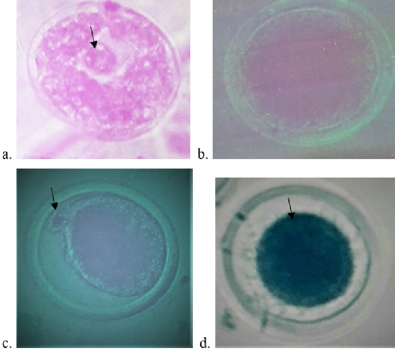
Photomicrographs of fully grown Nili Ravi buffalo oocytes during in vitro culture: (a) An intact germinal vesicle (indicated by arrow) with a single prominent nucleus arrested in dictyate stage of the first meiotic prophase. (b) Oocyte undergoing GVBD showing complete dissolution of the GV with only a small remnant of the nucleolus. (c) MII oocyte arrested at metaphase II stage and omitted a polar body (indicated by arrow). (d) Degenerated oocyte showing shrinkage of ooplasm. Photographs were taken from an inverted phase contrast (Nikon) microscope (objectives: a-d, X20).
References
- Abdoon, A.S.S., Kandil, O.M., Otoi, T. and Suzuki, T., 2001. Influence of oocyte quality, culture media and gonadotropins on cleavage rate and development of in vitro fertilized buffalo embryos. Anim. Reprod. Sci., 65: 215-223. https://doi.org/10.1016/S0378-4320(01)00079-3[CrossRef]
- Anisworth, L., Tsang, B.K., Downey, R.B., Marcus, G.J. and Armstrong, D.T., 1980. Interrelationship between follicular fluid steroid levels, gonadotrophic stimuli and oocytes maturation during preovulatory development of porcine follicles. Biol. Repord., 23: 621-627. https://doi.org/10.1095/biolreprod23.3.621[CrossRef]
- Bahrami, M., Morris, M.B. and Day, M.L., 2019. Amino acid supplementation of a simple inorganic salt solution supports efficient in vitro maturation (IVM) of bovine oocytes. Sci. Rep., 9: 11739. https://doi.org/10.1038/s41598-019-48038-y[CrossRef]
- Baruselli, P.S., Soares, J.G., Gimenes, L.U., Monteiro, B.M., Olazarri, M.J. and Carvalho, N.A.T., 2013. Control of buffalo follicular dynamics for artificial insemination, superovulation and in vitro embryo production. Buffalo Bull., 32: 160-176.
- Beker, A.R.C.L., Colenbrander, B. and Bevers, M.M., 2002. Effect of 17beta-estradiol on the in vitro maturation of bovine oocytes. Theriogenology, 58: 1663-1673. https://doi.org/10.1016/S0093-691X(02)01082-8[CrossRef]
- Conti, M. and Franciosi, F., 2018. Acquisition of oocyte competence to develop as an embryo: integrated nuclear and cytoplasmic events. Hum. Reprod. Update, 24: 245-266. https://doi.org/10.1093/humupd/dmx040[CrossRef]
- Cortvrindt, R., Hu, Y. and Smitz, J., 1998. Recombinant LH as survival and differentiation factor increases oocyte maturation in recombinant FSH supplemented mouse preantral follicle culture. Hum. Reprod., 13: 1292-1302. https://doi.org/10.1093/humrep/13.5.1292[CrossRef]
- Dell’Aquila, M.E., Albrizio, M., Maritato, F., Minoia, P. and Hinrichs, K., 2003. Meiotic competence of equine oocytes and pronucleus formation after intracytoplasmic sperm injection (ICSI) as related to granulosa cell apoptosis. Biol. Reprod., 68: 2065-2072. https://doi.org/10.1095/biolreprod.102.009852[CrossRef]
- Drost, M., 2007. Advanced reproductive technology in the water buffalo. Theriogenology, 68: 450-453. https://doi.org/10.1016/j.theriogenology.2007.04.013[CrossRef]
- El-Magd, M.A., Ghoniem, A.M., Helmy, N.M., Abdelfattah-Hassan, A., Saleh, A.A., Abd Allah, E.A., Essawi, W.M. and Kahilo, K.A., 2019. Effect of myostatin inhibitor (myostatin pro-peptide) microinjection on in vitro maturation and subsequent early developmental stages of buffalo embryo. Theriogenology, 126: 230-238. https://doi.org/10.1016/j.theriogenology.2018.12.027[CrossRef]
- Ferraz, M.L., Sá Filho, M.F., Batista, E.O.S., Watanabe, Y.F., Watanabe, M.R., Dayan, A. and Baruselli, P.S., 2015. Paradoxical effects of bovine somatotropin treatment on the ovarian follicular population and in vitro embryo production of lactating buffalo donors submitted to ovum pick-up. Anim. Reprod. Sci., 154: 1-7. https://doi.org/10.1016/j.anireprosci.2014.12.017[CrossRef]
- Ferré, L., Kjelland, M., Strøbech, L., Hyttel, P., Mermillod, P. and Ross, P., 2020. Recent advances in bovine in vitro embryo production: Reproductive biotechnology history and methods. Animal, 14: 991-1004. https://doi.org/10.1017/S1751731119002775[CrossRef]
- Galli, C., Duchi, R., Colleoni, S., Lagutina, I. and Lazzari, G., 2014. Ovum pick up, intracytoplasmic sperm injection and somatic cell nuclear transfer in cattle, buffalo and horses from the research laboratory to clinical practice. Theriogenology, 81: 138-151. https://doi.org/10.1016/j.theriogenology.2013.09.008[CrossRef]
- Gasparrini, B., 2013. In vitro embryo production in buffalo: Yesterday, today and tomorrow. Buffalo Bull., 32: 188-195.
- Gong, J.G., McBride, D., Bramley, T.A. and Webb, R., 1993. Effects of recombinant bovine somatotrophin, insulin-like growth factor-I and insulin on the proliferation of bovine granulosa cells steroidogenesis in vitro. J. Endocrinol., 143: 157-164. https://doi.org/10.1677/joe.0.1430157[CrossRef]
- Herrick, J.R., 2019. Assisted reproductive technologies for endangered species conservation: Developing sophisticated protocols with limited access to animals with unique reproductive mechanisms. Biol. Reprod., 100: 1158-1170. https://doi.org/10.1093/biolre/ioz025[CrossRef]
- Itoh, T., Kacchi, M., Abe, H., Sendai, Y. and Hoshi, H., 2002. Growth, antrum formation, and estradiol production of bovine preantral follicles cultured in a serum-free medium. Biol. Reprod., 67: 1099-1105. https://doi.org/10.1095/biolreprod67.4.1099[CrossRef]
- Khan, M.I., Jalali, S., Shahid, B., Shami, S.A. and Ikramullah, 2009. Effect of human recombinant follicle stimulating hormone on meiotic competence of in vitro grown Nili Ravi buffalo oocytes. Int. J. Biomed. Biol. Eng., 3: 493-496.
- Lima, G.L., Luz, V.B., Lima, L.F., Rocha, R.M.P., Castro, S.V., Castelo, T.S., Rodrigues, A.P.R., Figueiredo, J.R. and Silva, A.R., 2018. Interactions between different media and follicle-stimulating hormone supplementation on in vitro culture of preantral follicles enclosed in ovarian tissue derived from collared peccaries (Pecari tajacu Linneaus, 1758). Reprod. Domest. Anim., 53: 880-888. https://doi.org/10.1111/rda.13179[CrossRef]
- Lopes, J.S., Canha-Gouveia, A., Paris-Oller, E. and Coy, P., 2019. Supplementation of bovine follicular fluid during in vitro maturation increases oocyte cumulus expansion, blastocyst developmental kinetics, and blastocyst cell number. Theriogenology, 126: 222-229. https://doi.org/10.1016/j.theriogenology.2018.12.010[CrossRef]
- Maksura, H., Akon, N., Islam, M.N., Akter, I., Modak, A.K., Khatun, A., Alam, M.H., Hashem, M.A., Amin, M.R. and Moniruzzaman, M., 2021. Effects of estradiol on in vitro maturation of buffalo and goat oocytes. Reprod. Med. Biol., 20: 62-70. https://doi.org/10.1002/rmb2.12350[CrossRef]
- Newton, H., Wang, Y., Groome, N.P. and Illingworth, P., 2002. Inhibin and activin secretion during murine preantral follicle culture and following hCG stimulation. Hum. Reprod., 17: 38-43. https://doi.org/10.1093/humrep/17.1.38[CrossRef]
- Ocaña-Quero, J.M., Pinedo-Merlín, M., Ortega-Mariscal, M., and Moreno-Millán, M., 1998. Influence of human and bovine insulin on in vitro maturation, fertilization and cleavage rates of bovine oocytes. Arch. Zootec., 47: 85-93.
- Ohash, O.M., Nogueira, N., Cordeiro, S., Filho, T.R., Francisco, H., Ribeiro, L. and Guimarães, T.V., 2017. Produção in vitro de embrião (PIVE) na espécie bubalina. Rev. Bras. Reprod. Anim., 14: 195-200.
- Paulson, R.J., Fauser, B., Vuong, L.T.N. and Doody, K., 2016. Can we modify assisted reproductive technology practice to broaden reproductive care access? Fertil. Steril., 105: 1138-1143. https://doi.org/10.1016/j.fertnstert.2016.03.013[CrossRef]
- Pereira, B.A., Zangeronimo, M.G., Martín, M.C., Gadani, B., Chaves, B.R., Rodríguez-Gil, J.E., Bonet, S. and Yeste, M., 2019. Supplementing maturation medium with insulin growth factor I and vitrification-warming solutions with reduced glutathione enhances survival rates and development ability of in vitro matured vitrified-warmed pig oocytes. Front. Physiol., 9: 1894. https://doi.org/10.3389/fphys.2018.01894[CrossRef]
- Pioltine, E.M., Costa, C.B., Latorraca, L.B., Franchil, F.F., dos Santos, P.H., Mingoti, G. Z., de Paula-Lopes, F.F., and Nogueira, M.F.G., 2021. Treatment of in vitro-matured bovine oocytes with tauroursodeoxycholic acid modulates the oxidative stress signaling pathway. Front. Cell Dev. Biol., 9: 623852. https://doi.org/10.3389/fcell.2021.623852[CrossRef]
- Saliba, W., Gimenes, L., Drumond, R., Bayão, H., Alvim, M., Baruselli, P. and Gasparrini, B., 2013. Efficiency of OPU-IVEP-ET of fresh and vitrified embryos in buffaloes. Buffalo Bull., 32: 385-388.
- Shahid, B., Jalali, S., Khan, M.I., and Shami, S.A., 2014. Different methods of oocytes recovery for in vitro maturation in Nili Ravi buffalo’s oocytes. APCBEE Procedia, 8: 359-363. https://doi.org/10.1016/j.apcbee.2014.03.054[CrossRef]
- Shahid, B., Jalali, S., Khan, M.I., and Shami, S.A., 2015. Effect of gonadotrophins, oestradiol and insulin on cumulus expansion of Nili Ravi buffalo oocytes. Pak. J. Pharm. Sci., 28: 95-101.
- Spicer, L.J., Alpizar, E., and Echternkamp, S.E., 1993. Effects of insulin, insulin-like growth factor I, and gonadotropins on bovine granulosa cell proliferation, progesterone production, estradiol production, and(or) insulin-like growth factor I production in vitro. J. Anim. Sci., 71: 1232-1241. https://doi.org/10.2527/1993.7151232x[CrossRef]
- Sun, M.H., Li, X.H., Xu, Y., Xu, Y., Pan, Z.N. and Sun, S.C., 2020. Citrinin exposure disrupts organelle distribution and functions in mouse oocytes. Environ. Res., 185: 109476. https://doi.org/10.1016/j.envres.2020.109476[CrossRef]
- Tremoleda, J.L., Tharasanit, T., van Tol, H.T.A., Stout, T.A.E., Colenbrander, B. and Bevers, M.M., 2003. Effects of follicular cells and FSH on the resumption of meiosis in equine oocytes matured in vitro. Reproduction, 125: 565-577. https://doi.org/10.1530/rep.0.1250565[CrossRef]
- Ullah, I., Jalali, S., Shami, S.A., Farooq, K. and Khan, M.I., 2006. Effect of the 2-mercaptoethanol on Nili Ravi buffalo oocytes during in vitro maturation. J. Anim. Vet. Adv., 5: 380-385. https://medwelljournals.com/abstract/?doi=javaa.2006. 380.385
- van Tol, H.T.A., van Eijk, M.J.T., Mummery, C.L., van den Hurk, R. and Bevers, M.M., 1996. Influence of FSH and hCG on the resumption of meiosis of bovine oocytes surrounded by cumulus cells connected to membrana granulosa. Mol. Reprod. Dev., 45: 218-224. https://doi.org/10.1002/(SICI)1098-2795(199610)45:2<218::AID-MRD15>3.0.CO;2-X[CrossRef]
- Viana, J., 2019. Statistics of embryo production and transfer in domestic farm animals. International Embryo Transfer Society, Champaign, IL, USA, pp. 38.
- Widayati, D.T., and Pangestu, M., 2020. Effect of follicle-stimulating hormone on Bligon goat oocyte maturation and embryonic development post in vitro fertilization. Vet. World, 13: 2443-2446. https://doi.org/10.14202/vetworld.2020.2443-2446[CrossRef]
- Woudenberg, A.R.B., van Tol, H.T.A., Roelen, B.A.J., Colenbrander, B. and Bevers, M.M., 2004. Estradiol and its membrane-impermeable conjugate (estradiol-bovine serum albumin) during in vitro maturation of bovine oocytes: Effects on nuclear and cytoplasmic maturation, cytoskeleton, and embryo quality. Biol. Reprod., 70: 1465-1474. https://doi.org/10.1095/biolreprod.103.025684[CrossRef]
- Wrenzycki, C. and Stinshoff, H., 2013. Maturation environment and impact on subsequent developmental competence of bovine oocytes. Reprod. Domest. Anim., 48: 38-43. https://doi.org/10.1111/rda.12204[CrossRef]
- Younis, A.I., Brackett, B.G. and Fayrer-Hosken, R.A., 1989. Influence of serum and hormones on bovine oocytes maturation and fertilization in vitro. Gamete Res., 23: 189-201. https://doi.org/10.1002/mrd.1120230206[CrossRef]
Services
Article usage statistics
Loading…