
The reproductive performance of Egyptian buffalo might be determined by genetic variants, but it is also affected by environmental factors. The possible action of L-carnitine (LC) on the maturation of Egyptian buffalo oocytes has not been determined yet. This study aims to estimate the effects of supplementing LC on the in-vitro fertilization (IVF) of Egyptian buffaloes. We evaluated blastocyst and apoptosis rates in order to substantiate the potential development of cumulus oocytes–complexes (COC) and determine the effect of LC in reducing the occurrence of apoptosis under in vitro maturation (IVM) conditions for buffalo blastocysts. The tissue culture media included a control culture medium and other three LC supplemented media (2, 3 or, 4 mM LC). All LC treatments had higher COCs percentages at the metaphase II (MII) compared to the control group. The 2 Mmol LC treatment had the highest percentage of oocytes maturation rate at metaphase ΙΙ (47%) compared to the control group (33.3%). The same treatment had a higher percentage of cleaved embryos (75.4% vs 64.9%) and blastocyst rates (43,4% vs. 31,5%, respectively). Furthermore, the same 2 Mmol LC treatment had a lower apoptotic rate of blastocyst compared to the control group (35,8% vs 58,3%). In conclusion, LC could increase the maturation and blastocyst rates of oocytes and reduce apoptosis in Egyptian buffalo embryos.
Research Article · Volume 56, Issue 5, 13 July 2024 · Open Access
L-Carnitine Supplementation During In vitro Maturation of Egyptian Buffalo Oocytes Improved Embryo Yield and Decreased Blastocyst Apoptotic Rate
· · · · · ·
Affiliations & Notes ⌄
- 1Animal Production Department, Faculty of Agriculture, Kafrelshiekh, University, Kafrelsheikh, Egypt 2Biotechnology Research Department, Animal Production Research Institute, Agriculture Research Center, Egypt 3Animal Production Department, Faculty of Agriculture, Menoufia University, Menoufia, Egypt 4Animal Production Department, Faculty of Agriculture, Ain Shams University, Cairo, Egypt 5Animal Production Department, Faculty of Agriculture, Al-Azhar University, Nasr City, Cairo, Egypt * Corresp
Article Info ⌄
- Journal:
- Pakistan Journal of Zoology
- Published:
- 13 July 2024

From the article
- The results indicated that all LC treatments had higher percentages of oocytes of maturation rate undergoes at metaphase I and ΙΙ, compared to the control group.
- Effect of L-carnitine concentrations added to maturation medium on embryonic development of Egyptian buffalo oocytes.
- Based on the current results, adding LC during in vitro oocyte maturation with concentration of 2mM could enhance the cumulus expansion.
- Furthermore, LC could increase blastocyst rates of oocytes and reduced the apoptosis in embryos of Egyptian buffalo.
Selected from the article's own text.
Abstract
Introduction
With a population in excess of 4 million Egyptian buffalo heads, this particular species produces a large volume of milk in numerous countries, such as Egypt. On a national estimate, this animal produces nearly %70 of the annual dairy produce in Egypt, making it one of the most valuable livestock ; .
Several physiological characteristics determine a lower reproductive performance for Egyptian buffalo including silent ovulation, delayed maturity, breeding seasonality, prolonged generation interval, low conception rate, low population of primordial follicles, and inactivated ovaries. These problems cause low efficiency of reproductive performance.
Biotechnological applications such as in vitro embryonic production (IVEP) can accelerate genetic improvement in livestock . Both in vitro fertilization (IVF), and in vitro maturation (IVM) of oocytes are applied to improve reproductive efficiency, and genetic potential in other Bovidae species . For instance, IVM and IVF techniques have been developed in cattle to produce embryos. Many studies are designed to estimate different parameters responsible for oocyte maturation, fertilization and subsequently embryonic development. There are some crucial factors affecting in vitro production of embryos including culture media type, media supplementations, and seasons of the year, ; . The poor embryonic in vitro culture development is major problems recorded by investigators in buffaloes. In vitro oocyte maturation is a significant element in the in vitro production system. The success of embryo production mainly depends on development rates of in vitro oocyte maturation. Therefore, many studies have been conducted to enhance the efficiency of in vitro oocyte maturation ; .
In vitro production embryos possess limited quality compared to their in vivo-produced counterparts. Oocyte quality is the main regulator to blastocyst rate, whereas embryo quality is affected by culture factors ; . For instance, in vitro production of embryo depends on ambient gases, incubating temperature, protein content, growth promoting factors and other handling processes ; . For instance, oxidative stress results from the unbalanced amount of reactive oxygen species (ROS) and antioxidants. Many factors increase ROS production, reduce availability of antioxidants or both actions. Oxidative stress leads to limited oocyte maturation and embryo’s growth ; . Also, energy production pathways in mitochondria consume the most cellular oxygen sources and are the main origin of ROS .
L-carnitine (LC) is a molecule which boosts fatty acids movement to the mitochondria through for ATP production. Thus, LC possesses a vital action in lipid metabolism. Furthermore, LC protects the live cells through free-radical-scavenger, and different anti-apoptotic mechanisms ; . Adding LC in maturation or culture media improves oocyte’ maturation of oocytes Sovernigo et al. (2017).
Cumulus cells and oocytes possess limited capacity to biosynthesize L-carnitine from precursor amino acids as they lack the metabolic machinery ; . There are many trials that evaluated the effects of LC during IVM in different animal species including mouse , porcine , ovidea ; , bovine ; , and Chinese swamp buffaloes . While, the possible action of LC on oocyte maturation of Egyptian buffalo cows is not determined.
The herein study aims to evaluate the effects of supplementing LC on the IVF of Egyptian buffaloes. L-carnitine could improve the embryonic developmental rate and decrease the apoptotic rate during in vitro maturation of oocyte.
Materials and Methods
This current trial was carried out under the Seminar Ethical Committee of Animal Production Department, Faculty of Agriculture, Kafrelshiekh University (No. 2018-11-47-1-SHO).
Unless otherwise indicated, we acquired chemicals from Sigma Chemical Company, MO, USA.
Experimental design
The herein study included determination of developmental potential of cumulus oocytes–complexes (COCs) IVM and to evaluate the blastocyst formation of cleaved embryos resulting from IVF under the effect of LC. Then, we investigated the effect of LC, added to the IVM medium, in reducing apoptosis in buffalo blastocysts by the terminal-deoxynucleotidyl transferasemediated dUTP nick-end labelling (TUNEL) method.
Ovary collection and oocyte selection
Ovaries were collected from adult Egyptian buffaloes at local abattoir at Menoufia Governorate. After slaughter, the ovaries were transferred within 3-4 h to the laboratory in a thermos contains Dulbecco’s phosphate buffered saline (DPBS), at temperature ranging from 35 to 37 ºC.
In the laboratory, extraneous tissues were removed and ovaries were washed several times with warmed sterile distilled water and gently dried with paper towels. Oocytes were recovered from follicles 2-8 mm in diameter by aspiration technique, using 10 mL disposal plastic syringe attached to 18-gauge needle.
The syringe contents were placed in a 15 mL falcon tube and submerged in a water bath at 38 °C for 10 min to settle. Later, the follicular fluid (supernatant) was slightly removed, and the sedimentation of follicular granulose cells and oocytes was gradually transferred to a 60 mm sterile plastic petri dish for further studies.
The aspirated oocytes were counted and washed several times with (TL-HEPES washing medium) under a stereomicroscope and were sorted into four grades, based on compaction, the number of cumulus cell layers, and homogeneity of ooplasm according to Gupta et al. (2001).
Briefly, grade A: oocytes with compact cumulus cells ≥ 3 layers with homogenous ooplasm COCs. Grade B: oocytes with compact 1 to 2 layers of cumulus cells with less homogeneity ooplasm. Grade C: oocytes with expanded cumulus cells (expanded). Grade D: oocytes completely devoid of cumulus cells (denuded oocytes) with heterogeneous ooplasm. We only selected Grade A oocytes for in vitro maturation.
In vitro maturation
In the herein study, we used TCM-199 as basic tissue culture which contained 10% (v/v) fetal calf serum (FCS) and 50 µg/mL gentamycin. We added Gentamycin with PMSG 20 IU /mL, LH 10 µg/mL, 1µg/mL Ε2 (17β-estradiol) and 50 μg/mL gentamicin (control medium). The media pH was adjusted at 7.2-7.3 and osmolarity at 280-300 mOsm/kg and filtered by 0.22 μm Millipore filter (control medium). The control maturation medium was supplemented with 2, 3 or, 4 mM LC as indicated by Held-Hoelker et al. (2017) and Knitlova et al. (2017). Three droplets (50 µL in size) of prepared media placed into sterile disposable plastic Petri dishes (3.5 cm in diameter) and covered with sterile mineral oil, the Petri dishes were incubated in CO2 incubator at 38.5 °C, 5% CO2 and 95% relative humidity for at least 1 h for equilibration. Oocytes were matured in vitro for 24 h in maturation medium droplets.
Assessment of nuclear maturation
Matured oocytes were denuded, fixed in for examining the rate of nuclear maturation. After maturation, the cultured oocytes of each group were directly denuded by repeated pipetting for 2 min. The denuded oocytes were washed two times in washing medium, loaded on glass slide, and were covered by a slip. Then, the slides were transferred into fixation solution (3 Ethanol: 1 Glacial acetic acid v/v) for overnight. Oocytes were stained with 1 % orcein in 45 % glacial acetic acid and examined under phase contrast microscope according to Faheem et al. (2011). Briefly, we sorted matured oocytes into germinal vesicle (GV), metaphase I (MI). Also, we sorted and considered immature oocytes with a nuclear membrane in germinal vesicle stage. Finally, we sorted and considered mature, metaphase II oocytes with a metaphase plate and a first polar body.
In vitro fertilization (IVF) and culture (IVC)
Three droplets (50 µL in volume) of prepared IVF-TALP media were placed into sterile disposable plastic Petri dishes (3.5 cm in diameter) and covered with sterile mineral oil. Petri dish was incubated in CO2 incubator at 38.5 °C, 5% CO2 and 95% relative humidity at least 1 h for equilibration before used. We performed IVF according to Imai (2006). Briefly, at the end of IVM, matured oocytes were rinsed three times without damage cumulus cells in TL- HEPES medium and twice in fertilization medium (IVF-TALP). Then 8-10 oocytes were pipetted in each droplet). After that 2-3 µL of sperm suspension were added to achieve a final concentration in the micro droplet of 1.5×10⁶/mL. Co-culture of oocytes and sperm in fertilization media was conducted for 22 h at 38.5 °C, 5 % CO2 in air with 95% relative humidity. At the end of insemination, putative zygotes were completely denuded from cumulus cells and spermatozoa by gentle pipetting with a fine glass pipette in pre-incubated IVC medium. Groups of 15–20 embryos were separately placed in culture drops. Fertilized oocytes were placed into another 4 well Petri dish containing 0.05 mL per well of TCM–199 medium supplemented with 22 µL/mL Na- pyruvate, 3mg/mL BSA, 10µL/mL NEAA (100 X), 20 µL/mL EAA (50X) and 50 µL/mL gentamycin sulphate (culture medium) and covered with sterile mineral oil and incubated at 38.5 °C and 5% CO2 with 95% humidity. Half of the medium was replaced every 48 h with the same medium. We calculated the cleavage rates and blastocyst formation rates on day 7 from the total numbers of oocytes that were subjected to IVF.
Evaluation of apoptosis in blastocysts by TUNEL method
We determined the numbers of inner cell masses (ICM) and trophectoderm (TE) cells and assessed apoptosis with TUNEL (terminal deoxynucleotidyl transferase dUTP nick end labeling) labeling of nuclei as depicted by Fouladi-Nashta et al. (2005) with slight modifications. In brief, blastocysts were treated with 0.2% (v/v) Triton X-100 for 20 s at 38.5°C, and phosphate buffer solution (PBS) and 0.02 mg/ mL polyvinyl pyrrolidone (PBS-PVP) with 3mg/ mL propidium iodide (PI) for 5 min at 38.5°C. Embryos were washed twice in PBS-PVP and then treated with PBS-PVP with 4% (w/v) paraformaldehyde and 0.01 mg/mL Hoechst 33258 for 5 min at room temperature. Embryos were washed twice in PBS-PVP and treated with PBS containing 0.1% (v/v) Triton X-100 and 0.1% (w/v) sodium citrate for 5 min on ice. Then, such embryos were washed twice in PBSPVP and incubated in a 15µL microdrop of TUNEL reaction mixture (containing enzyme solution and label solution in 1: 9 v/v ratio) according to the manufacturer’s instructions for 45 min at humectation in the dark. Embryos were washed in PBS-PVP medium, carefully mounted on glass slides in a glycerol-based antifade solution (Molecular Probes, Eugene, OR, USA), flattened by coverslips, and examined under UV light first with excitation at 330–385 nm and emission at 420 nm to visualize ICM/TE cell nuclei. Then the samples were examined with excitation of 480 nm and emission at 510 nm using an epifluorescence microscope (Eclipse E-600; Nikon) to evaluate TUNEL positive cell nuclei. Cleavage rates, blastocyst rates and incidence of apoptosis in blastocysts were recorded by Abramoff et al. (2004), Huang et al. (2019).
Statistical analysis
All data were analyzed using ASSISTAT software (version 7.7, beta freeware) to carryout statistical analysis. The mean between groups for maturation, embryonic development and embryonic apoptotic cells were compared by analysis of variance (ANOVA). Data was presented in the percentage form as relation with total oocytes cultured. Percentage values were arcsine-transformed before analysis. p < 0.05 was considered as significant. The post-hoc test was performed according to Duncan (1955).
Yijk = µ + Ti + eijk
Where µ: general mean; Yijk: observations; Ti: treatments type effects (LC concentrations), eijk: error.
RESULTS
The descriptive statistics of evaluated traits are depicted in Supplementary Table S1. The effect of LC concentration on in vitro oocyte maturation at different stages is depicted in Table I and Figure 1. The results indicated that all LC treatments had higher percentages of oocytes of maturation rate undergoes at metaphase I and ΙΙ, compared to the control group. The treatment of 2 mM LC had the highest percentages of oocytes undergo metaphase ΙΙ (maturation rate) 47.1% compared to the control 33.3% (p<0.01). This result indicated that 2 mM LC improved the maturation rate of Egyptian buffalo.
The result of the effect of LC on in vitro maturation and embryonic development of L-carnitine during both maturation and post-fertilization medium is shown in Table III. The higher percentage of fertilization rates was observed in 2 and 3mM of L-carnitine treatments (p<0.01). These data indicated that the treatments of 2 or 3mM of LC to maturation medium enhanced the fertilization rate (75.4 and 70.3%) compared to the control group (64.9%).
Table I. Effect of L-carnitine concentrations on in vitro maturation of Egyptian buffalo oocytes.
| L-carnitine (Mm) | Total incubated oocytes (n) | Maturation phases | ||||
| GV n (% ± SEM) | GVBD n (% ± SEM) | M1 n (% ± SEM) | M11 n (% ±SEM) | DEG n (% ±SEM) | ||
| Control | 117 | 15(12.82±1.77)b | 18(15.38±2.11)b | 31(26.49±2.07)b | 39 (33.33±2.09)b | 14 (11.96 ±1.17) |
| 2mmol | 136 | 12 (8.82±1.98)b | 13 (9.95±2.27)b | 33 (24.26±2.66)a | 64 (47.05±2.18)a | 14 (10.29±2.35) |
| 3mmol | 137 | 14 (10.21±1.53)a | 15 (10.94±2.40)a | 55 (40.14±2.92)b | 44 (32.11±2.33)b | 9 (6.56±2.09) |
| 4mmol | 137 | 26 (18.97±1.47)b | 23(16.78± 1.49)ab | 36 (26.27±1.66)b | 41 (29.92±2.75)b | 11 (8.02±1.82) |
| p-value | < 0.001 | 0.052 | < 0.001 | < 0.001 | 0.442 | |
GV, germinal vesicle; GVBD, germinal vesicle breakdown; M1, metaphase 1; M II, Metaphase II; DEG, Degenerated.
a b and c means denoted within the same column with different superscripts are significantly different (p<0.05).
Data in Table II and Figure 2 revealed that the highest fertilization rate was noted in 2mM L-carnitine (75.4%), followed by 3mM (70.3%), 4mM (64%) and the control group (64.9%) (p<0.0001). All treated groups had higher developmental rate of oocytes following IVF compared to the control group (Table III). At the blastocyst stage, the treatment of 2mM L-carnitine had the higher developmental competence than those of 3, 4mM and control (43.5, 39.2 and 37.5% vs. 20.5 %; p<0.01).
Blastocyst stage was increased in all LC treated groups (43.4 and 39.2% vs control 31.5%; p<0.05) as depicted in Table III. However, the percentage of apoptotic one was higher in the control (58.3%) than the treated groups (35.8, 51.5 and 57.1%) as depicted in Table IV.
Table II. Effect of LC concentrations on fertilization rate of Egyptian buffalo oocytes.
| L-carnitine (Mm) | Total incubated oocytes (n) | Cleaved oocytes (n) | Fertilization rate (Means±SEM) % |
| Control | 117 | 76 | (64.95±0.45) c |
| 2 | 122 | 92 | (75.40±0.95) a |
| 3 | 145 | 102 | (70.34±0.77) b |
| 4 | 125 | 80 80 | (64±0.74) c |
| p-value | < | < 0.001 |
a, b and c means denoted within the same column with different superscripts are significantly different (p<0.001).
DISCUSSION
In this study, supplementation LC during IVM led to increase maturation rate of oocytes. Furthermore, LC improved both cleavage and blastocysts rates, and greatly affected total number of blastocysts.
The grade A cumulus expansion was significantly increased after adding 2 mM LC maturation medium. During this maturation Phase, oocytes are sensitive to oxidative stress ; , which can be avoided by using antioxidants. In the current study, adding 2mM/mL LC to maturation medium increased the maturation rate. Placidi et al. (2022) indicated that LC decreases reactive oxygen species (ROS) level in maturation medium. This result agrees with some previous studies ; . LC possesses an antioxidative action and removes many free radicals ; . The protective effects of LC reduce ROS intracellular levels, that lead to protect micro-organelles such as mitochondria ; . Additionally, LC boosts β-oxidation of fatty acids and ATP generation ; . Such oxidation assists both nuclear and cytoplasmic maturation and leads to oocyte developmental competence . In the herein study, proportion of oocytes was higher in LC treatments at MII stage. Furthermore, LC decreased degenerated oocytes rate. Thus, adding LC during IVM had an obvious impact on nuclear maturation by enhancing both meiotic competence and cytoplasmic maturation biological processes. All nuclear, cytoplasmic and molecular maturation stages of oocyte can markedly affect developmental competence and embryo quality ; .
Lorenzo et al. (1994) and Kumar et al. (2020) reported that the cumulus expansion is related to fluctuation of gonadotropins, steroids, secretions of the oocyte, growth factors, and unidentified compounds, in bovine oocytes. As, most authors we used the LC during IVM process, and we did not repeatedly add LC during in vitro culture ; . In the same way, our results agree with Knitlova et al. (2017) who indicated that the embryo production rate is increases by adding 2.5 mM of LC from meiotically more competent oocytes. In mouse, LC supplementation improves developmental competence oocytes in terms by improving metabolism of fatty acids ; .
Table III. Effect of L-carnitine concentrations added to maturation medium on embryonic development of Egyptian buffalo oocytes.
| L-carnitine (mM) | Total cleaved oocytes (n) | Developmental stage | |||
| 4 cells n (% ± SEM) | 8-16 cells n (%± SEM) | Morula n (% ± SEM) | Blastocyst n (%± SEM) | ||
| Control | 76 | 11(14.47±0.9) | 11(14.47±1.91) | 19(25±2.00) | 24 (31.57±2.34)c |
| 2 | 92 | 12(13.04±1.26) | 11(11.95±1.10) | 18(19.56±1.95) | 40(43.47±2.19)a |
| 3 | 102 | 17 (16.66±0.97) | 18(17.64±1.47) | 18(17.64±1.60) | 40(39.21±1.88)ab |
| 4 | 80 | 14(17.50±0.90) | 16 (20.0±2.21) | 18 (22.5±1.60) | 30(37.5±0.74)b |
| p-value | 0.289 | 0.324 | 0.631 | < 0.001 | |
a b and c means denoted within the same column with different superscripts are significantly different (p<0.001).
Table IV. Effect of L-carnitine concentrations on embryonic and apoptotic blastocysts.
| L-carnitine (Mm) | number of blastocysts (n) | Apoptotic blastocysts n (% ± SEM) |
| control | 24 | 14 (58.33±0.17) |
| 2 | 39 | 14 (35.89±0.167) |
| 3 | 33 | 17 (51.51±0 .26) |
| 4 | 28 | 16 (57.14± 0.22) |
| p-value | 0.083 |
These effects were not observed in the current investigation. the origin of LC is related to the variation between mouse and buffalo (LC hydrochloride, or LC inner salts) . Such discrepancies are sometimes related differences in quality of oocytes. In the herein investigation, LC was used in maturating COCs of buffalo at early- stage embryos.
Furthermore, excessive LC concentration may decrease lipid density which leads developmental se biological processes . Hashimoto (2009) and Somfai et al. (2011) recorded that LC improves the meiotic performance of oocytes throughout retardation the apoptosis of granulosa cells. In this study, adding LC boosted blastocyst formation its character as LC treatments had a higher total cell count. Therefore, LC was enhanced embryonic progress rather completion of 1st meiosis.
The results of the present study showed that the LC affects the cleavage rate and the 4-cell embryo stage, and this finding in agreement with previous reports by Held-Hoelker et al. (2017) and Zolini et al. (2019). Also, adding LC during IVM boosted the current blastocysts rate. On the other hand, Held-Hoelker et al. (2017) added 2.5 mM of LC had no remarkable effect on the embryonic production rate during in vitro culture. In our study, the LC led to improve embryo development rates d. This result is similar to Ghanem (2015), who found a significant increase in embryo production rates using LC at 1.5 mM during in vitro culture. Almost trials used LC during IVM, but they did not repeatedly add LC during in vitro culture ; ; ; ; . Phongnimitr et al. (2013) found that 0.6 mg/mL of LC enhanced the blastocyst production rate. While, no available trials were performed in transferring produced embryos into recipient bovine under LC supplementation. Our finding of improving blastocyst in the 2mM L-carnitine group agrees with the results of Agarwal et al. (2018) in mice, while Zolini et al. (2019), found unclear differences between treatment with 0.75mM LC compared to a control group during the whole in vitro culture. Our data disagree with Carrillo-Gonzalez et al. (2019) who found no effect for L-carnitine in maturating medium on embryonic developmental rate.
In the current study, blastocyst rates of LC treatments had higher values (43.4, 39.2 and 37.5% respectively) compared to the values indicated by Rasmussen et al. (2013) (30%) and Cavalieri et al. (2018) (37%). On a different trend, LC had a similar a blastocyst rate to that indicated by Pontes et al. (2009). We found that 2 mM of LC enhanced the embryonic rate production in oocytes from small follicles (<5 mm).
While, oocytes derived from medium follicles (6-10 mm) and sorted as meiotically more competent had no significant difference . In our investigation, the inclusion criteria to follicle diameter were ranged from 2 to 8 mm, according to Knitlova et al. (2017). The blastocyst rate in 2Mm treatment (43.4%) is higher than the results of Ghanem (2015) (22.5%), Phongnimitr et al. (2013) (25.1%), and Knitlova et al. (2017) (group <5 mm = 25.8%). Thus, LC treatments improved oocytes with high developmental competence. This improvement was limited with oocytes with high competence properties.
Programmed cell death involves many signs including cellular shrinkage, condensation of chromatin, membrane damage, and other indications . There are many other factors that can cause cell apoptosis including chromosomal abnormalities ; , imbalance of growth factors ; , reactive oxygen species and suboptimal culture conditions . Therefore, suboptimal in vitro culture conditions may lead to embryonic cell death. The rate of apoptotic cells in embryos is utilized to estimate embryo quality ; . Anywhere, the impact of LC to promote higher oocyte character indicates low apoptotic rates in embryos. Addition of antioxidants during IVM or IVC reduces the number of apoptotic cells in blastocysts. A higher incidence of apoptosis is related to fragmentation arrest of embryos, as embryos with a lower incidence of apoptosis and higher number of cells are more likely to alive and to develop ; . Hassan et al. (2022) indicated that LC declines apoptotic rates that induced by mitochondria under both in vivo and in vitro cases. Li et al. (2021) and Pillich et al. (2005) found that L-carnitine is not only stabilizing mitochondrial membranes, but also can improve the energy source to the organelle and protect cells against incidence of apoptosis. In our study, supplementation of 2mM of LC reduced the apoptotic rate in blastocysts of buffalo using IVM medium.
This study needs further comprehensive in measuring oxidative stress and antioxidant enzymes. Further investigations are required to study the molecular mechanisms of LC in oocyte maturation, and the embryonic development.
Conclusion
Based on the current results, adding LC during in vitro oocyte maturation with concentration of 2mM could enhance the cumulus expansion. Furthermore, LC could increase blastocyst rates of oocytes and reduced the apoptosis in embryos of Egyptian buffalo.
Acknowledgement
The authors would like to thank the member of Anatomy Lab of Faculty of Veterinary, Kafrelsheikh University for facilitating this work. The authors also are grateful to Dr. Shrief Shamiah from Animal Production Research Institute (APRI) for his valuable and continuous support.
Funding
The study received no external funding.
IRB approval
This work was approved by the Animal Production Department, Faculty of Agriculture, Kafrelshiekh University and The Animal Production Research Institute (APRI).
Ethical statement
This current trial was carried out under the Seminar Ethical Committee of Animal Production Department, Faculty of Agriculture, Kafrelshiekh University (No. 2018-11-47-1-SHO).
Supplementary material
There is supplementary material associated with this article. Access the material online at: https://dx.doi.org/10.17582/journal.pjz/20220811160819
Statement of conflict of interest
The authors have declared no conflict of interest.
Figures
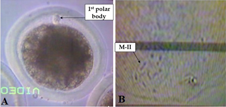
Nuclear maturation. (A) Fresh oocyte where first polar body was extruded in previtilline space (metaphase II). (B) Oocyte stained by orcein stain and showed that chromosomes at metaphase II.
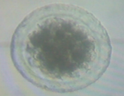
Morula stage embryo. The formed embryos were examined using stereomicroscope after seven days of culture by inverted microscope for kinetic cleavage stage (blastocyst stages).
References
- Abou-Bakr, S., 2009. Genetic and phenotypic trends of 305-day milk yield of Holstein cows raised at commercial farm in Egypt. Egypt. J. Anim. Prod., 46: 85-92. https://doi.org/10.21608/ejap.2009.94031[CrossRef]
- Abramoff, M.D., Magalhães, P.J., and Ram, S.J., 2004. Image processing with ImageJ. Biophotonics Int., 11: 36 - 42.
- Agarwal, A., Sengupta, P., and Durairajanayagam, D., 2018. Role of l-carnitine in female infertility. Reprod. Biol. Endocrinol., 16. https://doi.org/10.1186/s12958-018-0323-4[CrossRef]
- Bennemann, J., Grothmann, H., and Wrenzycki, C., 2018. Reduced oxygen concentration during in vitro oocyte maturation alters global DNA methylation in the maternal pronucleus of subsequent zygotes in cattle. Mol. Reprod. Dev., 85: 849-857. https://doi.org/10.1002/mrd.23073[CrossRef]
- Bezerra, F.T.G., Dau, A.M.P., Van Den Hurk, R., and Silva, J.R.V., 2021. Molecular characteristics of oocytes and somatic cells of follicles at different sizes that influence in vitro oocyte maturation and embryo production. Domest. Anim. Endocrinol., 74: 106485. https://doi.org/10.1016/j.domaniend.2020.106485[CrossRef]
- Bucktrout, R.E., Ma, N., Aboragah, A., Alharthi, A.S., Liang, Y., Lopreiato, V., Lopes, M.G., Trevisi, E., Alhidary, I.A., Fernandez, C., and Loor, J.J., 2021. One-carbon, carnitine, and glutathione metabolism-related biomarkers in peripartal Holstein cows are altered by prepartal body condition. J. Dairy Sci., 104: 3403-3417. https://doi.org/10.3168/jds.2020-19402[CrossRef]
- Carrillo-Gonzalez, D., Rodr, guez-Osorio, N., lida, Long, C., squez-Araque, N., and Maldonado-Estrada, J., 2019. Effect of l-carnitine supplementation during in vitro maturation and in vitro culture on oocyte quality and embryonic development rate of bovines. Asian Pac. J. Reprod., 8: 289-296. https://doi.org/10.4103/2305-0500.270107[CrossRef]
- Cavalieri, F.L.B., Morotti, F., Seneda, M.M., Colombo, A.H.B., Andreazzi, M.A., Emanuelli, I.P., and Rigolon, L.P., 2018. Improvement of bovine in vitro embryo production by ovarian follicular wave synchronization prior to ovum pick-up. Theriogenology, 117: 57-60. https://doi.org/10.1016/j.theriogenology.2017.11.026[CrossRef]
- de Camargo, J., Rodrigues, R., Valente, R.S., Muller, D.B., Vireque, A.A., Belaz, K.R.A., Bohrer, R.C., Basso, A.C., Eberlin, M.N., Fontes, P.K., Nogueira, M.F.G., and Sudano, M.J., 2022. Evaluation of a serum-free culture medium for the enhanced vitrification cryosurvival of bovine in vitro derived embryos. Livest. Sci., 260: 104922. https://doi.org/10.1016/j.livsci.2022.104922[CrossRef]
- Duncan, D.B., 1955. Multiple range and multiple F tests. Biometrics, 11: 1-42. https://doi.org/10.2307/3001478[CrossRef]
- Dunning, K.R., and Robker, R.L., 2012. Promoting lipid utilization with l-carnitine to improve oocyte quality. Anim. Reprod. Sci., 134: 69-75. https://doi.org/10.1016/j.anireprosci.2012.08.013[CrossRef]
- Eldawy, M.H., Lashen, M.E.-S., Badr, H.M., and Farouk, M.H., 2021. Milk production potential and reproductive performance of Egyptian buffalo cows. Trop. Anim. Hlth. Prod., 53: 282. https://doi.org/10.1007/s11250-021-02722-2[CrossRef]
- Elgebaly, M.M., Hazaa, A.B.M., Amer, H.A., and Mesalam, A., 2022. L-Cysteine improves bovine oocyte developmental competence in vitro via activation of oocyte-derived growth factors BMP-15 and GDF-9. Reprod. Domest. Anim., 57: 734-742. https://doi.org/10.1111/rda.14113[CrossRef]
- Fabian, D., Koppel, J., and Maddox-Hyttel, P., 2005. Apoptotic processes during mammalian preimplantation development. Theriogenology, 64: 221-231. https://doi.org/10.1016/j.theriogenology.2004.11.022[CrossRef]
- Faheem, M.S., Carvalhais, I., Chaveiro, A., and Moreira da Silva, F., 2011. In vitro oocyte fertilization and subsequent embryonic development after cryopreservation of bovine ovarian tissue, using an effective approach for oocyte collection. Anim. Reprod. Sci., 125: 49-55. https://doi.org/10.1016/j.anireprosci.2011.02.011[CrossRef]
- Fakruzzaman, M., Ghanem, N., Bang, J.I., Ha, A.N., Lee, K.L., Sohn, S.H., Wang, Z., Lee, D.S., and Kong, I.K., 2015. Effect of peroxiredoxin II on the quality and mitochondrial activity of pre-implantation bovine embryos. Anim. Reprod. Sci., 159: 172-183. https://doi.org/10.1016/j.anireprosci.2015.06.015[CrossRef]
- Fouladi-Nashta, A.A., Gutierrez, C.G., Garnsworthy, P.C., and Webb, R., 2005. Effects of dietary carbohydrate source on oocyte/embryo quality and development in high-yielding, lactating dairy cattle. Biol. Reprod. Abstr., M244: 135-136.
- Ghanem, N., 2015. L-carnitine improved bovine blastocyst rate and quality when supplemented at different preimplantation stages. Egypt. J. Anim. Prod., 52: 89-99. https://doi.org/10.21608/ejap.2015.93625[CrossRef]
- Giorgi, V.S.I., Da Broi, M.G., Paz, C.C.P., Ferriani, R.A., and Navarro, P.A., 2016. N-acetyl-cysteine and L-carnitine prevent meiotic oocyte damage induced by follicular fluid from infertile women with mild endometriosis. Reprod. Sci., 23: 342-351. https://doi.org/10.1177/1933719115602772[CrossRef]
- Gordon, I., 2003. Laboratory production of cattle embryos. CABI, Wallingford, UK. https://doi.org/10.1079/9780851996660.0000[CrossRef]
- Gülçin, İ., 2006. Antioxidant and antiradical activities of L-carnitine. Life Sci., 78: 803-811. https://doi.org/10.1016/j.lfs.2005.05.103[CrossRef]
- Gupta, P.S., Nandi, S., Ravindranatha, B.M., and Sarma, P.V., 2001. Effect of commercially available PMSG on maturation, fertilization and embryo development of buffalo oocytes in vitro. Reprod. Fertil. Dev., 13: 355-360. https://doi.org/10.1071/RD01026[CrossRef]
- Hardy, K., and Spanos, S., 2002. Growth factor expression and function in the human and mouse preimplantation embryo. J. Endocrinol., 172: 221-236. https://doi.org/10.1677/joe.0.1720221[CrossRef]
- Hashimoto, S., 2009. Application of in vitro maturation to assisted reproductive technology. J. Reprod. Dev., 55: 1-10. https://doi.org/10.1262/jrd.20127[CrossRef]
- Hassan, N., Rashad, M., Elleithy, E., Sabry, Z., Ali, G., and Elmosalamy, S., 2022. L-carnitine alleviates hepatic and renal mitochondrial-dependent apoptotic progression induced by letrozole in female rats through modulation of Nrf-2, Cyt c and CASP-3 signaling. Drug Chem. Toxicol., pages1-12. https://doi.org/10.1080/01480545.2022.2039180[CrossRef]
- Hassanat, F., Gervais, R., and Benchaar, C., 2017. Methane production, ruminal fermentation characteristics, nutrient digestibility, nitrogen excretion, and milk production of dairy cows fed conventional or brown midrib corn silage. J. Dairy Sci., 100: 2625-2636. https://doi.org/10.3168/jds.2016-11862[CrossRef]
- Held-Hoelker, E., Klein, S.L., Rings, F., Salilew-Wondim, D., Saeed-Zidane, M., Neuhoff, C., Tesfaye, D., Schellander, K., and Hoelker, M., 2017. Cryosurvival of in vitro produced bovine embryos supplemented with L-carnitine and concurrent reduction of fatty acids. Theriogenology, 96: 145-152. https://doi.org/10.1016/j.theriogenology.2017.03.014[CrossRef]
- Huang, C.H., Wang, F.T., and Chan, W.H., 2019. Enniatin B1 exerts embryotoxic effects on mouse blastocysts and induces oxidative stress and immunotoxicity during embryo development. Environ. Toxicol., 34: 48-59. https://doi.org/10.1002/tox.22656[CrossRef]
- Imai, K., 2006. The efficiency of embryo production by ovum pick-up and in vitro fertilization in cattle. J. Reprod. Dev., 52: S19-S29.
- Jeulin, C., 1996. Role of free L-carnitine and acetyl-L-carnitine in post-gonadal maturation of mammalian spermatozoa. Hum. Reprod. Update, 2: 87-102. https://doi.org/10.1093/humupd/2.2.87[CrossRef]
- Knitlova, D., Hulinska, P., Jeseta, M., Hanzalova, K., Kempisty, B., and Machatkova, M., 2017. Supplementation of L-carnitine during in vitro maturation improves embryo development from less competent bovine oocytes. Theriogenology, 102: 16-22. https://doi.org/10.1016/j.theriogenology.2017.06.025[CrossRef]
- Kononov, S.U., Meyer, J., Frahm, J., Kersten, S., Kluess, J., Meyer, U., Huber, K., and Dänicke, S., 2020. Effects of dietary L-carnitine supplementation on platelets and erythrogram of dairy cows with special emphasis on parturition. Dairy, 2: 1-13. https://doi.org/10.3390/dairy2010001[CrossRef]
- Kumar, S., Singla, S.K., Manik, R., Palta, P., and Chauhan, M.S., 2020. Effect of basic fibroblast growth factor (FGF2) on cumulus cell expansion, in vitro embryo production and gene expression in buffalo (Bubalus bubalis). Reprod. Biol., 20: 501-511. https://doi.org/10.1016/j.repbio.2020.08.003[CrossRef]
- Levy, R., 2001. Genetic regulation of preimplantation embryo survival. In: Int. Rev. Cytol., Academic Press, pp. 1-37. https://doi.org/10.1016/S0074-7696(01)10002-1[CrossRef]
- Li, P., Xia, Z., Kong, W., Wang, Q., Zhao, Z., Arnold, A., Xu, Q., and Xu, J., 2021. Exogenous L-carnitine ameliorates burn-induced cellular and mitochondrial injury of hepatocytes by restoring CPT1 activity. Nutr. Metab. (Lond.), 18: 65. https://doi.org/10.1186/s12986-021-00592-x[CrossRef]
- Lorenzo, P.L., Illera, M.J., Illera, J.C., and Illera, M., 1994. Enhancement of cumulus expansion and nuclear maturation during bovine oocyte maturation in vitro by the addition of epidermal growth factor and insulin-like growth factor I. Reproduction, 101: 697-701. https://doi.org/10.1530/jrf.0.1010697[CrossRef]
- Mario, T., Yvonne, D., Veronica, S., Alejandro, D., Juan, R.M., Diana, F., Edmundo, B., Eduardo, C., Mario, A., Alma, L., Ivan, B., Concepcion, G., Fahiel, C., and Miguel, B., 2022. Effects of perfluorooctanoic acid in oxidative stress generation, DNA damage in cumulus cells, and its impact on in vitro maturation of porcine oocytes. Environ. Toxicol., 37: 1394-1403. https://doi.org/10.1002/tox.23492[CrossRef]
- McCann, M.R., George De la Rosa, M.V., Rosania, G.R., and Stringer, K.A., 2021. L-carnitine and acylcarnitines: Mitochondrial biomarkers for precision medicine. Metabolites, 11: 51. https://doi.org/10.3390/metabo11010051[CrossRef]
- Mevorach, D., Trahtemberg, U., Krispin, A., Attalah, M., Zazoun, J., Tabib, A., Grau, A., and Verbovetski-Reiner, I., 2010. What do we mean when we write senescence, apoptosis, necrosis, or clearance of dying cells? Annls N. Y. Acad. Sci., 1209: 1-9. https://doi.org/10.1111/j.1749-6632.2010.05774.x[CrossRef]
- Mishra, A., Reddy, I., Gupta, P., and Mondal, S., 2016. Developmental regulation and modulation of apoptotic genes expression in sheep oocytes and embryos cultured in vitro with L-carnitine. Reprod. Domest. Anim., 51: 1020-1029. https://doi.org/10.1111/rda.12789[CrossRef]
- Montjean, D., Entezami, F., Lichtblau, I., Belloc, S., Gurgan, T., and Menezo, Y., 2012. Carnitine content in the follicular fluid and expression of the enzymes involved in beta oxidation in oocytes and cumulus cells. J. Assist. Reprod. Genet., 29: 1221-1225. https://doi.org/10.1007/s10815-012-9855-2[CrossRef]
- Mostagir, A., Elmetwally, M., Montaser, A., and Zaabel, S., 2019. Effects of L-carnitine and cryoprotectants on viability rate of immature buffalo oocytes in vitro after vitrification. Alex. J. Vet. Sci., 62: 45-52. https://doi.org/10.5455/ajvs.57457[CrossRef]
- Natarajan, R., Shankar, M.B., and Munuswamy, D., 2010. Effect of α-tocopherol supplementation on in vitro maturation of sheep oocytes and in vitro development of preimplantation sheep embryos to the blastocyst stage. J. Assist. Reprod. Genet., 27: 483-490. https://doi.org/10.1007/s10815-010-9430-7[CrossRef]
- Nguyen, T.T.M., Gillet, G., and Popgeorgiev, N., 2021. Caspases in the developing central nervous system: Apoptosis and beyond. Front. Cell Dev. Biol., 9: 702404. https://doi.org/10.3389/fcell.2021.702404[CrossRef]
- Otoi, T., Yamamoto, K., Horikita, N., Tachikawa, S., and Suzuki, T., 1999. Relationship between dead cells and DNA fragmentation in bovine embryos produced in vitro and stored at 4°C. Mol. Reprod. Dev., 54: 342-347. https://doi.org/10.1002/(SICI)1098-2795(199912)54:4<342::AID-MRD4>3.0.CO;2-7[CrossRef]
- Paczkowski, M., Schoolcraft, W.B., and Krisher, R.L., 2014. Fatty acid metabolism during maturation affects glucose uptake and is essential to oocyte competence. Reproduction, 148: 429-439. https://doi.org/10.1530/REP-14-0015[CrossRef]
- Phongnimitr, T., Liang, Y., Srirattana, K., Panyawai, K., Sripunya, N., Treetampinich, C., and Parnpai, R., 2013. Effect of L-carnitine on maturation, cryo-tolerance and embryo developmental competence of bovine oocytes. Anim. Sci. J., 84: 719-725. https://doi.org/10.1111/asj.12067[CrossRef]
- Pillich, R.T., Scarsella, G., and Risuleo, G., 2005. Reduction of apoptosis through the mitochondrial pathway by the administration of acetyl-L-carnitine to mouse fibroblasts in culture. Exp. Cell Res., 306: 1-8. https://doi.org/10.1016/j.yexcr.2005.01.019[CrossRef]
- Placidi, M., Di Emidio, G., Virmani, A., D’Alfonso, A., Artini, P.G., D’Alessandro, A.M., and Tatone, C., 2022. Carnitines as mitochondrial modulators of oocyte and embryo bioenergetics. Antioxidants, 11: 745. https://doi.org/10.3390/antiox11040745[CrossRef]
- Pontes, J.H.F., Nonato-Junior, I., Sanches, B.V., Ereno-Junior, J.C., Uvo, S., Barreiros, T.R.R., Oliveira, J.A., Hasler, J.F., and Seneda, M.M., 2009. Comparison of embryo yield and pregnancy rate between in vivo and in vitro methods in the same Nelore (Bos indicus) donor cows. Theriogenology, 71: 690-697. https://doi.org/10.1016/j.theriogenology.2008.09.031[CrossRef]
- Rasmussen, S., Block, J., Seidel, G.E., Brink, Z., McSweeney, K., Farin, P.W., Bonilla, L., and Hansen, P.J., 2013. Pregnancy rates of lactating cows after transfer of in vitro produced embryos using X-sorted sperm. Theriogenology, 79: 453-461. https://doi.org/10.1016/j.theriogenology.2012.10.017[CrossRef]
- Reader, K.L., Cox, N.R., Stanton, J.A.L., and Juengel, J.L., 2015. Effects of acetyl-L-carnitine on lamb oocyte blastocyst rate, ultrastructure, and mitochondrial DNA copy number. Theriogenology, 83: 1484-1492. https://doi.org/10.1016/j.theriogenology.2015.01.028[CrossRef]
- Rizos, D., Ward, F., Duffy, P., Boland, M.P., and Lonergan, P., 2002. Consequences of bovine oocyte maturation, fertilization or early embryo development in vitro versus in vivo: Implications for blastocyst yield and blastocyst quality. Mol. Reprod. Dev., 61: 234-248. https://doi.org/10.1002/mrd.1153[CrossRef]
- Samad, H., and Raza, A., 1999. Factors affecting recovery of buffalo follicular oocytes. Pakistan Vet. J., 19: 56-59.
- Shi, D., Lu, F., Wei, Y., Cui, K., Yang, S., Wei, J., and Liu, Q., 2007. Buffalos (Bubalus bubalis) cloned by nuclear transfer of somatic cells. Biol. Reprod., 77: 285-291. https://doi.org/10.1095/biolreprod.107.060210[CrossRef]
- Sirard, M.A., Richard, F., Blondin, P., and Robert, C., 2006. Contribution of the oocyte to embryo quality. Theriogenology, 65: 126-136. https://doi.org/10.1016/j.theriogenology.2005.09.020[CrossRef]
- Somfai, T., Kaneda, M., Akagi, S., Watanabe, S., Haraguchi, S., Mizutani, E., Dang-Nguyen, T.Q., Geshi, M., Kikuchi, K., and Nagai, T., 2011. Enhancement of lipid metabolism with L-carnitine during in vitro maturation improves nuclear maturation and cleavage ability of follicular porcine oocytes. Reprod. Fertil. Dev., 23: 912-920. https://doi.org/10.1071/RD10339[CrossRef]
- Sovernigo, T.C., Adona, P.R., Monzani, P.S., Guemra, S., Barros, F.D.A., Lopes, F.G., and Leal, C.L.V., 2017. Effects of supplementation of medium with different antioxidants during in vitro maturation of bovine oocytes on subsequent embryo production. Reprod. Domest. Anim., 52: 561-569. https://doi.org/10.1111/rda.12946[CrossRef]
- Stamperna, K., Giannoulis, T., Dovolou, E., Kalemkeridou, M., Nanas, I., Dadouli, K., Moutou, K., Mamuris, Z., and Amiridis, G.S., 2021. Heat shock protein 70 improves in vitro embryo yield and quality from heat stressed bovine oocytes. Animals, 11: 1794. https://doi.org/10.3390/ani11061794[CrossRef]
- Sutton-McDowall, M.L., Feil, D., Robker, R.L., Thompson, J.G., and Dunning, K.R., 2012. Utilization of endogenous fatty acid stores for energy production in bovine preimplantation embryos. Theriogenology, 77: 1632-1641. https://doi.org/10.1016/j.theriogenology.2011.12.008[CrossRef]
- Takahashi, T., Inaba, Y., Somfai, T., Kaneda, M., Geshi, M., Nagai, T., and Manabe, N., 2013. Supplementation of culture medium with carnitine improves development and cryotolerance of bovine embryos produced. Reprod. Fertil. Dev., 25: 589-599. https://doi.org/10.1071/RD11262[CrossRef]
- Veshkini, A., Mohammadi-Sangcheshmeh, A., Ghanem, N., Abazari-Kia, A.H., Mottaghi, E., Kamaledini, R., Deldar, H., Ozturk, I., and Gastal, E.L., 2018. Oocyte maturation with royal jelly increases embryo development and reduces apoptosis in goats. Anim. Reprod., 15: 124-134. https://doi.org/10.21451/1984-3143-2017-AR986[CrossRef]
- Wu, G.Q., Jia, B.Y., Li, J.J., Fu, X.W., Zhou, G.B., Hou, Y.P., and Zhu, S.E., 2011. L-carnitine enhances oocyte maturation and development of parthenogenetic embryos in pigs. Theriogenology, 76: 785-793. https://doi.org/10.1016/j.theriogenology.2011.04.011[CrossRef]
- Xu, H.Y., Yang, X.G., Lu, S.S., Liang, X.W., Lu, Y.Q., Zhang, M., and Lu, K.H., 2018. Treatment with acetyl-L-carnitine during in vitro maturation of buffalo oocytes improves oocyte quality and subsequent embryonic development. Theriogenology, 118: 80-89. https://doi.org/10.1016/j.theriogenology.2018.05.033[CrossRef]
- Yang, M., Rito, T., Metzger, J., Naftaly, J., Soman, R., Hu, J., Albertini, D.F., Barad, D.H., Brivanlou, A.H., and Gleicher, N., 2021. Depletion of aneuploid cells in human embryos and gastruloids. Nat. Cell Biol., 23: 314-321. https://doi.org/10.1038/s41556-021-00660-7[CrossRef]
- Yoneda, A., Suzuki, K., Mori, T., Ueda, J., and Watanabe, T., 2004. Effects of delipidation and oxygen concentration on in vitro development of porcine embryos. J. Reprod. Dev., 50: 287-295. https://doi.org/10.1262/jrd.50.287[CrossRef]
- You, J., Lee, J., Hyun, S.-H., and Lee, E., 2012. L-carnitine treatment during oocyte maturation improves in vitro development of cloned pig embryos by influencing intracellular glutathione synthesis and embryonic gene expression. Theriogenology, 78: 235-243. https://doi.org/10.1016/j.theriogenology.2012.02.027[CrossRef]
- Zhang, Y., Xu, Y., Chen, B., Zhao, B., and Gao, X.J., 2021. Selenium deficiency promotes oxidative stress-induced mastitis via activating the NF-κB and MAPK pathways in dairy cow. Biol. Trace Elem. Res., https://doi.org/10.1007/s12011-021-02882-0[CrossRef]
- Zolini, A.M., Carrascal-Triana, E., Ruiz de King, A., Hansen, P.J., Alves Torres, C.A., and Block, J., 2019. Effect of addition of l-carnitine to media for oocyte maturation and embryo culture on development and cryotolerance of bovine embryos produced in vitro. Theriogenology, 133: 135-143. https://doi.org/10.1016/j.theriogenology.2019.05.005[CrossRef]
Supplementary material
Services
Article usage statistics
Loading…