
Endocrine-disrupting chemicals e.g. bisphenol-A can alter fish reproduction. Vitellogenin (vtg) is the egg yolk precursor and used as a biomarker for estrogenic endocrine disruption. To elucidate the endocrine-disrupting effect of bisphenol-A, juvenile Catla catla was exposed to graded concentrations of bisphenol-A (10,100,1000μg/l) for 14 days. BPA exposure strongly elevates vtg mRNA level in fish exposed to 100μg/l but at 1000 μg/l exposure of BPA, vtg level decreased compared to 100µg/l exposed fish. These results showed that BPA has estrogenic action and cause endocrine disruption in juvenile C. catla at environmentally relevant concentration. Moreover, these results also depict the non-monotonic, biphasic dose response to BPA.
Short Communication · Volume 49, Issue 4, 30 July 2017 · Open Access
Non-Monotonic Endocrine-Disrupting Effects of Bisphenol-A on Vitellogenin Expression in Juvenile Freshwater Cyprinid, Catla catla
· ·
Affiliations & Notes ⌄
- 1Department of Zoology, Government College University, Lahore, Pakistan 2Department of Physiology and Cell Biology, University of Health Sciences, Lahore, Pakistan , * Corresponding author: mehwishfaheem@gcu.edu.pk
- 2mehwish_faheem@hotmail.com
Article Info ⌄
- Journal:
- Pakistan Journal of Zoology
- Published:
- 30 July 2017

From the article
- Fold change in vtg concentration exposed to 10, 100 and 1000µg/l BPA is shown in Figure 1.
- At 100µg/l vtg expression increased many thousand folds compared to control, whereas at 1000 μg/l, vtg expression increased only 6 fold compared to control group.
- It appears from these results that 100µg/l is the optimum dose for vtg induction in juvenile C. catla in our experimental conditions.
- Bisphenol-A exerts an estrogenic action and at environmentally relevant concentrations can induce vtg synthesis that cause potential harm to fish reproduction.
Selected from the article's own text.
Abstract
Materials and methods
Juvenile Catla catla were purchased from a commercial fish farm located at suburbs of Lahore, Pakistan. Fish were acclimatized in cement ponds for two weeks under natural photoperiods. After acclimatization, fish were divided into four groups (10 fish per group). Three groups were exposed to graded concentration (10, 100, 1000µg/l) of BPA for 14 days, and the fourth group was vehicle control. BPA stock solution was prepared in ethanol and control group was exposed to maximum level of ethanol used for BPA dilutions. A fresh toxicant solution was added every other day after renewal of 75 % water. Experiments and fish handling was performed according to OECD guidelines for fish toxicity (OECD, 1992). After 14 days, fish was anesthetized with clove oil and length and weight of fish measured. Fish were humanly sacrificed, liver was removed and snap frozen in liquid nitrogen and stored at -80 0C.
For insolating RNA tissue samples were ground in liquid nitrogen and total RNA was extracted from 100 mg of tissue using Trizol reagent (Sigma-Aldrich, USA) following manufacturer’s instructions. Quantity and quality of RNA was checked using nanodrop spectrophotometer (Thermo Fisher Scientific, Waltham, MA, USA) and ethidium bromide stained agarose gel respectively. Total RNA (1µg) was reverse transcribed with oligo-dT primers using Revert aid MMLV cDNA synthesis kit (Thermo-scientific). cDNA was diluted 1:10 for use in qRT-PCR.
Primers were designed using Primer3plus software. Primer sequence, annealing temperature and product size are listed in Table I. Validation of primer specificity was performed by conventional PCR and electrophoresing the PCR product on agarose gel to confirm a single band with the desired product size. Real-time PCR was performed using CFX 96 (Bio-Rad) with Syber green fluorescent label. In order to ensure amplification specificity, the melt curve of the PCR product was evaluated by heating from 60°C to 95 °C at the end of each reaction.
Ct value generated by software (CFX Manager Software, Version 3.1) at the end was used for further analysis. Baseline and threshold values were automatically set by the software. The Ct values for each of the gene were transformed into relative expression using the 2-ΔΔCt method .
Data are expressed as means ± standard error of the mean. Data were evaluated by one-way analysis of variance (ANOVA) followed by Tukey’s test using IBM SPSS (Version: 20) to examine the effects of BPA exposure on vtg mRNA expression relative to the control group. The level of significance was set at p < 0.05.
Geometric mean of three most stable reference genes should be used as a reference control to accurately estimate mRNA transcript abundance . Mean of three most stable reference genes, gapdh, tbp, and 18S was used as internal control as described by Faheem et al. Unpublished.
Results and discussion
No mortality was observed during 14 day period in any treatment groups. Fold change in vtg concentration exposed to 10, 100 and 1000µg/l BPA is shown in Figure 1. vtg mRNA expression increased in fish liver exposed to 10, 100 and 1000μg/l BPA. At 100µg/l vtg expression increased many thousand folds compared to control, whereas at 1000 μg/l, vtg expression increased only 6 fold compared to control group. It appears from these results that 100µg/l is the optimum dose for vtg induction in juvenile C. catla in our experimental conditions.
Vitellogenin is a female-specific protein synthesized in the liver, transported through the blood to growing oocytes and accumulated in yolky eggs as a food reserve for embryos and early larval stages of fish. The analysis of vtg mRNA expression in the liver is a promising approach to monitoring estrogenic exposure ; . In particular, levels of mRNA rise rapidly after vtg gene induction, revealing recent exposure to
Table I.- Primer sequences, amplicon lengths and annealing temperature of selected genes.
| Genes | Primer sequence 5’to 3’ | Amplicon size | Annealing temperature (oC) |
| gapdh | ATCA-CAGCCACGCAGAAGACC CAGGAATGACTTTGCCCACAGC | 126 | 60 |
| 18S | CGGTGAACCTTGGTGACTCT CT TGGATGTGGTAGCCGTTT | 189 | 60 |
| tbp | AACAGCTTGTCCCTCCTGGA TCCAGGAGGGACAAGCTGTT | 213 | 60 |
| vtg | GTTGCTCTCCAGACCTTTGC GCAGAGCCTCCACCTTGTAG | 180 | 60 |
estrogenic pollutants with elevated sensitivity ; ; and it is one of the most studied estrogen-dependent processes in the reproduction of oviparous species.
In the present study, the levels of vtg mRNA altered in juvenile fish exposed to 10, 100 and 1000μg/l BPA for 14 days. However, the increase was only statistically significant in100 µg/l treated fish. Significant increase in vtg level was reported in liver of juvenile Atlantic salmon exposed to BPA, 25 and 125 mg/kg body weight . Exposure of 50 μg/l of BPA for 21 days cause significant induction of vtg in Atlantic cod . Similarly, BPA induced significant increase in vtg level of both male and female fathead Minnow (Pimephales promelas) exposed at concentrations of 160 and 640µg/l .
Induction of vtg at 10, 100 and 1000 μg/l BPA observed in the present study also suggests estrogenic activity of BPA at environmentally relevant concentrations. Other studies have shown vtg induction in a dose- and time-dependent manner. Dose dependent increase was observed in liver vtg mRNA level of male Oryzias sinensis exposed to BPA for six days in a range of 0.02 to 2 mg/l . vtg level increased significantly in common carp exposed to range of BPA (1,10,100,1000μg/l) for 14 days and a significant increase in medaka exposed to 1000μg/l of BPA for 21 days . In rainbow trout exposure of 500μg/l of BPA for 12 days resulted in significant induction of vtg . All these studies reported that BPA is capable of vtg induction; however difference among studies can be due to difference in fish species used as model, species-specific estrogen receptor binding, water temperature and exposure time ; .
Mandich et al. (2007) reported dose dependent increase of vitellogenin in male and female Cyprinus carpio exposed for 14 days to gradient concentrations of BPA (1-1000µg/l). Our results are interesting as we observed the optimum increase in mRNA expression of vtg at 100 µg/l of BPA exposure, while at 1000µg/l of BPA increase in vtg mRNA expression is not significant compared to control. This means that BPA becomes less effective and toxic at concentrations above 100 µg/l. This is sometimes referred as a biphasic response. Our results at higher dose of BPA are contradictory to the findings of Mandich et al. (2007) who reported an increase of vitellogenin in cyprinus carpio exposed to 1000 µg/l. Recent studies from Zhang et al. (2016) showed similar inverted U-shaped response of BPA in rare minnow. BPA exposure of 1 and 15 µg/l significantly up-regulated vtg levels (1.09 and 1.13 folds, respectively) while BPA exposure at 225 µg/l causes down-regulation of vtg level. Virk et al. (2014) reported that common carp, a species related to C. catla, exposed to 100 µg/l of BPA had significantly higher plasma concentration of vitellogenin, while fish exposed to 1000µg/l BPA has lower plasma vitellogenin concentration. BPA showed inverted U-shaped kinetics regarding vitellogenin levels in common carp which is also observed in the present study. Down-regulation of vtg observed at higher dose of BPA is due to the fact that at higher doses BPA become toxic to liver cells. In an earlier study, we evaluated the histopathological effects of BPA and found that 1000µg/l BPA exposure caused degenerative effects in liver and other vital organs of juvenile C.catla (Faheem et al., 2016a). NIEHS expert panel in 2007 also concluded that BPA can produce non-monotonic dose response curves (vom . Vandenberg (2014) reported that non-monotonic dose-response curves are common with BPA and around 24% of in-vitro experiments with BPA showed non-monotonic response. In vivo studies with rodents also support the notion that BPA produce non-monotonic, biphasic responses ; ; .
Conclusion
Bisphenol-A exerts an estrogenic action and at environmentally relevant concentrations can induce vtg synthesis that cause potential harm to fish reproduction.
Statement of conflict of interest
The authors declare no conflict of interest regarding this paper.
Figures
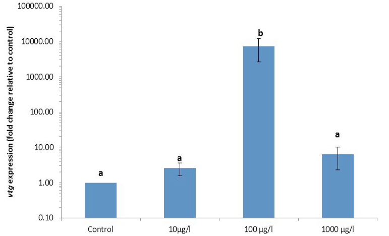
Relative gene expression of the biomarker gene vitellogenin (vtg) normalized to mean of three selected genes (gapdh, 18s, tbp), in livar of juvenile C. catla after 14 days of exposure to graded concentrations of BPA. Different letters indicate significant differences among group. P<0.05.
References
- Angle, B.M., Do, R.P., Ponzi, D., Stahlhut, R.W., Drury, B.E., Nagel, S.C., Welshons, W.V., Besch-Williford, C.L., Palanza, P., Parmigiani, S., vom Saal. F.S. and Taylor, J.A., 2013. Reprod. Toxicol., 42: 256-68. https://doi.org/10.1016/j.reprotox.2013.07.017[CrossRef]
- Arukwe, A., Celius, T., Walther, B. T. and Goksøyr, A., 2000. Aquat. Toxicol., 49: 159–170. https://doi.org/10.1016/S0166-445X(99)00083-1[CrossRef]
- Bowman, C.J., Kroll, K.J., Hemmer, M.J., Folmar, L.C. and Denslow, N.D., 2000. Gen. Comp. Endocrinol., 120: 300–313. https://doi.org/10.1006/gcen.2000.7565[CrossRef]
- Bhandari, R.K., Deem, S.L., Holliday, D.K., Jandegian, C.M., Kassotis, C.D., Nagel, S.C., Tillitt, D.E., vom Saal, F.S. and Rosenfeld, C.S., 2015. Gen. Comp. Endocrinol., 214: 195–219. https://doi.org/10.1016/j.ygcen.2014.09.014[CrossRef]
- Crain, D.A., Eriksen, M., Iguchi, T., Jobling, S., Laufer, H., LeBlanc, G.A. and Guillette Jr., L.J., 2007. Reprod. Toxicol., 24: 225–239. https://doi.org/10.1016/j.reprotox.2007.05.008[CrossRef]
- Craft, J.A., Brown, M., Dempsey, K., Francey, J., Kirby, M., Scott, A.P., Katsiadaki, I., Robinson, C.D., Davies, I.M., Bradac, P. and Moffat, C.F., 2004. Mar. Environ. Res., 58: 419–423. https://doi.org/10.1016/j.marenvres.2004.03.025[CrossRef]
- Diamanti-Kandarakis, E., Bourguignon, J.P., Giudice, L.C., Hauser, R., Prins, G.S., Soto, A.M., Zoeller, R.T. and Gore, A.C., 2009. Endocr. Rev., 30: 293–342. https://doi.org/10.1210/er.2009-0002[CrossRef]
- Eggen, R.I.L., Bengtsson, B.E., Bowmer, C.T., Gerritsen, A.A.M., Gibert, M., Hylland, K., Johnson, A.C., Leonards, P., Nakari, T., Norrgren, L., Sumpter, J.P., Suter, M.J.F., Svenson, A. and Pickering, A.D., 2003. Pure appl. Chem., 75: 2445–2450. https://doi.org/10.1351/pac200375112445[CrossRef]
- Faheem, M., Jahan, N. and Lone, K.P., 2016a. J. Anim. Pl. Sci., 26: 514-522.
- Goksøyr, A., 2006. J. Toxicol. environ. Hlth. A. 69: 175–184.
- Harries, J.E., Sheahan, D.A. and Jobling, S., 1997. Environ. Toxicol. Chem., 16: 534-541. https://doi.org/10.1002/etc.5620160320[CrossRef]
- Ishibashi, H., Watanabe, N., Matsumura, N., Hirano, M., Nagao, Y., Shiratsuchi, H., Kohra, S., Yoshihara, S. and Arizono, K., 2005. Life Sci., 77: 2643-2655. https://doi.org/10.1016/j.lfs.2005.03.025[CrossRef]
- Jenkins, S., Wang, J., Eltoum, I., Desmond, R. and Lamartiniere, C.A., 2011. Environ. Hlth. Perspect, 119: 1604-1609. https://doi.org/10.1289/ehp.1103850[CrossRef]
- Kang, I., Yokota, H., Oshima, Y., Tsuruda, Y., Oe, T., Imada, N., Tadokoro, H. and Honjo, T., 2002. Environ. Toxicol. Chem., 21: 2394–2400. https://doi.org/10.1897/1551-5028(2002)021<2394:EOBAOT>2.0.CO;2[CrossRef]
- Kime, D.E., 1999. Sci. Total Environ., 225: 3-11. https://doi.org/10.1016/S0048-9697(98)00328-3[CrossRef]
- Larsen, B.K., Bjornstad, A., Sundt, R.C., Taban, I.C., Pampanin, D.M. and Andersen, O.K., 2006. Aquat. Toxicol., 78: 25–33. https://doi.org/10.1016/j.aquatox.2006.02.026[CrossRef]
- Livak, K.J. and Schmittgen, T.D., 2001. Methods, 25: 402–408. https://doi.org/10.1006/meth.2001.1262[CrossRef]
- Lindholst, C., Pedersen, K.L. and Pedersen, S.N., 2000. Aquat. Toxicol., 48: 87–94. https://doi.org/10.1016/S0166-445X(99)00051-X[CrossRef]
- Lee, C., Park, M., Kim, H.M., Kim, H.J. and Choi, K., 2007. Mol. cell. Toxicol., 3: 185-189.
- Matozzo, V., Gagne, F., Marin, M. G., Ricciardi, F. and Blaise, C., 2008. Environ. Int., 34: 531-545. https://doi.org/10.1016/j.envint.2007.09.008[CrossRef]
- Mandich, A., Bottero, S., Benfenati, E., Cevasco, A., Erratico, C., Maggioni, S., Massari, A., Pedemonte, F. and Viganò, L., 2007. Gen. Comp. Endocrinol., 153: 15–24. https://doi.org/10.1016/j.ygcen.2007.01.004[CrossRef]
- Organization for Economic Cooperation and Development, 1992. Fish acute toxicity test. Test Guideline 203. OECD Guidelines for the Testing of Chemicals, Paris, France.
- Scholz, S., Kordes, C. and Hamann, J., 2004. Mar. Environ. Res., 57: 235–244. https://doi.org/10.1016/S0141-1136(03)00082-5[CrossRef]
- Sohoni, P., Tyler, C.R., Hurd, K., Caunter, J., Hetheridge, M., Williams, T., Woods, C., Evans, M., Toy, R., Gargas, M. and Sumpter, J.P., 2001. Environ. Sci. Technol., 35: 2917–2925. https://doi.org/10.1021/es000198n[CrossRef]
- Staples, C.A., Dorn, P.B., Klecka, G.M., O’Block, S.T. and Harris, L.R., 1998. Chemosphere, 36: 2149–2173. https://doi.org/10.1016/S0045-6535(97)10056-X[CrossRef]
- Staples, C.A., Woodburn, K., Caspers, N., Hall, A.T. and Klecka, G.M., 2002. Hum. Ecol. Risk Assess., 8: 1083-1105. https://doi.org/10.1080/1080-700291905837[CrossRef]
- Sumpter, J.P. and Jobling, S., 1995. Environ. Hlth. Perspect., 103 (Suppl 7): 173–178. https://doi.org/10.1289/ehp.95103s7173[CrossRef]
- Thomas-Jones, E., Thorpe, K., Harrison, N., Thomas, G., Morris, C., Hutchinson, T., Woodhead, S. and Tyler, C., 2003. Environ. Toxicol. Chem., 22: 3001–3008. https://doi.org/10.1002/etc.5620220506[CrossRef]
- Vandenberg, L.N., 2014. Dose-Response, 12: 259–276. https://doi.org/10.2203/dose-response.13-020.Vandenberg[CrossRef]
- Virk, P., Ali, A., Al-Sakran, M. and Elobeid, M.A., 2014. Trop. J. pharm. Res., 13: 1107-1112. https://doi.org/10.4314/tjpr.v13i7.14[CrossRef]
- Vandesompele, J., Preter, D.K., Pattyn, F., Poppe, B., Roy, V.N., Paepe, D.A. and Speleman, F., 2002. Genome Biol., 3: 34. https://doi.org/10.1186/gb-2002-3-7-research0034[CrossRef]
- vom Saal, F.S., Akingbemi, B.T., Belcher, S.M., Birnbaum, L.S., Crain, D.A, Eriksen, M., Farabollini, F., Guillette, L.J. Jr., Hauser, R., Heindel, J.J., Ho, S.M., Hunt, P.A., Iguchi, T., Jobling, S., Kanno, J., Keri, R.A., Knudsen, K.E., Laufer, H., LeBlanc, G.A., Marcus, M., McLachlan, J.A., Myers, J.P., Nadal, A., Newbold, R.R., Olea, N., Prins, G.S., Richter, C.A., Rubin, B.S., Sonnenschein, C., Soto, A.M., Talsness, C.E., Vandenbergh, J.G., Vandenberg, L.N., Walser-Kuntz, D.R., Watson, C.S., Welshons, W.V., Wetherill, Y. and Zoeller, R.T., 2007. Reprod. Toxicol., 24: 131-138. https://doi.org/10.1016/j.reprotox.2007.07.005[CrossRef]
- Xu, X.H., Zhang, J., Wang, Y.M., Ye, Y.P. and Luo, Q.Q., 2010. Horm. Behav., 58: 326-333. https://doi.org/10.1016/j.yhbeh.2010.02.012[CrossRef]
- Zhang, Y., Tao, S., Yuan, C., Liu, Y. and Wang, Z., 2016. Chemosphere, 144: 304-311. https://doi.org/10.1016/j.chemosphere.2015.10.083[CrossRef]
Services
Article usage statistics
Loading…