
Bisphenol A (BPA) is an aquatic contaminant, exerting adverse effects on aquatic organisms especially fish. Therefore, current research was conducted to assess the deleterious effects of bisphenol A on antioxidant system, oxidative stress content, genotoxicity and histological alterations in Labeo rohita. Four fish groups (A–D) were setup. Group A was control and groups B–D were exposed to three different amounts of BPA for twenty-one days. Results showed significant increase in oxidative stress content such as TBARS (thiobarbituric acid reactive substance) and ROS (reactive oxygen species) and decreased antioxidant enzymes such as POD (peroxidase), GSH (reduced glutathione), SOD (superoxidase dismutase) and CAT (catalase) in exposed fish. Comet assay showed DNA damage in erythrocytes. Histological examination revealed that BPA causes various degenerative effect in soft tissues of liver, kidney and gills of L. rohita. The current investigation concluded that bisphenol A induces harmful effects by disrupting the physiology of some important organs and histological alteration in various tissues of exposed fish.
Research Article · Volume 57, Issue 5, 12 August 2025 · Open Access
Assessing the Impact of the Environmental Stressor (Bisphenol A) on Labeo rohita: Insights into Histological Alterations, Genotoxic Effects, and Antioxidant Defense Mechanisms
· · · ·
Affiliations & Notes ⌄
- 1Department of Zoology, University of Okara, Okara 56130 Pakistan 2Department of Zoology, Government College University, Faisalabad 37251, Pakistan * Corresponding author: dr.khurram@uo.edu.pk
Article Info ⌄
- Journal:
- Pakistan Journal of Zoology
- Published:
- 12 August 2025

From the article
- In the current investigation, significant increase in TBARS and ROS contents were observed in the kidney, liver and gills of the BPA exposed fish.
- The current study concluded that bisphenol A causes lethal effects on erythrocytes and different important tissues of Labeo rohita.
- Histological examination showed different degenerative effects in the soft tissues of the kidney, liver and gills of fish after BPA exposure.
- Furthermore, bisphenol A causes oxidative damage by increasing ROS content and TBARS level and decreasing the amount of antioxidant enzymes (peroxidase, catalase, reduced glutathione, and superoxide dismutase) in the kidney, liver and gills of L. rohita in a dose dependent way.
Selected from the article's own text.
Abstract
Introduction
Bisphenol A (BPA) is an environmental stressor and is a plastic making chemical that has been extensively used to make different kinds of plastic materials . People of all ages are continuously exposed to BPA because of its widespread use in the synthesis of plastic beverage and food products and food can coatings . It is one of the world’s most manufactured compounds due to rising plastic product demand. BPA is a toxic compound and a number of studies concerning its toxicological effects on vertebrates and invertebrates have been reported . Therefore, the use of BPA in manufacturing different kinds of materials has now been banned in many developed countries . Because of this, several functional analogues of BPA have introduced the market across the world but it is still the most widely used chemical in developing countries . BPA was found in more than 90% of urine samples from humans, indicating widespread exposure. The liver quickly conjugates and excretes it in the urine . Numerous adverse effects including a rise in body weight, accumulation of lipids in different tissues, leptin variations, alterations in estradiol and osteocalcin levels, as well as pancreatic functioning were reported. It also causes diabetes, cardiovascular abnormalities, and liver enzyme variations . BPA poses a potential threat to humans and other animals, especially aquatic organisms. Urban sewage, discharge from the petrochemical industries, and leachate from landfills are all potential sources of BPA exposure . The amount of BPA in water samples has been found to be between 4.4 and 8000 ng/L in Europe, Asia and North America . So, it is problematic for aquatic animals to escape the toxic effects of BPA. Among all aquatic animals, fish are extremely susceptible to various kinds of pollutants, including bisphenol A, which enters the body through various absorbing sites such as gills, skin, and the oral cavity and gets accumulated into different body organs, especially the liver, kidney, and gills, causing disruption in biochemical and physiological processes . Because of the hazardous effects of BPA on aquatic life including fish, it has been widely used in recent years for testing aquatic toxicity. Fish are frequently used as biological markers to assess the condition of aquatic environments because of their biological responses. Earlier studies about the sublethal effects of BPA on morphological characters, growth, hematobiochemical , genotoxicity, oxidative stress , and histopathological alterations in fish were reported .
Very few studies are available about the sublethal toxicity of environmental stressor (bisphenol A) and its effects on stress content, antioxidant mechanism and genotoxic damage in L. rohita. Therefore, current research work was designed to evaluate the sublethal toxic effects of BPA on freshwater teleost L. rohita by studying the effects of BPA on antioxidant mechanism, genotoxic alterations and histology of some vital body organs. As mentioned above, the present study was conducted to explore BPA’s effects on Labeo rohita. L. rohita is the most common and commercially important tropical freshwater carp and good for farming in Pakistan and other countries. It constitutes about 60% of the cultured fish in Pakistan . Therefore, we selected L. rohita as a model organism to evaluate BPA toxicity at low levels.
Materials and Methods
Animal collection and placement
The current study was conducted in Aquaculture Lab of Department of Fisheries and lab of Department of Zoology, University of Okara, Pakistan with approval from the Office of Research and Innovative Commercialization (ORIC). Healthy and active Labeo rohita were taken from fish farm at Head Balloki, District Kasur, Punjab, Pakistan and transported to the university aquaculture laboratory. No mortality was found during transport. Fish with an average size of 18.38 ± 0.30 cm, weighing 32.37±5.73 g were placed in a large glass aquarium having dimensions of 35ʺ L × 40ʺ W × 30ʺ H with 80 liters of water. The fish were rinsed with a 0.1% KMnO4 solution and acclimatized in glass aquariums for seven days.
Chemicals
Bisphenol A (98% purity) was acquired from MACLIN, China. For preparation of the stock solution, an appropriate concentration of solid BPA was dissolved in ethanol and kept in glass containers at 4 °C.
Experimental design
After acclimatization, for the experimental trial, four groups (A, B, C, and D) were designed as control and three treatment groups, each with three replicas. Group A was treated as a chemical free freshwater. Groups B, C and D were exposed to three different doses of BPA, as 400, 800, and 1600 µg/L, respectively for 21 days. Doses were made on the basis of earlier research . Every second day, about 90% of water in each glass aquarium was changed. Every day, fish were fed with commercial feed that contained 22% protein. At the beginning, middle, and end of the trial, the physicochemical parameters of water were measured as presented in Table I.
Table I. Physicochemical parameters of water for trial animals. The values are shown as mean ± SD.
| Variables (mg/l) | Values |
| Temperature (0C) | 27.0 ± 0.12 |
| DO | 8.09 ± 0.14 |
| Total dissolved solid | 182.11 ± 3.02 |
| Total hardness | 171.3 ± 0.13 |
| Magnesium | 13.9 ± 0.21 |
| Chlorides | 7.3 ± 0.02 |
| pH | 8.93 ± 0.17 |
| Sodium | 12.6 ± 0.16 |
| Potassium | 1.6 ± 0.09 |
| Calcium | 37.3 ± 0.13 |
| Sulphates | 39.6 ± 0.12 |
Comet assay
On day 21, fish were removed from treated and control groups. Blood was collected by means of a BD syringe from the caudal vein and stored for further processes. This examination was carried out in a laboratory using specific experimental techniques. Fish were anesthetized by using clove oil. For the estimation of genotoxicity in erythrocytes, comet assay approach was used following the Singh et al. (1988) method under alkaline environment. The ethidium bromide was used for staining microscopic slides and examined with a fluorescent microscope at 400 × magnification. Comet IV computer software was used to score microscopic pictures of the comet .
Oxidative and antioxidant estimation
After blood collection, fish were dissected humanely. The kidney, liver and gills were removed from each fish for oxidant and antioxidant enzyme estimation. A teflon homogenizer was used to homogenize all of the samples in phosphate buffer saline. After filtering and centrifuging at 1600 × g at 4°C for 10 min, the supernatant was obtained and stored at -20 °C for more study. The activities of antioxidant enzymes were assessed using commercially available diagnostic reagent kits according to the given instructions (ELISA Kits Manufacturer, USA). According to earlier investigations, several antioxidant enzymes and oxidative stress indicators were identified. Using the techniques described by Hayashi et al. (2007) and Ohkawa et al. (1979), ROS, GSH and TBARS were evaluated, in the kidney, liver, and gills of each BPA exposed fish. Several antioxidant enzymes, POD (peroxidase), SOD (superoxidase dismutase), and CAT (catalase) were assessed by the methods of Kakkar et al. (1984) in kidney, liver and gills of BPA exposed fish.
Histology
After dissection, liver, kidney, and gills were removed. All organs were preserved in a 10% formalin solution, as described by Troyer (1980). Before preservation, samples were dehydrated in ethanol, cleaned in xylene, and then soaked with wax. Sections (5µm thick) were cut using automatic microtome (Leica RM 2255). Tissue samples were stained with hematoxylin, eosin, and PAS stains following the method of Joseph et al. (2006). Slides of all tissues were examined and photographed using a camera fitted microscope (Leica, Japan).
Statistical analysis
Statistical analysis and graphical representations were performed on GraphPad Prism (V 9.5.1) software. Significant difference between the means of control and experimental groups was determined by one-way ANOVA test by applying Dunnett multiple comparison test. Asterisks represent different significant levels, * = p<0.05, ** = p<0.01, *** = p<0.001.
Results
Oxidative damage and antioxidant enzymes
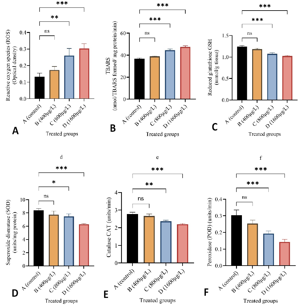
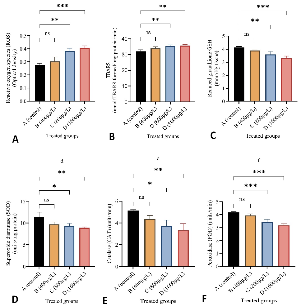
In current study, oxidative stress content and antioxidant enzymes levels in liver, gills and kidney were observed (Figs. 1, 2 and 3). Current results showed a significant rise in TBARS and ROS content in kidney, liver and gills of L. rohita exposed with 800 µg/L and 1600 µg/L BPA as compared with the group A. No significant rise was observed in TBARS and ROS contents between the control group and group B (400 µg/L). A significant decrease was observed in POD, GSH, SOD, and CAT activities in the kidney, liver and gills of L. rohita exposed with 800 µg/L and 1600 µg/L BPA as compared with control group. No significant decrease was observed in POD, GSH, SOD and CAT activities in all examined tissues between the control group and group B (400 µg/L).
Histology
Histological alterations in soft tissues (kidney, liver and gills) of BPA exposed L. rohita were observed (Figs. 4-6). No alterations were observed in gills, kidney and liver of control fish. The effects of the dose-dependent treatment included necrosis, pyknotic nuclei formation, sinusoidal spaces, cluster nuclei, dilated renal tubules, edema, and the rupturing of gill filaments in fish of group B, C and D. The results of histological investigation of gill sections, which revealed cell necrosis, swelling (edema), and moderate to severe rupturing of gill filaments, including primary and secondary gill lamellae as shown in Figure 4. In the tissue histology of the liver depicted in Figure 5, pyknotic nuclei formation, sinusoidal spaces, cell necrosis and cluster nuclei formation were observed. Fish exposed to BPA had kidney tissue sections with altered histology that revealed dilated or elongated renal tubules, cluster nuclei development, sinusoidal gaps, and cell swelling (edema) (Fig. 6).
Genotoxic examination
A comet test was used under alkaline conditions to measure DNA damage in erythrocytes of fish after exposure to BPA (Fig. 7). BPA exposure increased DNA damage concentration dependently compared to unexposed group. A highly significant rise was observed in % DNA damage at a high dose of BPA (1600 g/L). A significant change was observed throughout the increase in BPA doses from A (0 µg/L), B (400 µg/L), C (800 µg/L), and D (1600 µg/L). It was noticed that the olive tail moment reached its maximum level as the dosage of BPA increased. A significant change was observed between unexposed group and BPA exposed groups.
Discussion
In toxicological studies, exposing organisms to specific dosages at different acute or sublethal concentrations helps to better understand the hazardous levels of chemicals, including bisphenol A. The extent to which environmental contaminants harm aquatic life is crucial . Pollutants may harm fish fauna via physiological, biochemical and histological alterations . Over the last few decades, there has been a worldwide rise in efforts to monitor and record the impacts of toxins such as pesticides, environmental stressors and industrial effluents. Many pollutants from different kinds of sources readily and instantly enter water bodies. Therefore, aquatic organisms are more vulnerable to damage than terrestrial animals . Many of these synthetic substances, such as bisphenol A, are endocrine disruptors that damage fish tissues (gills, heart, kidney and liver) . Therefore, to minimize the public health concerns associated with bisphenol A, it is important to conduct ongoing monitoring and evaluation of its toxicological impacts at low levels.
Investigation of activities of some important antioxidant enzymes (POD, GSH, CAT and SOD) and oxidative content such as ROS are important biomarkers for inflammatory responses and suitable tools to determine the harmful effects of environmental pollutants and protect tissues from damage triggered by free oxide (O¯) radicals . In the current investigation, significant increase in TBARS and ROS contents were observed in the kidney, liver and gills of the BPA exposed fish. A higher level of oxidative stress content may be due to the imbalance and reduction of antioxidant enzymes . Previous research showed that the same results were reported in other fishes such as common carp and Aristicthys nobilis . Various organisms exposed to different toxicants including BPA showed increased ROS content due to detoxification processes. BPA may affect oxidative homeostasis directly or indirectly by elevating lipid peroxidation and hydrogen peroxide while decreasing antioxidant enzymes, triggering mitochondrial malfunction, altering cell signaling pathways, and causing apoptosis in vital organs of the body . Increased formation of ROS is associated with peroxidation of lipids, which in turn disrupts cell membranes and increases TBARS synthesis . BPA increases lipid peroxidation and decreases antioxidant enzyme production in target organisms. In the current experimental study, a significant reduction was observed in levels of antioxidant enzymes including SOD, POD, GSH, and CAT in kidney, liver and gills of BPA exposed fish. Our results are supported by previous literature reported by Afzal et al. (2022). In the current study, low levels of antioxidant enzymes in different organs could be due to the excessive formation of free radicals caused by BPA exposure, leading to abnormal functioning and disturbing the antioxidant mechanism. There is a decline in the activity of antioxidant enzymes, which is related to tissue dysfunction and the increased consumption of energy to deal with oxidative stresses .
The histological examination is an excellent biomarker to observe the impact of different environmental stressors on aquatic and terrestrial animals ; . Gills are an excellent biomonitoring organ for evaluating the impacts of pollutants in aquatic environments due to their high absorption rate and direct interaction with water. It is essential organ for regulating ions and respiration . According to the current study, histological changes in gill tissues, as rupturing of primary and secondary gill lamellae, cell swelling, as well as necrosis were observed in BPA treated groups. No changes were observed in BPA free group. Previously reported the same findings on gill changes in fish exposed to BPA . Similar observations were reported on Catla catla and Cyprinus carpio . The liver of fish serves as the primary organ responsible for the process of detoxification of environmental pollutants, including bisphenol A. Therefore, alterations observed in the liver of aquatic organisms, such as fish, can serve as excellent indicator of aquatic pollution . In this study, L. rohita exposed to bisphenol A showed moderate to severe changes in liver, including pyknotic nuclei formation, cluster nuclei formation, cell necrosis and sinusoidal spaces, while no changes were seen in control fish. Changes in liver tissues with sinusoidal spaces and cluster nuclei formation are due to the destruction of structural proteins. The hepatic vein and hepatic artery are responsible for supplying blood to the dorsal aorta. However, the presence of obstruction in the hepatic vein may block the flow of blood. The blockage of the hepatic vein may lead to cell damage and necrosis. Similar changes were also described by Faheem et al. (2016). Same changes were reported in the liver tissues of Oreochromus spilurus after BPA exposure (Abdulla Bin-Dohaish, 2012). The kidneys of fish are the primary osmoregulatory and hematopoietic organs. Histological changes in kidney tissues can serve as an indication of environmental contamination . Histological examination showed alterations in the kidneys of L. rohita after exposure to BPA, such as dilated and elongated renal tubules, cluster nuclei formation, sinusoidal spaces, and cell swelling (edema). Faheem et al. (2016) reported the same findings in fish exposed to BPA.
Genotoxic examination via comet assay is a reliable, suitable and extensively used method to measure DNA damage in various cells of aquatic and terrestrial animals . According to current study, results showed significant rise in % DNA damage in blood erythrocytes of L. rohita under alkaline conditions. Akram et al. (2021) reported the same findings in fish exposed to BPA, describing the genotoxic potential of BPA in bighead carp and zebrafish . In this investigation, a significant rise in olive tail moment (OTM) was observed in erythrocytes of exposed fish. The basic molecular and cellular pathways of the genotoxic potential of PBA are still unknown. However, the genotoxicity caused by BPA has been primarily related to oxidative damage and lipids peroxidation which may directly induce DNA changes .
Conclusion
The current study concluded that bisphenol A causes lethal effects on erythrocytes and different important tissues of Labeo rohita. Histological examination showed different degenerative effects in the soft tissues of the kidney, liver and gills of fish after BPA exposure. Bisphenol A also causes genotoxic alterations in erythrocytes of L. rohita. Furthermore, bisphenol A causes oxidative damage by increasing ROS content and TBARS level and decreasing the amount of antioxidant enzymes (peroxidase, catalase, reduced glutathione, and superoxide dismutase) in the kidney, liver and gills of L. rohita in a dose dependent way.
Declarations
Acknowledgements
Funding
This research did not receive any specific grant from funding agencies in the public, commercial, or not for profit sectors.
Ethical statement
Ethical approval was taken from the University of Okara ‘s Ethical Committee for the current research work (Reference no. UO/ETH/2023/misc.)
Statement of conflict of interest
Figures
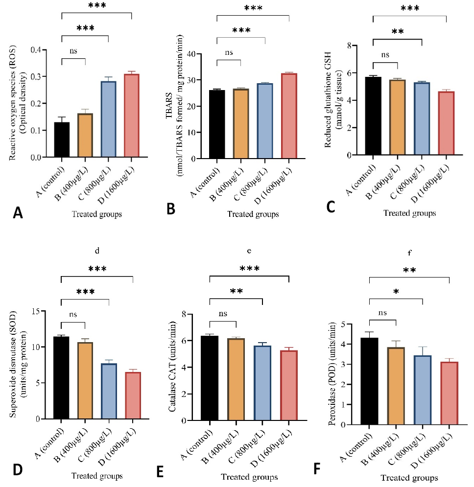
Effect of sub-lethal doses of BPA on the level of (A) ROS, (B) TBARS, (C) GSH, (D) SOD, (E) CAT and (F) POD in gills of L. rohita. Results are shown as mean ± SD. * = p<0.05, ** = p<0.01, *** = p<0.001
Effect of sub-lethal doses of BPA on the level of (A) ROS, (B) TBARS, (C) GSH, (D) SOD, (E) CAT and (F) POD in kidney of L. rohita. Results are shown as mean ± SD. * = p<0.05, ** = p<0.01, *** = p<0.001
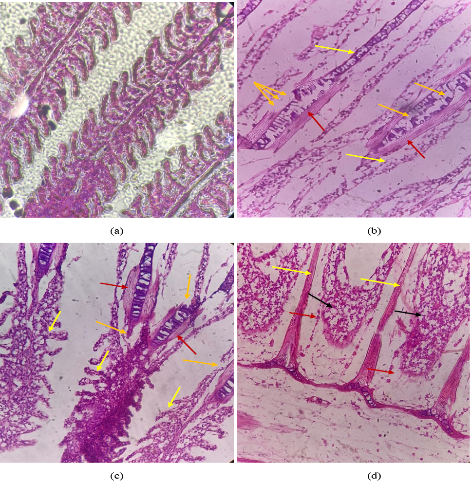
Effect of BPA on histological structure of gills of Labeo rohita (a) showing normal gill filament arrangement including primary lamellae and secondary lamellae. (b), (c) and (d) showing variations in gill tissues as cell damage (orange arrow), edema (red arrow), rupturing of gill lamellae (yellow arrow), necrotic cells (black arrow).
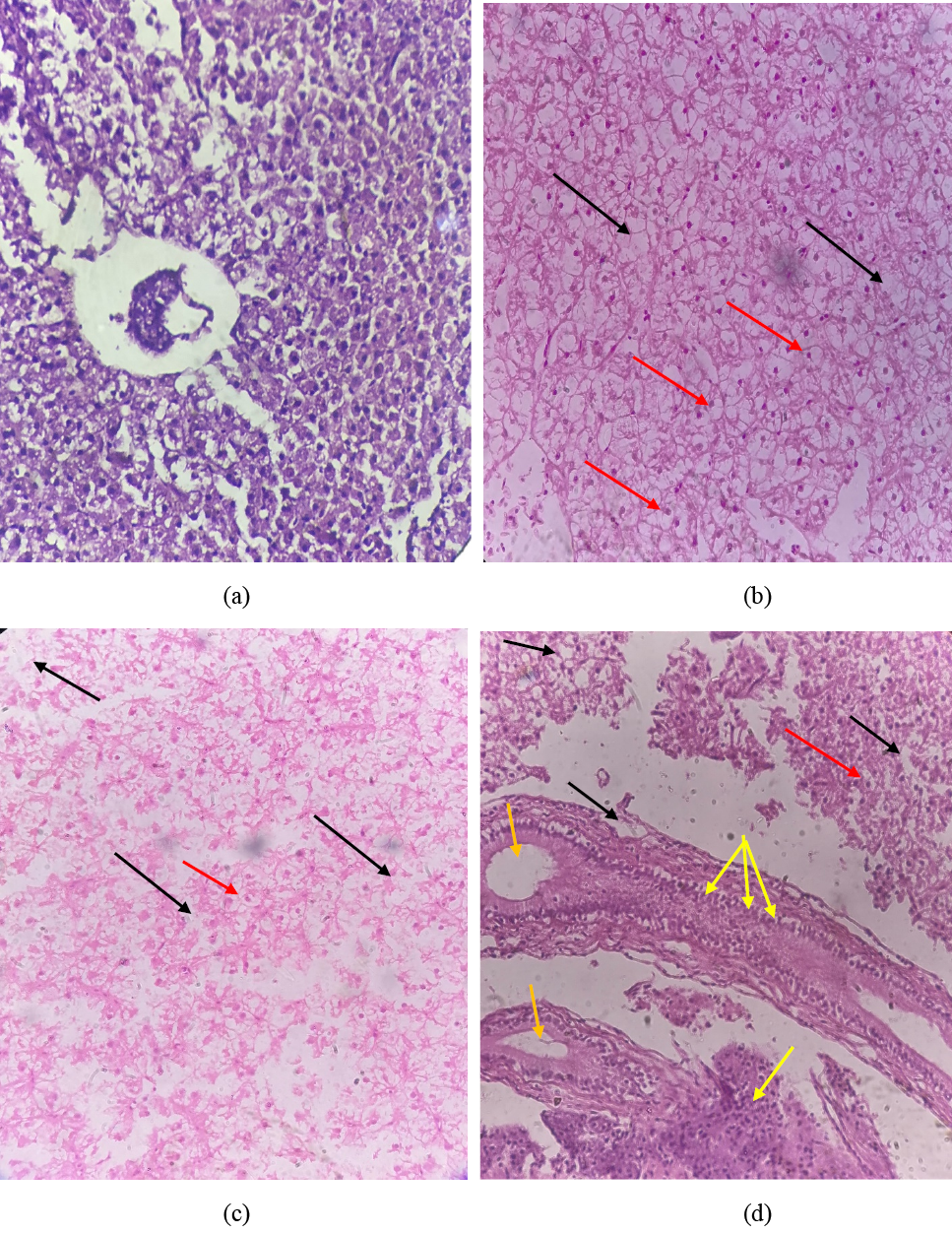
Effect of BPA on histological structure of liver of Labeo rohita as (a) showing normal arrangement of hepatic cells in the control. (b), (c) and (d) showing necrosis (black arrow), pyknotic nuclei (red arrow), cluster nuclei formation (yellow arrow) and sinusoidal spaces (orange arrow).
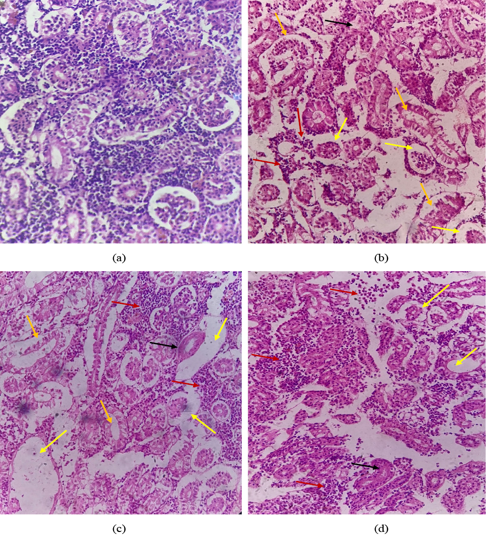
Effect of BPA on histological structure of kidney of Labeo rohita tissues as (a) showing normal arrangement of renal cells. (b), (c) and (d) showing variations, such as edema (red arrow), dilated renal tubules (orange arrow), cluster nuclei formation (red arrow), and sinusoidal spaces (yellow arrow).
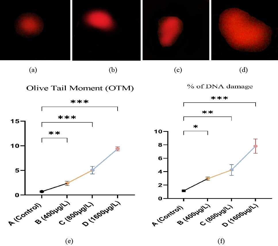
Genotoxic potential of BPA in erythrocytes. photographs of comet assay showing (a) normal cell. (b), (c) and (d) showing nuclear material damage. (e) olive tail moment and (f) % DNA damage. * = p<0.05, ** = p<0.01, *** = p<0.001
References
- Abdulla-Bin-Dohaish, E.J., 2012. The effects of 4-nonylphenol contamination on livers of Tilapia fish (Oreochromus spilurs) in Jeddah. Biol. Res., 45: 15-20. https://doi.org/10.4067/S0716-97602012000100002[CrossRef]
- Afzal, G., Ahmad, H.I., Hussain, R., Jamal, A., Kiran, S., Hussain, T. and Saeed, S., 2022. Bisphenol A induces histopathological, hematobiochemical alterations, oxidative stress, and genotoxicity in common carp (Cyprinus carpio L.). Oxidative Med. Cell. Long., https://doi.org/10.1155/2022/5450421[CrossRef]
- Akram, R., Iqbal, R., Hussain, R., Jabeen, F. and Ali, M., 2021. Evaluation of oxidative stress, antioxidant enzymes and genotoxic potential of bisphenol A in fresh water bighead carp (Aristichthys nobilis) fish at low concentrations. Environ. Pollut., 268: 115896. https://doi.org/10.1016/j.envpol.2020.115896[CrossRef]
- Almeida, S., Raposo, A., Almeida-González, M. and Carrascosa, C., 2018. Bisphenol A: Food exposure and impact on human health. Compreh. Rev. Fd. Sci. Fd. Saf., 17: 1503-1517. https://doi.org/10.1111/1541-4337.12388[CrossRef]
- Andújar, N., Gálvez-Ontiveros, Y., Zafra-Gómez, A., Rodrigo, L., Álvarez-Cubero, M.J., Aguilera, M., Monteagudo, C. and Rivas, A., 2019. Bisphenol A analogues in food and their hormonal and obesogenic effects: A review. Nutrients, 11: 2136. https://doi.org/10.3390/nu11092136[CrossRef]
- Asenuga, E.R., Olajuyigbe, F.M., Akinmoladun, A.C. and Fasakin, E.A., 2022. Deeper insights into the biochemical, hematological and histological changes induced by bisphenol a exposure in adult male African catfish (Clarias gariepinus). https://doi.org/10.21203/rs.3.rs-1701247/v1[CrossRef]
- Braz-Mota, S., Sadauskas-Henrique, H., Duarte, R.M., Val, A.L. and Almeida-Val, V.M., 2015. Roundup® exposure promotes gills and liver impairments, DNA damage and inhibition of brain cholinergic activity in the Amazon teleost fish Colossoma macropomum. Chemosphere, 135: 53-60. https://doi.org/10.1016/j.chemosphere.2015.03.042[CrossRef]
- Cengiz, E.I., 2006. Gill and kidney histopathology in the freshwater fish Cyprinus carpio after acute exposure to deltamethrin. Environ. Toxicol. Pharmacol., 22: 200-204. https://doi.org/10.1016/j.etap.2006.03.006[CrossRef]
- Chaubey, R., 2005. Computerized image analysis software for the comet assay. Mol. Toxicol. Protoc., pp. 97-106.
- Eid, J.I., Eissa, S.M. and El-Ghor, A.A., 2015. Bisphenol A induces oxidative stress and DNA damage in hepatic tissue of female rat offspring. J. Basic appl. Zool., 71: 10-19. https://doi.org/10.1016/j.jobaz.2015.01.006[CrossRef]
- Eladak, S., Grisin, T., Moison, D., Guerquin, M.J., N’Tumba-Byn, T., Pozzi-Gaudin, S., Benachi, A., Livera, G., Rouiller-Fabre, V. and Habert, R., 2015. A new chapter in the bisphenol A story: Bisphenol S and bisphenol F are not safe alternatives to this compound. Fertil. Steril., 103: 11-21. https://doi.org/10.1016/j.fertnstert.2014.11.005[CrossRef]
- Faheem, M., Jahan, N. and Lone, K., 2016. Histopathological effects of bisphenol-A on liver, kidneys and gills of Indian major carp, Catla catla (Hamilton, 1822). J. Anim. Pl. Sci., 26: 514-522.
- Gassman, N.R., 2017. Induction of oxidative stress by bisphenol A and its pleiotropic effects. Environ. Mol. Mutagen., 58: 60-71. https://doi.org/10.1002/em.22072[CrossRef]
- Ghaffar, A., Hussain, R., Abbas, G., Khan, R., Akram, K., Latif, H., Ali, S., Baig, S., Du, X. and Khan, A., 2021. Assessment of genotoxic and pathologic potentials of fipronil insecticide in Labeo rohita (Hamilton, 1822). Toxin Rev., 40: 1289-1300. https://doi.org/10.1080/15569543.2019.1684321[CrossRef]
- Gupta, A., Gupta, S.K., Priyam, M., Siddik, M.A., Kumar, N., Mishra, P.K., Gupta, K.K., Sarkar, B., Sharma, T.R. and Pattanayak, A., 2021. Immunomodulation by dietary supplements: A preventive health strategy for sustainable aquaculture of tropical freshwater fish, Labeo rohita (Hamilton, 1822). Rev. Aquacult., 13: 2364-2394. https://doi.org/10.1111/raq.12581[CrossRef]
- Hayashi, I., Morishita, Y., Imai, K., Nakamura, M., Nakachi, K. and Hayashi, T., 2007. High-throughput spectrophotometric assay of reactive oxygen species in serum. Mutat. Res. Genet. Toxicol. Environ. Mutagen., 631: 55-61. https://doi.org/10.1016/j.mrgentox.2007.04.006[CrossRef]
- Jabeen, G., Manzoor, F., Arshad, M. and Barbol, B., 2021. Effect of cadmium exposure on hematological, nuclear and morphological alterations in erythrocyte of fresh water fish (Labeo rohita). Continent. Vet. J., 1: 20-24.
- Joseph, J., Vemuganti, G.K., Garg, P. and Sharma, S., 2006. Histopathological evaluation of ocular microsporidiosis by different stains. BMC Clin. Pathol., 6: 1-8. https://doi.org/10.1186/1472-6890-6-6[CrossRef]
- Kakkar, P., Das, B. and Viswanathan, P., 1984. A modified spectrophotometric assay of superoxide dismutase. Ind. J. Biochem. Biophys., 21: 130-132.
- Khoshnood, Z., Khodabandeh, S., Mosafer, S. and Khoshnood, R., 2010. Effects of cortisol on gill chloride cells in persian sturgeon Acipenser persicus fry. Cell J. (Yakhteh), 11: 424-431.
- Lemly, A.D., 2002. Symptoms and implications of selenium toxicity in fish: The Belews Lake case example. Aquat. Toxicol., 57: 39-49. https://doi.org/10.1016/S0166-445X(01)00264-8[CrossRef]
- Lombó, M., Getino-Álvarez, L., Depincé, A., Labbé, C. and Herráez, M.P., 2019. Embryonic exposure to bisphenol a impairs primordial germ cell migration without jeopardizing male breeding capacity. Biomolecules, 9: 307. https://doi.org/10.3390/biom9080307[CrossRef]
- Mahmood, A., Ahmad, S., Akmal, H. and Shahzad, K., 2023. Evaluation of hemotoxic, hepatotoxic and nephrotoxic potential of profenofos-based insecticide in freshwater Labeo rohita fish at low concentrations: Evaluation of profenofos-based insecticide’s potential. Pak. BioMed. J., 6: 32-40. https://doi.org/10.54393/pbmj.v6i11.920[CrossRef]
- Mantecca, P., Vailati, G. and Bacchetta, R., 2006. Histological changes and micronucleous induction in the zebra mussel dreissena polymorpha after paraquat exposure. Histol. Histopathol., 21: 829-840.
- Maradonna, F., Nozzi, V., Dalla Valle, L., Traversi, I., Gioacchini, G., Benato, F., Colletti, E., Gallo, P., Pisciottano, I.D.M. and Mita, D.G., 2014. A developmental hepatotoxicity study of dietary bisphenol A in Sparus aurata juveniles. Comp. Biochem. Physiol. C Toxicol. Pharmacol., 166: 1-13. https://doi.org/10.1016/j.cbpc.2014.06.004[CrossRef]
- Meli, R., Monnolo, A., Annunziata, C., Pirozzi, C. and Ferrante, M.C., 2020. Oxidative stress and BPA toxicity: An antioxidant approach for male and female reproductive dysfunction. Antioxidants, 9: 405. https://doi.org/10.3390/antiox9050405[CrossRef]
- Moon, M.K., Kim, M.J., Jung, I.K., Do Koo, Y., Ann, H.Y., Lee, K.J., Kim, S.H., Yoon, Y.C., Cho, B.J. and Park, K.S., 2012. Bisphenol A impairs mitochondrial function in the liver at doses below the no observed adverse effect level. J. Korean med. Sci., 27: 644-652. https://doi.org/10.3346/jkms.2012.27.6.644[CrossRef]
- Moreman, J., Lee, O., Trznadel, M., David, A., Kudoh, T. and Tyler, C.R., 2017. Acute toxicity, teratogenic, and estrogenic effects of bisphenol A and its alternative replacements bisphenol S, bisphenol F, and bisphenol AF in zebrafish embryo-larvae. Environ. Sci. Technol., 51: 12796-12805. https://doi.org/10.1021/acs.est.7b03283[CrossRef]
- Ohkawa, H., Ohishi, W. and Yagi, K., 1979. Colorimetric method for determination of MDA activity. Biochemistry, 95: 351. https://doi.org/10.1016/0003-2697(79)90738-3[CrossRef]
- Singh, N.P., McCoy, M.T., Tice, R.R. and Schneider, E.L., 1988. A simple technique for quantitation of low levels of DNA damage in individual cells. Exp. Cell Res., 175: 184-191. https://doi.org/10.1016/0014-4827(88)90265-0[CrossRef]
- Stefan-van Staden, R.I., Gugoaşă, L.A., Calenic, B., van Staden, J.F. and Legler, J., 2014. Screening of children saliva samples for bisphenol A using stochastic, amperometric and multimode microsensors. Anal. Chem. Res., 1: 1-7. https://doi.org/10.1016/j.ancr.2014.06.001[CrossRef]
- Tavakkoli, A., Abnous, K., Hassani, F.V., Hosseinzadeh, H., Birner-Gruenberger, R. and Mehri, S., 2020. Alteration of protein profile in cerebral cortex of rats exposed to bisphenol a: A proteomics study. Neurotoxicology, 78: 1-10. https://doi.org/10.1016/j.neuro.2020.01.013[CrossRef]
- Troyer, H., 1980. Principles and techniques of histochemistry, 1st ed. Little Brown. https://cir.nii.ac.jp/crid/1130282268834792320
- Tsai, W.-T., 2006. Human health risk on environmental exposure to Bisphenol-A: A review. J. Environ. Sci. Hlth., 24: 225-255. https://doi.org/https://doi.org/10.1080/10590500600936482[CrossRef]
- Vandenberg, L.N., Chahoud, I., Heindel, J.J., Padmanabhan, V., Paumgartten, F.J. and Schoenfelder, G., 2010. Urinary, circulating, and tissue biomonitoring studies indicate widespread exposure to bisphenol A. Environ. Hlth. Perspect., 118: 1055-1070. https://doi.org/10.1289/ehp.0901716[CrossRef]
- Verma, G., Khan, M.F., Akhtar, W., Alam, M.M., Akhter, M. and Shaquiquzzaman, M., 2018. Molecular interactions of bisphenols and analogs with glucocorticoid biosynthetic pathway enzymes: An in silico approach. Toxicol. Mech. Methods, 28: 45-54. https://doi.org/10.1080/15376516.2017.1356415[CrossRef]
- Vigliano, F., Aleman, N., Quiroga, M. and Nieto, J., 2006. Ultrastructural characterization of gills in juveniles of the Argentinian silverside, Odontesthes bonariensis (Valenciennes, 1835) (Teleostei: Atheriniformes). Anatom. Histol. Embryol., 35: 76-83. https://doi.org/10.1111/j.1439-0264.2005.00640.x[CrossRef]
- Wang, Q., Yang, H., Yang, M., Yu, Y., Yan, M., Zhou, L., Liu, X., Xiao, S., Yang, Y. and Wang, Y., 2019. Toxic effects of bisphenol A on goldfish gonad development and the possible pathway of BPA disturbance in female and male fish reproduction. Chemosphere, 221: 235-245. https://doi.org/10.1016/j.chemosphere.2019.01.033[CrossRef]
- Xu, J., Zhou, L., Wang, S., Zhu, J., Liu, T., Jia, Y., Sun, D., Chen, H., Wang, Q. and Xu, F., 2018. Di-(2-ethylhexyl)-phthalate induces glucose metabolic disorder in adolescent rats. Environ. Sci. Pollut. Res., 25: 3596-3607. https://doi.org/10.1007/s11356-017-0738-z[CrossRef]
- Zhang, H., Yang, R., Shi, W., Zhou, X. and Sun, S., 2022. The association between bisphenol A exposure and oxidative damage in rats/mice: A systematic review and meta-analysis. Environ. Pollut., 292: 118444. https://doi.org/10.1016/j.envpol.2021.118444[CrossRef]
Services
Article usage statistics
Loading…